http://crossmark.crossref.org/dialog/?doi=10.14474/ptrs.2020.9.3.140&domain=pdf&date_stamp=2020-9-25
Received: 9 August, 2020 Revised: 27 August, 2020 Accepted: 28 August, 2020 Corresponding author: Dae-sung Park (ORCID https://orcid.org/0000-0003-4258-0878)
Department of Physical Therapy, Konyang University, 158 Gwanjeodong-ro, Seo-gu, Daejeon 35365, Republic of Korea Tel: 82-42-600-8456 Fax: 82-41-600-1098 E-mail: [email protected]
This is an Open-Access article distributed under the terms of the Creative Commons Attribution Non-Commercial License (http://creativecommons.org/licenses/ by-nc/4.0) which permits unrestricted non-commercial use, distribution, and reproduction in any medium, provided the original work is properly cited.
Copyright © 2020 Korean Academy of Physical Therapy Rehabilitation Science https://doi.org/10.14474/ptrs.2020.9.3.140
pISSN 2287-7576 eISSN 2287-7584
Phys Ther Rehabil Sci 2020, 9 (3), 140-148 www.jptrs.org
Effects of hamstring self-stretches on pelvic mobility in
persons with low back pain
Dae-sung Parka , Seung-hwa JungbaDepartment of Physical Therapy, Konyang University, Daejeon, Republic of Korea
bDepartment of Physical Therapy, The Graduate School of Medical Science, Konyang University, Daejeon, Republic of Korea
Objective: The purpose of this study was to examine the effectiveness of pelvic displacement when self-hamstring muscle
stretches were applied to persons with low back pain.
Design: Three-group pretest-posttest design.
Methods: Forty persons with low back pain participated in this study. Pelvic tilt angle, hamstring flexibility, Quadruple Visual
Analogue Scale (QVAS), Fear Avoidance Beliefs Questionnaire (FABQ), Korea version of the Oswestry Disability Index (KODI) and pelvic mobility were measured at pre-post. All participants were divided into either the pevic anterior tilt group (PAT group, n=12), pelvic midrange group (PMR group, n=18), or the pelvic posterior tilt group (PPT group, n=10). Self-stretching was per-formed using the pilates ring three times a week for a total of four weeks and the post-test was conducted and compared with the pre-test.
Results: Hamstring flexibility, QVAS, KODI scores were significantly different compared to before the intervention (p<0.05).
The changes in hamstring flexibility of the three groups were significantly different (p<0.05). Changes in KODI and FABQ results of the three groups were not significant. The pelvic posterior tilt range were significantly different in the PMR and PPT groups (p<0.05). The pelvic anterior tilt ranges showed significant differences after stretching in the PPT group (p<0.05).
Conclusions: A large amount of change of hamstring flexibility, pelvic mobility of anterior and posterior tilt test in the PPT
group was observed. Furthermore, therapists should consider pelvic displacement of the participant when applying intervention, which may, accordingly, have different effectiveness.
Key Words: Hamstring muscles, Low back pain, Muscle stretching exercises, Pelvis
Introduction
Anatomically, the spine and the pelvis are connected as a complex, and pelvic displacements related to low back pain include pelvic anterior and posterior inclination [1]. It is a typical pelvic displacement that leads to low back pain, and it plays a role of rotating the pelvis forward through having the lower back extensor muscles acting as cocontractors. On the contrary, the posterior inclination is caused by the cocon-traction of the rectus abdominis and external oblique mus-cles with the gluteus maximus and hamstring musmus-cles, caus-ing the pelvis to rotate backwards. Secondary problems
caused by such pelvic displacement include swayback, flat-back syndrome, kyphosis, lordosis, and scoliosis. Such an abnormal posture causes a decrease in flexibility due to ten-sion in the spinal column, sacrum fascia, ligaments and joint pockets, and a limitation of pelvic mobility due to tension in the hip flexors and extensors, which can be considered as a major cause of low back pain [2].
The fascia meridian theory is a theory stating that muscles move as a single functional unit across the whole body as a long tension band through the connected fascia by combin-ing adjacent muscle tissues equivalent to a spider web. The sacral nodular ligament is located between the sacrum and
the ishcial tuberosity, and is present in one fascial line along the erector spinae and occipital bone to the orbital ridge, and to the sole of the foot along the posterior femur and calf. The length and tension of the muscles for posture maintenance influence each other, and are classified into a superficial posterior path [3]. Based on this, hamstring muscle short-ening and decreased flexibility causes back pain [4]. Tension in the hamstring muscles that are attached to the posterior is-chial tuberosities of the pelvis can influence the pelvic posi-tion [5]. Decreased hamstring flexibility is a major cause of injury and is a very important factor in preventing and man-aging low back pain because it causes non-specific low back pain or changes in lumbar pelvic rhythm [6-8].
Unbalanced postures, including lumbar pelvic hyper-tension and mobility limitations, reduces excessive stress on the spine, muscle strength, endurance, and flexibility, and in order to prevent such problems, exercise or stretching to strengthen the abdominal and lumbar muscles and improve flexibility is essential [2,9]. Among them, the effect of ham-string stretches is a topic of constant interest to researchers. Increasing the movement of the torso and legs can occur by releasing muscle tension through stretching the hamstrings, leading to increases hamstring flexibility, straight leg raise test range, and sit-and-reach test range, which aides in re-lieving and recovery from back pain [10-13].
In patients with low back pain, static stretching of the hamstrings did not show a significant difference in the meas-urement of the pelvic inclination, but it assisted in increasing the range of the straight leg raise [14]. Some previous studies reported that there was no significant difference in lumbar lordosis between the low back pain group and the non-low back pain group [15,16]. Another study reported that lumbar lordosis was decreased in the low back pain group and that the female patients had greater lordosis than male patients [17]. Another study reported that the low back pain group showed increased lumbar lordosis compared to the non-low back pain group [18].
Based on these results, arguments on the correlation be-tween low back pain and pelvic displacement are varied and are not clear. Therefore, it should be considered that even pa-tients with low back pain with the same symptoms may have different pelvic displacements. Therefore, this study was conducted to compare the effectiveness of hamstring strentch-ing on low back pain between the neutral pelvic, anterior pel-vic and posterior pelpel-vic groups.
Methods
Research participants
This study included 40 patients who visited the physical therapy room department of Phil hospital of Korean medi-cine located in Daejeon Metropolitan City through out-patient treatment for chronic low back pain. The criteria for selection of the study participants were those who had expe-rienced back pain for the first time and had been experienc-ing for more than 3 months, those who had a Quadruple Visual Analogue Scale (QVAS) of 40 or more for their cur-rent low back pain, and those without radiating pain. Those who had experienced or currently fractured a vertebrae, those who had surgical experience of the vertebrae, those who had experienced or currently fractured their hip joint, and those diagnosed with lumbar disc herniation and steno-sis were excluded from the study.
Research procedure
This study was a comparative study based on three groups before and after 4 weeks of intervention. The participants’ pelvic inclination was compared and classified into ≤5° pos-terior inclination, 6°-10° neutral, and ≥11° anpos-terior inclina-tion. This subjects were classified into three groups based on the pelvic incidence, which is a representative anatomical index of the spine and pelvis, and the measured values of the previous studies were used to analyze the difference be-tween the sagittal spine indices [19].
The participants pelvic tilt (Anterior superior iliac spine- Posterior superior iliac spine, ASIS-PSIS), hamstrings flexi-bility test using an inclinometer, the 4 items QVAS, psycho-social level test (FABQ), and the low back pain dysfunction level test (KODI) was used to determine the degree of back pain, and pelvic mobility was measured using a Sensbalance Therapy Cushion (STC).
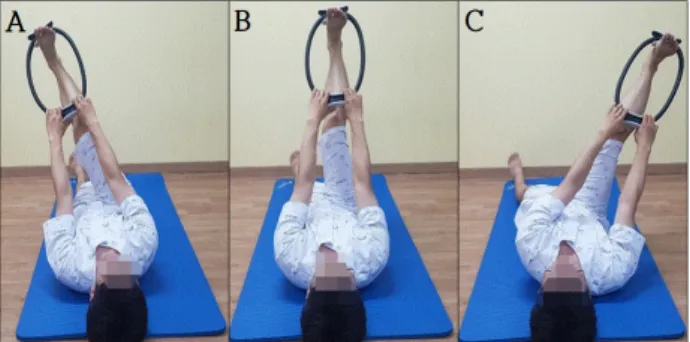
Then, starting from a supine hook-lying position, the knee joint was extended while the ankle joint was fixed using a pi-lates ring, and the exercise was performed to maintain the knee joint within the maximum range for 30 seconds. In or-der to selectively stretch the hamstrings, the stretches were performed for 3 sets of 5 times per leg in 3 directions of ad-duction, neutral, and abad-duction, and a rest period of 10 sec-onds was provided between each exercise set (Figure 1). Intervention was conducted three times a week for a total of 4 weeks, and post-tests were performed. Prior to the experi-ment, all participants fully understood the contents of this study and provided their informed consent. This study was
Figure 3. Measurement of hamstring flexibility. (A) Start. (B) Finish.
Figure 2. (A) Posturemeter. (B) Measurement of pelvic tilt angle. Figure 1. Hamstring stretching. (A) Biceps femoris. (B) Semitendi-nosus. (C) SemimembraSemitendi-nosus.
approved by the Institutional Bioethics Committee of the of Konyang University (IRB No. KYU-2019-332-01). Measurement and evaluation method
Pelvic tilt assessment
In order to group all participants according to the pelvic tilt measurements, the angle between the line connecting the right upper ASIS and the PSIS and the horizontal line was measured.
A posture meter (Posturemeter; Spomedic healthcare, Midvale, UT, 2004) was used to measure the pelvic tilt, which means a 2° inclination per scale (Figure 2). The nor-mal pelvic tilt range is considered to be 0° to 11.3°±4.3° [20].
Measurement of flexibility of the hamstring muscle
Active knee extension was assessed with the participants lying in supine position. To limit the movement of the pelvis, a belt was used to fix both ASISs.
With both knees flexed, the hip joint was flexed to 90 de-grees, and the legs were placed on a wooden box previously installed. In this position, the right knee was actively
extended. While stretching the knee, the ankle was in neutral position. For the measurement, a smartphone goniometer application (Clinometer + bubble level; Plaincode, Stephan-skirchen, Germany) was used, and it was fixed with a strap 2 cm above the malleolus of the ankle. With the goniometer set at 0°, the maximum active knee extension angle was measured 3 times and the average value was recorded (Figure 3). The intra-measure reliability of this test method wasreported as r=0.86-0.99, and the inter-measure reli-ability was r=0.76-0.89 [21,22].
Quadruple visual analogue scale
A 4-item QVAS was used to objectify the subjective back pain level reported by the patients. It consists of a total of 4 questions: current pain level, average pain level, mildest pain level, and most severe pain level [23]. Each question is marked with a ‘V’ on a 10 cm horizontal line. Each 1 cm is scored as 1 point, the scores of the 4 questions are summed and averaged, and the total score is calculated by multi-plying by 10.
In this test, the intra-measure reliability was reported as r=0.76-0.84 [24]. The higher the QVAS score, the more se-vere the pain.
Psychosocial level test
The FABQ level test used to evaluate the psychosocial level of the study participants. The FABQ is a questionnaire that evaluates the effect of fear related to pain on physical and occupational activities.
In this study, a questionnaire for fear avoidance response that was translated in Korean was used.
It consists of 5 items related to physical activity and 11 items related to occupational work, and out of a total of 16 items, 5 items (2, 8, 13, 14, 16) are not used when adding up after the questionnaire evaluation.This evaluation was cal-culated with a total score of 66, and the higher the score, the
Figure 4. (A) Sensbalance therapy cushion. (B) Setting for meas-uring pelvic mobility.
stronger the degree of fear avoidance reaction of the patient. The test retest reliability of this test was reported as r=0.95 [25].
Low back pain dysfunction level test
The KODI was used to evaluate the degree of dysfunction due to low back pain. The KODI consists of a total of 10 areas (pain level, personal hygiene, object lifting, walking, sitting, standing, sex life, sleeping, social activity, travel), and the level of discomfort or disability that the patient feels is indicated. The total score is 50 points, and the evaluation score of the patient was divided by the total score and con-verted into a percentage. The test-retest reliability of the KODI is high at r=0.9. The higher the score, the lower the level of functional performance due to back pain [26].
Pelvic mobility assessment
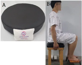
The STC (Sensbalance Therapy Cushion; Sensamove Inc., Groessen, Netherlands) was used in order to evaluate pelvic mobility. Data storage and processing were stored through the Pedalo-Sensamove-Balance-Test Pro software (Sensa-move Inc.). STC is a device that displays the position of the center of gravity on a monitor in real time. In this study, after placing the STC on a flat chair, the participant was instructed to sit comfortably, and a wooden box with an appropriate height was placed on the sole of the foot so that the leg could be bent 90°. While maintaining a comfortable sitting pos-ture, the Calibrate button was pressed to place the center of gravity displayed on the monitor in the center.
Next, in order to measure the range of motion of the pel-vis, the inclination of the pelvis in the front, back, left and right direction was instructed.
At this time, in order to minimize the movement of the trunk and induce pure movement of the pelvis, both hands were held at the bottom of the chair (Figure 4). The greater the mobility, the greater the distance of movement, and the measurement value increases.
This was performed with the monitor covered to prevent visual feedback. The test-retest reliability of this evaluation tool was reported as r=0.67-0.84 [27].
Statistical analysis
Descriptive statistics were used for the general character-istics of the study participants.
The Wilcoxon signed rank test was used to determine the pre- and post-intervention changes in the measurement vari-ables for each group, and it was judged that there was a
stat-istically significant difference when the p-value was 0.05. The Kruskal-Wallis test was used to compare the outcome variables between each group, and when the p-value was 0.05, it was judged as a statistically significant difference, and when the p-value was less than 0.01 using the Bonferroni correction by post hoc analysis, it was judged that there was one difference. The study analysis was performed using the PASW Statistics for Windows, Version 18.0 (SPSS Inc., Chicago, IL, USA).
Results
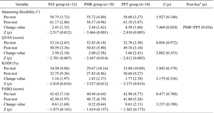
A total of 40 participants were classified into 12 pelvic an-terior tilt group, 18 pelvic midrange (PMR) group, and 10 pelvic posterior tilt (PPT) group. Among the general medi-cal characteristics of the participants, there was a significant difference between the three groups in mean pelvic incli-nation (p<0.01; Table 1). As a result of analyzing the differ-ences in hamstring flexibility, there was a difference in the amount of change between the groups, but the post-test re-sults were not statistically significant. The changes in ham-string flexibility before and after the intervention were sig-nificantly increased in all three groups (p<0.05).
As a result of analyzing the differences at each point of measurement of the QVAS, there was no significant differ-ence between the three groups, and in the analysis of changes within the group before and after the intervention, all three groups showed a significant decrease (p<0.05).
As a result of analyzing the differences in measurement time point of the KODI, there was no difference between
Table 2. Measurement of hamstring flexibility, QVAS, KODI, FABQ (N=40)
Variable PAT group (n=12) PMR group (n=18) PPT group (n=10) Z (p) Post-hoca (p)
Hamstring flexibility (°) Pre-test 58.75 (3.72) 55.72 (4.80) 58.60 (3.27) 3.927 (0.140) Post-test 61.17 (2.86) 58.17 (4.96) 63.10 (3.87) Change value 2.41 (1.35) 2.44 (1.62) 4.50 (1.60) 7.460 (0.024) PMR<PPT (0.026) Z (p) −2.517 (0.012) −3.466 (0.001) −2.810 (0.005) QVAS (score) Pre-test 53.16 (2.65) 52.83 (6.18) 52.70 (3.50) 0.056 (0.972) Post-test 50.59 (3.36) 50.83 (5.90) 49.10 (3.10) Change value 2.58 (2.10) 2.00 (2.58) 3.60 (2.41) 2.082 (0.353) Z (p) −2.701 (0.007) −2.447 (0.014) −2.812 (0.005) KODI (%) Pre-test 34.50 (9.80) 29.67 (10.16) 33.80 (10.09) 1.945 (0.378) Post-test 32.75 (9.26) 27.83 (8.86) 30.60 (9.27) Change value 1.16 (1.97) 1.83 (2.57) 1.77 (2.58) 2.179 (0.336) Z (p) −2.410 (0.016) −2.527 (0.012) −2.375 (0.018) FABQ (score) Pre-test 43.42 (7.14) 40.94 (6.64) 42.90 (4.77) 0.477 (0.788) Post-test 42.50 (5.97) 40.72 (6.79) 41.80 (5.26) Change value 0.61 (1.68) 0.22 (0.64) 0.61 (2.11) 3.237 (0.198) Z (p) −1.473 (0.141) −1.414 (0.157) −1.362 (0.173)
Values are presented as mean (SD).
PAT: pelvic anterior tilt, PMR: pelvic midrange PPT: pelvic posterior tilt, QVAS: quadruple visual analogue scale, KODI: Korean version of Oswestry disability index, FABQ: fear avoidance beliefs questionnaire.
aBonferroni correction.
Table 1. General characteristics of the participants (N=40)
Variable PAT group (n=12) PMR group (n=18) PPT group (n=10) Z (p) Post-hoca (p)
Sex (male/female) 6/6 14/4 6/4 2.524 (0.28)
Age (y) 34.25 (5.65) 36.00 (8.64) 35.20 (9.47) 0.317 (0.85)
Weight (kg) 62.41 (10.05) 63.88 (9.19) 64.00 (7.54) 0.585 (0.74)
Height (cm) 168.08 (8.88) 171.88 (9.84) 166.70 (6.70) 2.619 (0.27)
Pelvic tilt angle (°) 11.54 (0.58) 7.52 (1.26) 4.35 (0.88) 34.19 (0.001) PPT<PMR<PAT (0.007)
Values are presented as number only or mean (SD).
PAT: pelvic anterior tilt, PMR: pelvic midrange, PPT: pelvic posterior tilt.
aBonferroni correction.
groups. As a result of analyzing the changes within the groups before and after the intervention, all three groups showed a significant decrease (p<0.05).
As a result of analyzing the differences between groups at each point of measurement of FABQ and changes within the groups before and after intervention, there was no sig-nificant difference (Table 2).
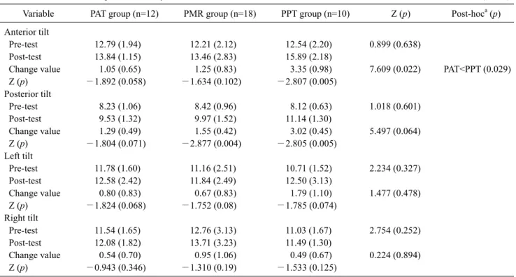
As a result of analyzing the difference between the groups in anterior pelvic tilt range, there was a difference between groups in the amount of change, but the post-test results
were not statistically significant. As a result of analyzing the changes in the anterior tilt range within the group before and after the intervention, there was a significant increase in the PPT group (p<0.05).
As a result of analyzing the differences between groups in PPT range, there was no significant difference. As a result of analyzing the changes within the groups before and after the intervention, there was a significant increase in the PMR and PPT groups (p<0.05).
Table 3. Measurement of pelvic mobility (N=40)
Variable PAT group (n=12) PMR group (n=18) PPT group (n=10) Z (p) Post-hoca (p)
Anterior tilt
Pre-test 12.79 (1.94) 12.21 (2.12) 12.54 (2.20) 0.899 (0.638)
Post-test 13.84 (1.15) 13.46 (2.83) 15.89 (2.18)
Change value 1.05 (0.65) 1.25 (0.83) 3.35 (0.98) 7.609 (0.022) PAT<PPT (0.029)
Z (p) −1.892 (0.058) −1.634 (0.102) −2.807 (0.005) Posterior tilt Pre-test 8.23 (1.06) 8.42 (0.96) 8.12 (0.63) 1.018 (0.601) Post-test 9.53 (1.32) 9.97 (1.52) 11.14 (1.30) Change value 1.29 (0.49) 1.55 (0.42) 3.02 (0.45) 5.497 (0.064) Z (p) −1.804 (0.071) −2.877 (0.004) −2.805 (0.005) Left tilt Pre-test 11.78 (1.60) 11.16 (2.51) 10.71 (1.52) 2.234 (0.327) Post-test 12.58 (2.42) 11.84 (2.49) 12.50 (3.13) Change value 0.80 (0.83) 0.67 (0.83) 1.79 (1.10) 1.477 (0.478) Z (p) −1.824 (0.068) −1.752 (0.08) −1.785 (0.074) Right tilt Pre-test 11.54 (1.65) 12.76 (3.13) 11.03 (1.67) 2.754 (0.252) Post-test 12.08 (1.82) 13.71 (3.23) 11.49 (1.30) Change value 0.54 (0.70) 0.95 (1.06) 0.49 (0.67) 0.224 (0.894) Z (p) −0.943 (0.346) −1.310 (0.19) −1.533 (0.125)
Values are presented as mean (SD).
PAT: pelvic anterior tilt, PMR: pelvic midrange, PPT: pelvic posterior tilt.
a
Bonferroni correction.
in left and right pelvic lateral tilt range and the changes with-in the groups before and after the with-intervention, there was no significant difference (Table 3).
Discussion
This study was conducted to compare the effectiveness of the hamstring muscle stretching according to the character-istics of pelvic displacement, and it was found that ham-string flexibility was significantly increased in all three groups.
A study by Handel et al. [28] showed that stretching the hamstrings increased the range of motion of the knee joint by 6.7°, and Bandy and Irion [29] stated that hamstring stretches performed 5 times a week for 6 weeks was effec-tive in increasing the range of motion of the knee joint. In a study by Kim and Kim [30], it was confirmed that the imme-diate effect of stretching the hamstring muscles caused de-creased muscle stiffness and a significant increase in the range of motion of the knee joint. According to a study by Jo et al. [31], the manual extension of the hamstring muscle was ef-fective in increasing the flexibility of the hamstrings while maintaining pelvic neutrality.
Evidence has been suggested through several studies on the mechanism of increasing the range of motion through stretching.
Bandy et al. [32] reported that the perception of the streth-ing was changed due to the decrease in the sensitivity of the pain receptor after stretching, resulting in an increase in tol-erance to stretching, and Halbertsma et al. [33] stated that this was due to increased tolerance to discomfort. Magnusson et al. [34] argued that the range of motion was increased due to changes in perception of the stretching and the effect of con-trolling pain through free nerve endings in muscles and joints. It is believed that this study also showed an increase in the range of motion of the knee joint as a result of these physiological changes.
It was confirmed that the QVAS for evaluating the sub-jective back pain level significantly decreased in all three groups. According to Lee’s study [35], the degree of ham-string flexibility is a factor that can affect the pain level of patients with chronic back pain by reducing pain. In a study by Han et al. [36], a significant increase in hamstring flexi-bility and a significant decrease in pain level were applied to workers suffering from low back pain after applying the hamstring stretches with pelvic control three times a week
for 6 weeks.
In this study, an increase of an average of 3.11° in ham-string flexibility was observed in all three groups. It is thought that the QVAS, which evaluates pain levels, was sig-nificantly reduced in this regard.
It was confirmed that the KODI, used for evaluating dys-function due to low back pain, significantly decreased in all three groups. In a study by Monga and Singh [37], it was found that there was a significant negative correlation as a result of confirming the relationship between KODI levels and the sit-and reach test in patients with chronic low back pain. According to previous studies, the V-sit and reach test was suggested as a representative assessment method for hamstring flexibility [38], and it was confirmed that the range of the sit-and reach test was significantly increased as an immediate effect of stretching the hamstrings [39]. In this study, it was confirmed that hamstring flexibilty was in-creased through stretching, and the results of previous stud-ies were reflected, and it is thought that the KODI score was significantly reduced. As a result of this study, there was a clear difference in the amount of change in pain levels and hamstring flexibility in the pelvic posterior inclination group. However, the changes in KODI levels was similar between the three groups.
Fritz and Irrgang [40] argued that there is no one-to-one correlation between pain and activity levels (disability in-dex) because disability level measures the difficulty of ac-tivities during daily life. It is believed that the difference in the amount of change between groups was not large because the participants that were recruited were familiar with daily living by activities by visiting the outpatient clinic of the hospital.
The FABQ used to assess psychosocial levels showed in-significant results in all three groups. This raises the ques-tion of whether the rehabilitaques-tion program for the recovery of the psychosocial level of patients with chronic low back pain should focus on hamstring flexibility. Similarly, a study by Marshall et al. [41] showed that there was no significant correlation between hamstring flexibilitys and the FABQ in patients with chronic low back pain. However, according to a study by Kim et al. [42], there was no significant correla-tion between the pain levels of patients low back pain and the FABQ-vocational activity item, but the FABQ-physical ac-tivity item showed a significant positive correlation. In addi-tion, according to a study by Nagrale et al. [43], the applica-tion of a rehabilitaapplica-tion program including the hamstring stretching in patients low back pain brought a significant
change in the FABQ level. As such, it is unclear whether hamstring flexibility should be considered in back pain re-habilitation programs to recover the psychosocial level, as claims on the association between the hamstring flexibility and FABQ have not yet been clear.
As a result of evaluating pelvic mobility, there was a sig-nificant difference in the anterior and posterior inclination ranges in the posterior tilt group, and a significant difference in the posterior tilt range in the pelvic neutral range group.
Overactivation of the hamstring muscle inhibits the activ-ity of the gluteus maximus muscle, thereby changing the muscle mobilization pattern and transforming the move-ment of the pelvis [44]. The long-term contraction of the hamstrings causes a change in the motion of the lumbar-pel-vic rhythm, among which, particularly, the anterior tilt of the pelvis is limited [45].
Based on these contents, in this study, it is thought that in-creasing flexibility through stretching the hamstring mus-cles had an effect on the increase in the range of the anterior and posterior pelvic tilt.
However, among the variables for evaluating pelvic mo-bility, the range of left and right pelvic tilt did not show any significant change.
This is thought to be the result of reflecting the difference in the motion surface in which the action occurs depending on the attachment site of the muscle. Since the action of the hamstrings occur in the sagittal plane and the anterior and posterior inclination of the pelvis also occurs in the sagittal plane, a significant result was shown, and since the left and right lateral inclination occurred in the frontal plane it was thought that an insignificant result was shown.
When the results of this study were combined, there was no statistically significant difference between the three groups in hamstring flexibility, QVAS, and pelvic anterior and posterior inclination variables, but a larger amount of change in the posterior pelvic inclination group was confirmed. This is thought that there was a larger change in the PPT group because the hamstring muscle shortening was strong due to the coupling action in the pelvis.
However, in the pretest of this study, the difference in the number of participants did not show a significant difference in hamstring flexibility between the three groups.
This study had the following limitation in explaining the results of this study. In evaluating pelvic mobility, the chair was held with both hands to minimize the movement of the trunk. At this time, there were many participants who had stated that their posture was unnatural because the arm
length of each participant was not taken into account, which could affect the evaluation result.
In the future, if a device that reliably fixes the trunk and does not interfere with pelvic movements by supplementing the limitations of this study is developed and added, the methods used render greater accuracy and be more widely applied.
In conclusion, this study was conducted to investigate for the effect of self-stretching of the hamstring muscles accord-ing to the pelvic inclination of patients with low back pain. As a result, although not statistically significant, there was a difference in the amount of change between the three groups. In particular, it was confirmed that the PPT group showed a relatively large amount of change in hamstring flexibility, QVAS, and pelvic anterior and posterior in-clination variables.
Through this, self-stretching of the hamstrings can be ex-pected to have a great effect when the pelvis is inclined posteriorly. Furthermore, therapists can expect different ef-fects even when applying interventions to patients with low back pain, and can be applied in consideration of the partic-ipant’s pelvic displacement.
Conflict of Interest
The authors declared no potential conflicts of interest with respect to the authorship and/or publication of this article.
References
1. Panjabi M, Yamamoto I, Oxland T, Crisco J. How does posture affect coupling in the lumbar spine? Spine 1989;14:1002-11. 2. Lee KW. Therapeutic exercise in low back pain. J Korean Acad
Rehabil Med 1995;19:1.
3. Myers TW. The superficial back line. In: Myers TW, editor. Anatomy trains. 2nd ed. Edinburgh: Churchill Livingstone/ Elsevier; 2009. p. 73-95.
4. Oh YT. Effect of hold-relax technique for college students with hamstring shortening. J Korean Soc Phys Med 2013;8:433-41. 5. Congdon R, Bohannon R, Tiberio D. Intrinsic and imposed
ham-string length influence posterior pelvic rotation during hip flexion. Clin Biomech (Bristol, Avon) 2005;20:947-51. 6. Hartig DE, Henderson JM. Increasing hamstring flexibility
de-creases lower extremity overuse injuries in military basic trainees. Am J Sports Med 1999;27:173-6.
7. Jones MA, Stratton G, Reilly T, Unnithan VB. Biological risk in-dicators for recurrent non-specific low back pain in adolescents. Br J Sports Med 2005;39:137-40.
8. Esola MA, McClure PW, Fitzgerald GK, Siegler S. Analysis of lumbar spine and hip motion during forward bending in subjects
with and without a history of low back pain. Spine 1996;21:71-8. 9. Rok S, Wytrazek M, Bilski B. Efficacy of therapeutic exercises in low back pain surveyed in a group of nurses. Med Pr 2005;56: 235-9.
10. Halbertsma JP, van Bolhuis AI, Göeken LN. Sport stretching: ef-fect on passive muscle stiffness of short hamstrings. Arch Phys Med Rehabil 1996;77:688-92.
11. López-Miñarro PA, Alacid Cárceles F, y Muyor Rodríguez JM. Comparison of spinal curvatures and hamstring extensibility be-tween paddlers and runners. Rev Int Med Cienc Act Fís Deporte 2009;9:379-92.
12. López-Miñarro PA, Alacid F, Rodríguez-García PL. Comparison of sagittal spinal curvatures and hamstring muscle extensibility among young elite paddlers and non-athletes. Int Sport Med J 2010;11:301-12.
13. López-Miñarro PA, Alacid F. Influence of hamstring muscle ex-tensibility on spinal curvatures in young athletes. Sci Sports 2010;25:188-93.
14. Shamsi M, Shahsavari S, Safari A, Mirzaei M. A randomized clinical trial for the effect of static stretching and strengthening exercise on pelvic tilt angle in LBP patients. J Bodyw Mov Ther 2020;24:15-20.
15. Hansson T, Bigos S, Beecher P, Wortley M. The lumbar lordosis in acute and chronic low-back pain. Spine 1985;10:154-5. 16. Evcik D, Yücel A. Lumbar lordosis in acute and chronic low
back pain patients. Rheumatol Int 2003;23:163-5.
17. Kim BG, Yi SJ, Kang JD, Park RJ. Difference of lumbar lordosis in patients with low back pain and controls. J Kor Soc Phys Ther 2000;12:185-90.
18. Christie HJ, Kumar S, Warren SA. Postural aberrations in low back pain. Arch Phys Med Rehabil 1995;76:218-24.
19. Kang KB, Ahn YJ, Kim YJ, Kim YB, Park SC. Changes in sag-ittal spinopelvic parameters according to pelvic incidence in asymptomatic old Korean men. J Korean Soc Spine Surg 2011; 18:223-9.
20. Levine D, Whittle MW. The effects of pelvic movement on lum-bar lordosis in the standing position. J Orthop Sports Phys Ther 1996;24:130-5.
21. Cameron DM, Bohannon RW. Relationship between active knee extension and active straight leg raise test measurements. J Orthop Sports Phys Ther 1993;17:257-60.
22. Reurink G, Goudswaard GJ, Oomen HG, Moen MH, Tol JL, Verhaar JA, et al. Reliability of the active and passive knee ex-tension test in acute hamstring injuries. Am J Sports Med 2013; 41:1757-61.
23. Yang JH. A study of pain, depression and self-efficacy according to the classifications of pain among chronic pain patients. J Korean Acad Adult Nurs 2004;16:202-10.
24. Boonstra AM, Schiphorst Preuper HR, Reneman MF, Posthumus JB, Stewart RE. Reliability and validity of the visual analogue scale for disability in patients with chronic musculos-keletal pain. Int J Rehabil Res 2008;31:165-9.
25. Joo MK., Kim TY, Kim JT, Kim SY. Reliability and validity of the Korean version of the fear-avoidance beliefs questionnaire. Phys Ther Korea 2009;16:24-30.
26. Kim DY, Lee SH, Lee HY, Lee HJ, Chang SB, Chung SK, et al. Validation of the Korean version of the oswestry disability index. Spine 2005;30:E123-7.
27. Jung SH, Park DS. Reliability and validity of the measurement of pelvic movement in low back pain patients using cushion sensor in sitting position. J Korean Soc Phys Med 2020;15:83-91. 28. Handel M, Horstmann T, Dickhuth HH, Gülch RW. Effects of
contract-relax stretching training on muscle performance in athletes. Eur J Appl Physiol Occup Physiol 1997;76:400-8. 29. Bandy WD, Irion JM. The effect of time on static stretch on the
flexibility of the hamstring muscles. Phys Ther 1994;74:845-50; discussion 850-2.
30. Kim JH, Kim TH. Immediate effects of stretching on hamstring stiffness. J Kor Soc Phys Ther 2010;22:1-7.
31. Jo M, Kwon N, Park S, Seo D, Jung J, Ha M, et al. Effects of the stretching exercise of hamstring muscle on flexibility and foot pressure in subjects with and without pelvis neutral position. J Korean Soc Integr Med 2016;4:31-9.
32. Bandy WD, Irion JM, Briggler M. The effect of static stretch and dynamic range of motion training on the flexibility of the ham-string muscles. J Orthop Sports Phys Ther 1998;27:295-300. 33. Halbertsma JP, Mulder I, Göeken LN, Eisma WH. Repeated
pas-sive stretching: acute effect on the paspas-sive muscle moment and extensibility of short hamstrings. Arch Phys Med Rehabil 1999; 80:407-14.
34. Magnusson SP, Simonsen EB, Aagaard P, Dyhre-Poulsen P, McHugh MP, Kjaer M. Mechanical and physical responses to stretching with and without preisometric contraction in human skeletal muscle. Arch Phys Med Rehabil 1996;77:373-8. 35. Lee MH. Effects of the low back exercise program including a
hamstring stretching on lumbar muscle strength and subjective pain in chronic low back pain patients [Master thesis]. Seoul: Korea National Sport University; 2007.
36. Han HI, Choi HS, Shin WS. Effects of hamstring stretch with
pelvic control on pain and work ability in standing workers. J Back Musculoskelet Rehabil 2016;29:865-71.
37. Monga A, Singh S. Impact of back muscle functions, spinal range of motion and fear avoidance beliefs on disability in chron-ic non-specifchron-ic low back pain. Hum Bio Rev 2013;2:46-55. 38. Attrey P, Yadav M, Singh S. Relationship between passive
straight leg raising test and V -sit and reach test in measuring the hamstring flexibility. Indian J Phys Educ Sports Appl Sci 2017; 7:23-30.
39. López-Miñarro PA, Muyor JM, Belmonte F, Alacid F. Acute ef-fects of hamstring stretching on sagittal spinal curvatures and pelvic tilt. J Hum Kinet 2012;31:69-78.
40. Fritz JM, Irrgang JJ. A comparison of a modified Oswestry Low Back Pain Disability Questionnaire and the Quebec Back Pain Disability Scale. Phys Ther 2001;81:776-88.
41. Marshall PW, Mannion J, Murphy BA. Extensibility of the ham-strings is best explained by mechanical components of muscle contraction, not behavioral measures in individuals with chronic low back pain. PM R 2009;1:709-18.
42. Kim S, Kim JH, Kim YL, Lee SM. Comparison of pain, disorder, back performance, and psychological factors in patients with low back pain and radicular pain. J Phys Ther Sci 2018;30:127-31. 43. Nagrale AV, Patil SP, Gandhi RA, Learman K. Effect of slump
stretching versus lumbar mobilization with exercise in subjects with non-radicular low back pain: a randomized clinical trial. J Man Manip Ther 2012;20:35-42.
44. Vogt L, Pfeifer K, Banzer W. Neuromuscular control of walking with chronic low-back pain. Man Ther 2003;8:21-8.
45. Kim GC, Hwang BG. Kinetic analysis on the lumbar at the trunk flexion according to the degree of hamstring flexibility of healthy adult. J Korean Soc Phys Med 2012;7:501-7.