Inactivation of Glycogen Synthase Kinase-3
 Is Required for
Osteoclast Differentiation
*
□SReceived for publication, May 2, 2011, and in revised form, September 19, 2011Published, JBC Papers in Press, September 23, 2011, DOI 10.1074/jbc.M111.256768 Hyun Duk Jang‡1,2, Ji Hye Shin‡1,3, Doo Ri Park‡3, Jin Hee Hong§, Kwiyeom Yoon‡, Ryeojin Ko‡3, Chang-Yong Ko¶, Han-Sung Kim¶, Daewon Jeong储, Nacksung Kim**, and Soo Young Lee‡ ‡‡4
From the‡Division of Life and Pharmaceutical Sciences, Center for Cell Signaling and Drug Discovery Research,‡‡Department of Bioinspired Science and the Department of Life Science, Ewha Womans University, Seoul 120-750, the§Center for Cell Dynamics and Department of Physics, Korea University, Seoul 136-701, the¶Department of Biomedical Engineering, College of Health Science, Institute of Medical Engineering, Yonsei University, Wonju 220-701, the储Department of Microbiology and the Aging-associated Disease Research Center, Yeungnam University College of Medicine, Daegu 705-717, and the **Medical Research Center for Gene Regulation, Chonnam National University Medical School, Gwangju 501-746, Korea
Background:Bone homeostasis is maintained by balancing the activities of bone-resorbing osteoclasts and bone-forming
osteoblasts.
Results: GSK-3 is inactivated by receptor activator of NF-B ligand stimulation via serine phosphorylation during
osteoclastogenesis.
Conclusion:GSK-3is crucial for receptor activator of NF-B ligand-mediated signaling as a negative regulator of osteoclast
differentiation.
Significance:GSK-3acts as a novel negative regulator of osteoclast biology.
Glycogen synthase kinase-3(GSK-3) is a serine/threonine
kinase originally identified as a regulator of glycogen
deposi-tion. Although the role of GSK-3in osteoblasts is well
charac-terized as a negative regulator of-catenin, its effect on
oste-oclast formation remains largely unidentified. Here, we show
that the GSK-3inactivation upon receptor activator of NF-B
ligand (RANKL) stimulation is crucial for osteoclast
differenti-ation. Regulation of GSK-3activity in bone marrow
macro-phages by retroviral expression of the constitutively active
GSK-3 (GSK3-S9A) mutant inhibits RANKL-induced
osteoclastogenesis, whereas expression of the catalytically
inac-tive GSK-3 (GSK3-K85R) or small interfering RNA
(siRNA)-mediated GSK-3 silencing enhances osteoclast
for-mation. Pharmacological inhibition of GSK-3 further
con-firmed the negative role of GSK-3in osteoclast formation. We
also show that overexpression of the GSK3-S9A mutant in
bone marrow macrophages inhibits RANKL-mediated NFATc1
induction and Ca2ⴙoscillations. Remarkably, transgenic mice
expressing the GSK3-S9A mutant show an osteopetrotic
phe-notype due to impaired osteoclast differentiation. Further, oste-oclast precursor cells from the transgenic mice show defects in expression and nuclear localization of NFATc1. These findings
demonstrate a novel role for GSK-3in the regulation of bone
remodeling through modulation of NFATc1 in RANKL signaling.
Bone homeostasis is maintained by balancing the activities of bone-resorbing osteoclasts and bone-forming osteoblasts, and imbalances in bone remodeling cause a variety of bone diseases (1, 2). Two cytokines are essential for osteoclast differentiation: receptor activator of NF-B ligand (RANKL),5which belongs to
the tumor necrosis factor (TNF) family, and macrophage colo-ny-stimulating factor (M-CSF) (3, 4). RANKL mediates its sig-nal transduction through binding to RANK, which is expressed by osteoclast precursors. RANK recruits the adapter molecule, TNF receptor-associated factor 6 (TRAF6), which activates NF-B and mitogen-activated protein kinases (MAPKs), including c-Jun N-terminal kinase and p38 (5, 6). RANKL-in-duced activation of NF-B and c-Fos is required for initial expression of the key transcription factor, NFATc1 (7, 8).
NFATc1 is an NFAT family member, which is activated by the Ca2⫹/calmodulin-regulated phosphatase calcineurin (9). In osteoclast precursors, calcium signaling activates the existing NFATc1, and an AP-1 complex containing c-Fos may cooper-ate with NFATc1 to trigger NFATc1 autoamplification (8). Autoamplification enables robust induction of NFATc1 on activation of RANKL signaling (10). Upon activation, NFATc1 proteins are dephosphorylated by calcineurin, and then they translocate from the cytoplasm into the nucleus, where they direct transcription of osteoclast-specific genes, such as TRAP,
cathepsin K, Oscar, and Atp6v0d2 (11, 12). Nuclear export of NFAT members is facilitated by phosphorylation, and several
*This work was supported by National Research Foundation of Korea (NRF) grants funded by the Korea Government (Ministry of Education, Science, and Technology; Grants R0A-2008-000-20001-0; R31-2008-000-10010-0; 2011-0006244; and 2010-0020577).
□S
The on-line version of this article (available at http://www.jbc.org) contains supplemental Figs. S1–S4.
1Both authors contributed equally to this work.
2Supported by Research Professor Grant 2010 of Ewha Womans University. 3Supported by the second stage of the Brain Korea 21 Project.
4To whom correspondence should be addressed: Division of Life and Phar-maceutical Sciences, Dept. of Bioinspired Science, Department of Life Sci-ence, Ewha Womans University, Seoul 120-750, Korea. Tel.: 82-2-3277-3770; Fax: 82-2-3277-3760; E-mail: [email protected].
5The abbreviations used are: RANKL, receptor activator of NF-B ligand; GSK-3, glycogen synthase kinase-3; NFAT, nuclear factor of activated T-cells; BMM, bone marrow macrophage; TRAP, tartrate-resistant acid phospha-tase; MNC, multinucleated cells; Tg, transgenic.
at Ewha Medical Library on July 6, 2016
http://www.jbc.org/
Downloaded from
at Ewha Medical Library on July 6, 2016
http://www.jbc.org/
Downloaded from
at Ewha Medical Library on July 6, 2016
http://www.jbc.org/
Downloaded from
at Ewha Medical Library on July 6, 2016
http://www.jbc.org/
Downloaded from
at Ewha Medical Library on July 6, 2016
http://www.jbc.org/
Downloaded from
at Ewha Medical Library on July 6, 2016
http://www.jbc.org/
Downloaded from
at Ewha Medical Library on July 6, 2016
http://www.jbc.org/
Downloaded from
at Ewha Medical Library on July 6, 2016
http://www.jbc.org/
kinases have been suggested to regulate NFAT function, including GSK-3 (13), CK1 (14), p38 (15), and JNK1 (16).
Glycogen synthase kinase-3 (GSK-3) is a serine/threonine kinase originally identified for its role in the regulation of gly-cogen deposition. GSK-3 has two isoforms, GSK-3␣ and GSK-3 (17), both of which are implicated in many different biological processes including metabolism, transcription, translation, cell growth, and apoptosis (18). With respect to transcription, GSK-3 regulates a wide variety of transcription factors, including cyclin D1, c-Jun, NFATc, and-catenin (13, 19, 20). In resting cells, GSK-3 is constitutively active, and its activity is inhibited by various kinases via phosphorylation of a serine residue, Ser-21 in GSK-3␣ and Ser-9 in GSK-3 in response to different stimuli (21). Serine phosphorylation on GSK-3 blocks the access of substrate to the GSK-3 catalytic domain, thus inhibiting substrate phosphorylation (22).
Of the two isoforms of GSK-3, GSK-3 is a more likely can-didate for being an NFATc1 kinase, influencing NFATc1 sub-cellular localization through phosphorylation (13). However, the significance of the ability of GSK-3 to regulate NFATc1 during osteoclastogenesis has not yet been demonstrated. In addition, because GSK-3-deficient mice die in utero (23), the
in vivorelevance of GSK-3 in osteoclast precursors has not been well characterized. Therefore we investigated the role of GSK-3 in RANKL-mediated osteoclast differentiation and also clarified the relevance of GSK-3 and NFATc1. In addi-tion, to understand the physiological role of GSK-3 in vivo, we generated transgenic mice that express a constitutively active GSK-3 (GSK3-S9A) mutant under the control of the mouse tartrate-resistant acid phosphatase (TRAP) gene promoter, which is specific to the osteoclast lineage. Our results demon-strate that GSK-3 suppresses osteoclastogenesis through reg-ulation of NFATc1 expression. We also show that GSK-3 is inactivated by RANKL stimulation via serine phosphorylation. Remarkably, transgenic mice expressing the GSK3-S9A mutant show a defect in NFATc1 and osteoclast-specific gene expression and therefore show a severe osteopetrotic pheno-type. Based on these results, we suggest that GSK-3 is crucial for RANKL-mediated signaling as a negative regulator of oste-oclast differentiation.
EXPERIMENTAL PROCEDURES
Cells and Reagents—Primary cells used in this study, includ-ing bone marrow macrophages (BMMs) and osteoblasts, were generated from murine bone marrow precursors of 4 – 6-week-old C57BL/6 mice (The Jackson Laboratory) and cultured in ␣-minimum essential medium (␣-MEM; HyClone, Logan, UT) supplemented with 10% fetal bovine serum (FBS) and antibiot-ics. RAW264.7 cells were maintained in Dulbecco’s modified Eagle’s medium (DMEM; HyClone) with 10% FBS and antibi-otics. To generate stable cell lines, RAW264.7 cells were infected with recombinant virus expressing GSK3-S9A and then selected with puromycin (2 g/ml) after 2 weeks of growth. Kenpaullone was purchased from Biomol International (Plymouth Meeting, PA). SB216763 and SB415286 were from Sigma.
Protein Analysis—For Western blot analysis, equal amounts of cell lysates harvested at the indicated conditions were
sub-jected to analysis using specific antibodies against NFATc1, phospho-NFATc1 (Santa Cruz Biotechnology), GSK-3␣/ (Invitrogen), GSK-3, phospho-GSK-3 (Cell Signaling), OSCAR (R&D), hemagglutinin (HA; Roche Applied Science), and-actin (Sigma). Anti-Atp6v0d2 (24) antibody was kindly provided by Y. Choi (University of Pennsylvania).
Retroviral Infection—HA epitope-tagged cDNAs of wild-type GSK-3 and two mutants, catalytically inactive GSK-3 (GSK3-K85R) and constitutively active GSK-3 (GSK3-S9A) mutant plasmids, were kindly provided by J. Chung (Seoul National University, Korea). V5 epitope-tagged cDNAs of GSK-3␣ and catalytically inactive GSK-3␣ (GSK3␣-K148A) plasmids were kindly provided by J. R. Woodgett (Samuel Lunenfeld Research Institute). cDNAs of wild-type and mutant GSK-3 or GSK-3␣ were cloned into the retroviral vector, pMX-puro, as described previously (25). The plasmids were transfected into PLAT-E cells using LipofectamineTM 2000
(Invitrogen), and the supernatant was collected 24 –36 h after transfection. The pMX-puro vector and PLAT-E cells were kindly provided by T. Kitamura (University of Tokyo, Japan). The supernatant including retroviruses was used to infect BMMs or RAW 264.7 cells as described previously (25). BMMs were cultured with virus supernatant for 1 day and then changed to the medium with M-CSF (100 ng/ml) and puromy-cin (2 g/ml) for 2 days. Puromycin-resistant BMMs were used for osteoclast differentiation and Ca2⫹ measurement experiments.
In Vitro Osteoclast Differentiation—Osteoclasts were pre-pared from bone marrow cells using a standard method (26). In brief, bone marrow cells were cultured with 30 ng/ml M-CSF (R&D Systems) for 3 days to obtain osteoclast precursor cells of the monocyte/macrophage lineage. The precursors were cul-tured with 30 ng/ml M-CSF and 100 ng/ml RANKL for the indicated time periods. The various GSK-3 inhibitors including kenpaullone, SB216763, and SB415286 were added at the time of RANKL addition. TRAP⫹cells with more than three nuclei or cells larger than 100m in diameter that contained more than 20 nuclei were counted as TRAP⫹ multinucleated cells (MNCs). Co-culture experiments were performed as described previously (27). In brief, primary calvarial osteoblast precursors were obtained from newborn C57BL/6 mice by digesting iso-lated calvariae with 0.8 units/ml dispase (Roche Applied Sci-ence, Penzberg, Germany) and 0.1% collagenase (Sigma) and incubating the dissociated cells in ␣-MEM (HyClone). The BMMs (2 ⫻ 105) infected with retrovirus expressing either
GSK-3 or its mutants were seeded on top of the calvarial osteoblast precursors (2⫻ 104) on each well of 48-well plates
and cultured for 7 days in the presence of 10 nM1,25(OH)2D3
(Sigma) and 1Mprostaglandin E2 (Sigma). Osteoclasts were
identified by TRAP staining and counted.
Transfection and Reporter Assay—Stable RAW264.7 cells were seeded at a density of 105cells/well in a 12-well plate 1 day
prior to transfection using LipofectamineTM2000 (Invitrogen)
according to the manufacturer’s protocol. Luciferase reporter constructs driven by the NFAT-responsive elements were described previously (28). Luciferase activity was measured by the luciferase assay system (Promega) and normalized relative to-galactosidase activity. The data were obtained from three
at Ewha Medical Library on July 6, 2016
http://www.jbc.org/
independent transfections and presented as the -fold induction in luciferase activity (mean⫾ S.D.) relative to the control.
GSK-3 Activity Measurements—For measurements of
GSK-3 activity, BMMs were stimulated by RANKL at the indi-cated times and immunoprecipitated by anti-GSK-3 antibody. The immunoprecipitated beads were washed with kinase buffer (20 mMTris, pH 7.5, 5 mMMgCl2, 1 mMdithiothreitol) and then
resuspended in 30l of kinase buffer containing 250 MATP,
1.4Ci of [␥-32P]ATP, and 50
Mphospho-glycogen synthase
peptide (Upstate Biotech Millipore, Charlottesville, VA). The samples were incubated at 30 °C for 30 min and spotted on P81 filter paper circles (Whatman International Ltd., Maidstone, UK). The filter paper was dried, washed three times in 0.5% phosphoric acid for 1 h, and finally washed with 95% ethanol for 10 min. The air-dried filter was counted by scintillation coun-ter, and activity was normalized to the amount of GSK-3, as determined by immunoblotting.
Immunofluorescence Staining—BMMs were plated on
poly-L-lysine-coated coverslips in 12-well plates and treated with RANKL and M-CSF for 3 days. Cells were then washed with phosphate-buffered saline (PBS), fixed with 3.8% paraformal-dehyde in PBS, and permeabilized by incubation for 10 min with 0.1% Triton in PBS. The cells were blocked in 4% bovine serum albumin (BSA)-PBS for 1 h at room temperature. The cells were incubated with anti-NFATc1 antibody overnight, washed, and stained with Alexa Fluor 488-conjugated anti-mouse immunoglobulin (IgG) antibody (Alexa Fluor 488 goat anti-mouse IgG, Invitrogen) for 1 h. The coverslips were washed three times with PBS and then incubated with DAPI (4⬘,6-diamidino-2-phenylindole; Sigma) for 5 min. After wash-ing three times, the coverslips were mounted on slides and visu-alized using an LSM 510 META (Carl Zeiss) confocal microscope.
RNA Interference—Custom SMARTpool plus small interfer-ing RNA (siRNA) to target mouse GSK-3 (catalogue number M-041080) was designed and synthesized by Dharmacon (Lafayette, CO). siRNA (10 nmol) was transfected into BMMs using LipofectamineTM 2000 (Invitrogen) according to the
manufacturer’s protocol. After transfection, BMMs were cul-tured with M-CSF and RANKL for 4 days and then differenti-ated into osteoclasts.
Measurement of Intracellular Ca2⫹—Ca2⫹ measurements
were performed as described previously (10). Infected BMMs were incubated with M-CSF and RANKL for 3 days and then incubated in the presence of 5Mfluo-4 acetoxymethyl
(fluo-4AM; Invitrogen), 5Mfura red AM (Invitrogen), and 0.05%
plutonic F127 for 30 min. Cells were washed twice and post-incubated in DMEM with 10 ng/ml M-CSF for 20 min. For measurement of [Ca2⫹]
c(cytosolic Ca
2⫹concentration), single
cells were viewed with a laser-scanning confocal system (Flu-oView 500, Olympus, Tokyo, Japan) attached to an upright microscope (BX51WI, Olympus). An argon laser (488 nm) was used for excitation, a green emission filter (505–525 nm) was used for fluo-4, and a red emission filter (⬍660 nm) was used for fura red to observe the fluorescent images. The ratio of the fluorescence intensity of fluo-4 to fura red was calculated. The maximum intensity of [Ca2⫹]
cwas obtained with the addition
of 10Mionomycin at the end of each experiment. The ratio of
increase from the basal level was expressed as the percentage of maximum ratio increase.
Generation of Transgenic Mice—The constitutively active GSK-3 (GSK3-S9A) mutant cDNA was fused to the mouse TRAP gene promoter as described previously (29, 30). For gen-erating transgenic mice, we used the standard pronuclear injec-tion method with C57BL/6 mice (The Jackson Laboratory). Genomic DNA isolated from the tail was analyzed by polymer-ase chain reaction (PCR) using the specific primers (GT-F, 5
⬘-TAGCCATCAACAGCCGTCAGT; GT-R, 5
⬘-CTTCTGCCC-CAGAGAATAAAG; GP-F, 5
⬘-CAGGGTACAGTTTAGAAT-GGG; GP-R, 5⬘-GTACTAGGCAGACTGTGTAAAG) to
detect the transgene. All the mouse experiments were per-formed with 4 – 6-week-old mice under the animal protocol approved by the Animal Care Committee of the Ewha Labora-tory Animal Genomics Center.
Bone Histomorphometry and Microcomputed Tomography Analysis—Bones were fixed in 10% formaldehyde, decalcified in 0.5MEDTA, pH 7.4, embedded in paraffin, and then cut into 4-m sections. Hematoxylin and eosin (H&E) or TRAP stain-ing was performed accordstain-ing to a standard protocol (24). The histomorphometric data were analyzed by Osteomeasure XP (OsteoMetrics Inc.). Quantitative microcomputed tomography was performed with Skyscan 1076 (Skyscan N.V.). The data from scanned slices were used for the three-dimensional anal-ysis to calculate femoral morphometric parameters by CT-AN 1.10 (Skyscan N.V.). The nomenclature and units were accord-ing to the recommendation of the Nomenclature Committee of the American Society for Bone and Mineral Research (31).
RANKL-induced Bone Loss—Five-week-old female mice were administered with a local calvarial injection of RANKL at 2 mg/kg of body weight. After 5 days, osteoclast number per millimeter of trabecular bone surface and the percentage of bone surface covered by osteoclasts (eroded surface) were measured as described (32).
Statistics—Data are expressed as mean⫾ S.D. from at least three independent experiments. Statistical analyses were per-formed using the two-tailed Student’s t test to analyze differ-ences among groups. p ⬍ 0.05 was considered statistically significant.
RESULTS
GSK-3 Is Inactivated upon RANKL Treatment—To
exam-ine the role of GSK-3 in RANKL-mediated osteoclast differ-entiation, we first assessed the time course of GSK-3 Ser-9 phosphorylation, which results in inhibition of GSK-3 activity in response to RANKL. Phosphorylation of GSK-3 was increased by RANKL stimulation in BMMs, indicating GSK-3 inactivation (Fig. 1A). Interestingly, the phosphorylation of GSK-3 was highly increased on day 3 after RANKL stimula-tion without any change in the level of GSK-3 protein during osteoclastogenesis. To confirm that RANKL stimulation in BMMs affects the kinase activity of GSK-3, the kinase activity was measured using a phospho-glycogen synthase peptide as a primed substrate. Cell lysates from 3 days after RANKL stimu-lation retained GSK-3 activity less than 20% when compared with the day 0 activity (Fig. 1B). Taken together, GSK-3 was inactivated by RANKL stimulation through phosphorylation.
at Ewha Medical Library on July 6, 2016
http://www.jbc.org/
GSK-3 Inhibits Osteoclast Differentiation—To better
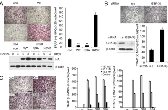
understand the role of GSK-3 in osteoclast differentiation, primary BMMs were infected with retroviruses expressing wild-type GSK-3 or its two mutants, constitutively active GSK-3 (GSK3-S9A) or catalytically inactive GSK-3 (GSK3-K85R). RANKL stimulation of control vector-infected BMMs increased the number of TRAP⫹MNCs, whereas over-expression of wild-type GSK-3 or GSK3-S9A mutant in BMMs suppressed the formation of TRAP⫹MNCs (⬎20 or ⬎3 nuclei) (Fig. 2A andsupplemental Fig. S1). Conversely, BMMs infected with GSK3-K85R showed a marked increase in the number of TRAP⫹ MNCs (Fig. 2A). Notably, the formation of large (⬎100 M) osteoclasts containing 20 nuclei was greatly
increased in BMMs expressing the GSK3-K85R mutant, suggest-ing that the terminal differentiation of osteoclasts, characterized by multinucleation, was regulated by GSK-3 activity. We further analyzed the effect of GSK-3 using an siRNA-mediated knock-down experiment. RANKL-induced osteoclast formation was enhanced by knocking down GSK-3, similar to the BMMs infected with the GSK3-K85R mutant (Fig. 2B). Taken together, these data suggest that GSK-3 plays a critical role in the RANKL-mediated osteoclast differentiation process.
To further confirm the role of GSK-3 in the differentiation of osteoclasts, we investigated the effects of pharmacological inhibition of GSK-3 on osteoclast formation. To augment the positive effect of GSK-3 inhibitors, kenpaullone, SB216763, or SB415286, BMMs were treated with a suboptimal concentra-tion of RANKL (20 ng/ml). Consistently, the inhibitors remark-ably enhanced the formation of TRAP⫹ MNCs (⬎3 or ⬎20 nuclei) in a dose-dependent manner (Fig. 2C). When BMMs expressing GSK3-S9A were co-cultured with primary osteo-blasts in the presence of 1,25(OH)2D3and prostaglandin E2, the
number of TRAP⫹MNCs was reduced when compared with empty vector control, whereas BMMs expressing GSK3-K85R showed increased TRAP⫹MNCs (supplemental Fig. S2). Col-lectively, these data point to the importance of GSK-3 in the differentiation process.
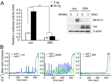
GSK-3 Blocks NFATc1 Transcription and Ca2⫹Oscillation—
Because it has been reported that GSK-3 causes phosphoryl-ation and nuclear export of NFATc proteins as proven by over-expression of GSK-3 in COS cells (13), we examined the
rele-vance of GSK-3 and NFATc1 in RANKL-stimulated
osteoclast formation. For this, we performed a reporter assay with a luciferase reporter plasmid driven by tandem NFAT
FIGURE 1. GSK-3 is inactivated during RANKL-induced
osteoclastogen-esis. A, phosphorylation of GSK-3 is increased upon RANKL treatment
dur-ing osteoclastogenesis. Cell lysates were analyzed by Western blottdur-ing with anti-phospho-GSK-3 (p-GSK-3), GSK-3, or NFATc1 antibody. -Actin was used as a loading control. B, the kinase activity of GSK-3 upon RANKL treat-ment was measured using a phospho-glycogen synthase peptide (P-GS). Cell lysates were immunoprecipitated with anti-GSK-3 antibody, and then the activity of GSK-3 was measured as described under “Experimental Proce-dures.” The activity of each sample was normalized to the amount of GSK-3. Data represent means⫾ S.D. *, p ⬍ 0.01.
FIGURE 2. GSK-3 activity regulates osteoclast formation. A, in vitro differentiation of osteoclasts from BMM cells infected with pMX-puro (con), HA-GSK3 (WT), HA-GSK3-S9A (S9A), or HA-GSK3-K85R (K85R), respectively. Because there was a large difference in numbers of large osteoclasts (⬎20 nuclei) among samples, we counted TRAP⫹MNCs containing more than 20 nuclei. Of note, TRAP⫹MNCs (more than three nuclei) were counted (supplemental Fig. S1). GSK-3 expression following infection was confirmed by Western blot analysis using the HA antibody and then reprobed with -actin as a loading control. Data represent means⫾ S.D. *, p ⬍ 0.05, **, p ⬍ 0.01. Scale bar, 200m. B, effect of siRNA against GSK-3 on RANKL-induced osteoclastogenesis. Nonspecific RNA duplex was used as a negative control. Knockdown of GSK-3 was confirmed by Western blot analysis. Data represent means ⫾ S.D. *, p ⬍ 0.01. Scale bar, 200 m. n.s., not significant. C, effects of the GSK-3 inhibitors kenpaullone (Ken), SB216763 (SB2), and SB415286 (SB4) on osteoclastogenesis. Data represent means⫾ S.D. Scale bar, 200m.
at Ewha Medical Library on July 6, 2016
http://www.jbc.org/
binding sites in the RAW264.7 cells stably expressing the GSK3-S9A mutant. The GSK3-S9A-expressing cells neither showed the NFAT transcriptional activity nor expressed a high level of NFATc1 protein after RANKL stimulation, whereas control cells effectively elevated transcriptional activity of NFATc1 and showed induction of NFATc1 at 3 day of RANKL treatment (Fig. 3A).
During osteoclastogenesis, RANKL stimulation induces sus-tained Ca2⫹oscillation, which is necessary for NFATc1
activa-tion and expression. To examine whether GSK-3 could regu-late the activation of Ca2⫹ oscillation induced by RANKL,
BMM cells were infected with retroviruses expressing the GSK3-S9A or GSK3-K85R mutants. Ca2⫹ oscillation was severely impaired in the BMMs infected with the GSK3-S9A mutant when compared with empty vector controls, whereas BMMs infected with the GSK3-K85R mutant were more sen-sitive to Ca2⫹ oscillation than control cells (Fig. 3B). Taken together, these data suggest that regulation of GSK-3 activity may modulate Ca2⫹oscillation on RANKL stimulation.
Transgenic Mice Expressing GSK3-S9A Show Osteopetrotic Phenotype due to Impaired Osteoclast Formation and NFATc1 Down-regulation—To investigate the in vivo physiological role of GSK-3, we generated transgenic mice expressing GSK3-S9A under control of the TRAP gene promoter, an enzyme that is highly expressed in osteoclasts (33) (supplemental Fig. S3, A and B). RT-PCR analysis indicated specific expression of the GSK3-S9A mutant in the osteoclast lineage from Tg mice (supplemental Fig. S3C). Consistent with the previous findings
FIGURE 3. GSK-3 inactivation is required for the induction of NFATc1
and Ca2ⴙoscillation by RANKL. A, left panel, inhibition of NFATc1 tran-scriptional activity by GSK-3. The stable RAW264.7 cells expressing HA-GSK3-S9A (S9A) or vector control (con) were transfected with an NFATc1 reporter construct, and then the transcriptional activity of NFATc1 was measured. Data represent means⫾ S.D. *, p ⬍ 0.01, **, p ⬍ 0.001. Right
panel, expression of NFATc1 in RAW264.7 cells stably expressing either
empty vector (con) or HA-GSK3-S9A (S9A). The stable RAW264.7 cells were stimulated with RANKL and analyzed by Western blotting with NFATc1 antibody. B, [Ca2⫹]ichanges were traced in BMMs infected with
empty vector (con), GSK-3 K85R (K85R), or GSK3-S9A (S9A) treated with RANKL and M-CSF for 3 days. Changes were estimated as the ratio of fluorescence intensity of fluo-4 to fura red. Each color indicates a different cell in the same field. Note that Ca2⫹oscillations are impaired in BMMs infected with S9A but that BMMs infected with K85R were more sensitive to the Ca2⫹oscillation than control cells. Max, maximum.
FIGURE 4. Impaired osteoclastogenesis and NFATc1 induction in BMMs of Tg mice expressing GSK3-S9A mutant. A, BMMs isolated from GSK3-S9A Tg (TG) mice or wild-type littermates (WT) were cultured for 4 days in the presence of M-CSF and RANKL as indicated. A representative TRAP staining is shown in the left panel, and TRAP⫹MNCs were counted in the right panel. Data represent mean⫾ S.D. *, p ⬍ 0.01. Scale bar, 100m. B, overexpression of GSK3-S9A in mice down-regulates induction of NFATc1 itself and the NFAT-dependent genes, Atp6v0d2 and Oscar. BMMs isolated from TG mice and WT littermates were cultured for 3 days with or without RANKL. Cell lysates were analyzed by Western blotting using the indicated antibodies specific for GSK-3␣/, NFATc1, Atp6v0d2, and Oscar. C, immunofluorescent images of NFATc1 protein in BMMs isolated from TG mice and WT littermates. BMM cells were stimulated with RANKL and M-CSF for 3 days and analyzed for the subcellular localization of NFATc1 protein as described under “Experimental Procedures.” Nuclei were stained with DAPI. Scale bar, 10m.
at Ewha Medical Library on July 6, 2016
http://www.jbc.org/
(Fig. 2), osteoclast formation of BMMs from Tg mice express-ing the GSK3-S9A mutant was markedly lower than that of wild-type littermates (Fig. 4A). Expression of the GSK3-S9A mutant was effectively induced by RANKL stimulation in Tg mice, which further confirmed successful control by the TRAP promoter in the Tg mice (Fig. 4B). Further, Western blot anal-ysis revealed that NFATc1 protein was decreased 3 days after RANKL stimulation of the BMMs from the Tg mice, and the protein levels of NFAT-dependent genes, Atp6v0d2 and Oscar, were also impaired (Fig. 4B).
We questioned whether the impairment of osteoclast forma-tion and the defect in NFAT expression in the Tg mice may
arise from NFATc1 phosphorylation by GSK-3, thereby caus-ing nuclear export of NFATc1. To answer this question, BMMs from Tg mice and wild-type littermates were incubated with M-CSF and RANKL for 3 days and then analyzed by immuno-fluorescence staining. Consistent with the Western blot data, NFATc1 immunoreactivity was barely detectable in the pre-osteoclasts from Tg mice, and further, most NFATc1 remained in the cytosol (Fig. 4C). In contrast, NFATc1 immunoreactivity in osteoclasts derived from wild-type littermates was readily detectable in the nuclei, but not the cytosol of multinucleated cells (Fig. 4C). Together, these results indicate that overexpres-sion of GSK3-S9A in Tg mice down-regulates NFATc1 expression and regulates nuclear localization of NFATc1, thereby suppressing osteoclast formation.
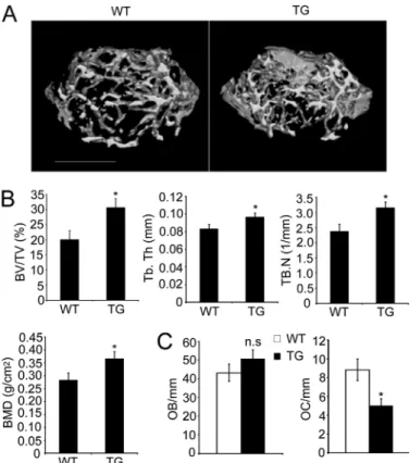
To evaluate the role of GSK-3 in vivo, we analyzed the bone phenotype of Tg mice by microcomputed tomography and his-tological analysis. Microcomputed tomography analysis of dis-tal femur metaphyses revealed a significant increase in bone volume due to increased trabecular thickness and number in the Tg mice (Fig. 5, A and B). Histomorphometric analysis indi-cated an increase in bone volume associated with a decrease in osteoclast number, but no significant difference in the number of osteoblasts (Fig. 5C).
To investigate the role of GSK-3 in a RANKL-induced model of bone destruction, we treated Tg mice expressing the GSK3-S9A mutant that were injected with RANKL or PBS (Control). The extent of bone erosion was notably reduced in the Tg mice, and the formation of TRAP⫹MNCs was greatly suppressed (Fig. 6, A and B), suggesting that overexpression of GSK3-S9A in Tg mice can inhibit RANKL-induced bone destruction. Thus, these results demonstrate that overexpres-sion of GSK3-S9A in Tg mice results in an osteopetrotic phe-notype due to a defect in osteoclast formation.
DISCUSSION
GSK-3 is a multitasking kinase that plays central roles in a diverse range of signaling pathways, including Wnts, hedgehog, growth factors, cytokines, and G protein-coupled ligands (34, 35). Specifically, in osteoblasts, it has been shown that GSK-3 acts as a negative regulator of the Wnt/-catenin signaling pathways required for osteoblast differentiation (1, 36, 37). However, the role of GSK-3 in RANKL-induced osteoclast formation is unknown. Here, we demonstrated for the first time a physiological role for GSK-3 in osteoclast biology. Our
find-FIGURE 5. Osteopetrotic phenotype of Tg mice expressing GSK3-S9A
mutant. A, three-dimensional microstructural analysis of the femurs of
wild-type (WT) and Tg mice (TG) by microcomputed tomography. B, histograms represent the three-dimensional trabecular structural parameters in femurs: bone volume fraction (BV/TV), trabecular thickness (Tb.Th), trabecular num-ber (Tb.N), and bone mineral densities (BMD). C, quantification of osteoclasts and osteoblasts form histological analysis of long bone of WT and Tg mice.
OC/mm, osteoclast number per bone surface; OB/mm, osteoblast number per
bone surface. Data represent means⫾ S.D. n ⫽ 6. *, p ⬍ 0.05. n.s., not significant.
FIGURE 6. Protective phenotype of Tg mice expressing GSK3-S9A mutant. The role of GSK-3 in RANKL-induced bone destruction in wild-type (WT) mice and Tg (TG) mice was studied. A, histology of the calvarial bone injected with PBS (Control) or RANKL in WT and Tg mice (TRAP and hematoxylin staining). Scale
bar, 0.02 mm. B, eroded surface (left) and the number of osteoclasts (right) were analyzed. Data represent means⫾ S.D. n ⫽ 5. *, p ⬍ 0.05.
at Ewha Medical Library on July 6, 2016
http://www.jbc.org/
ings through in vitro and in vivo analysis suggest a novel role for GSK-3 as a negative regulator of NFATc1 in RANKL-induced differentiation of monocytes/macrophages into osteoclasts.
Unlike other kinases, GSK-3 is constitutively active and is inactivated in response to cellular signal (34). We found that GSK-3 protein was expressed in osteoclast precursors and that the level of GSK-3 remained constant during the course of osteoclastogenesis. Remarkably, 3 days after RANKL treat-ment, GSK-3 was highly phosphorylated at Ser-9, causing its inactivation. We confirmed that the kinase activity was decreased at day 3 of RANKL treatment. It is likely that RANKL-induced GSK-3 inactivation is involved in osteoclast formation. Our findings in primary BMMs support this possi-bility in that overexpression of catalytically inactive GSK-3 mutant, pharmacological inhibition of GSK-3, or RNA inter-ference of GSK-3 accelerated efficient formation of oste-oclasts. Conversely, overexpression of constitutively active GSK-3 mutant markedly abolished formation of osteoclasts. However, overexpression of GSK-3␣ in BMMs had no effect on RANKL-mediated osteoclastogenesis (supplemental Fig. S4). These findings suggest that kinase activity of GSK-3 is critical for suppression of osteoclast formation.
It is notable that increase in NFATc1 protein level follows GSK-3 phosphorylation. Because NFATc1 represents a mas-ter switch that regulates mas-terminal differentiation of osteoclasts downstream of RANKL, induction of NFATc1 by RANKL is crucial for optimal differentiation of osteoclasts (7). In this con-text, it is likely that GSK-3 inactivation is a prerequisite for the NFATc1 amplification process. A previous study showed that GSK-3 causes phosphorylation and nuclear export of NFAT proteins in vitro (13), but the physiological importance of NFATc1 phosphorylation by GSK-3 has not yet been clarified. We found that overexpression of constitutively active GSK-3 showed a defect in NFATc1 expression and its transcriptional activity. Consistent with the finding from in vitro analysis, the formation of TRAP⫹MNCs induced by RANKL was markedly inhibited in the Tg mice expressing a constitutively active GSK-3 mutant. Protein levels of NFATc1 as well as NFATc1-dependent genes such as Atp6v0d2 and Oscar induced by RANKL in the Tg mice were also decreased at 3 days after RANKL stimulation. More importantly, RANKL-dependent nuclear localization of NFATc1 was severely impaired in the Tg mice. Based on our data, we propose that GSK-3 may promote nuclear export of NFATc1 through phosphorylation, thereby resulting in down-regulation of NAFTc1 induction.
Interestingly, our results also showed that overexpression of constitutively active GSK-3 in osteoclast precursors decreased Ca2⫹ oscillation. In contrast, catalytically inactive GSK-3 promoted Ca2⫹ oscillation. It is well known that
NFATc1 is activated by Ca2⫹signaling induced by the activa-tion of the immunoglobulin-like receptor signaling associated with immunoreceptor tyrosine-based activation motif-harbor-ing adaptors, which is known to provide co-stimulatory signal-ing (38). The long lastsignal-ing Ca2⫹ oscillation, which is evident
during osteoclastogenesis, may ensure the robust induction of NFATc1 through an autoamplification mechanism (10). We currently have no evidence that GSK-3 directly regulates Ca2⫹signaling cascades through its kinase activity. However,
we cannot exclude the possibility that GSK-3 may directly regulate RANKL- or co-stimulatory receptor-induced Ca2⫹ signaling cascades by unidentified mechanism(s). Otherwise, it seems likely that the defect in robust induction of NFATc1 by overexpression of the constitutively active GSK-3 may affect Ca2⫹oscillation. Specifically, our data showed that the protein
level of OSCAR was markedly decreased in RANKL-stimulated pre-osteoclasts from Tg mice. Consistent with this finding, it was reported that the OSCAR-FcR␥ (Fc receptor common ␥ subunit) complex regulates RANKL-mediated activation of Ca2⫹signaling in pre-osteoclasts, which leads to the amplifica-tion of NFATc1 (38). Further studies will be required to eluci-date mechanism(s) of how GSK-3 regulates Ca2⫹oscillation. Given the multiple processes regulated by GSK-3, GSK-3 has been implicated in various diseases such as diabetes, cancer, bipolar disorder, and Alzheimer disease, and GSK-3 inhibi-tors are being actively developed as drugs for the treatment of the various disorders (19, 39). Moreover, because GSK-3 is a negative regulator of Wnt--catenin signaling that is known to stimulate bone formation, modulating its activity with small molecules is a promising strategy for increasing bone mass (36). However, it is important to note that GSK-3 is not a special-ized repressor of-catenin as it can participate in RANKL sig-naling as shown in our study. In the context of skeletal health, given the potential risk of long term inhibition of GSK-3, a more comprehensive study with regard to targeting of drugs to the GSK-3 for the treatment of chronic disorders such as osteoporosis should be performed using animal models fol-lowed by human case study. Moreover, further mechanistic studies will be needed to understand the pathophysiological relevance of GSK-3 in bone remodeling and its disorders.
Acknowledgments—We thank T. Kitamura for providing PLAT-E cells and Y. Choi for the anti-Atp6v0d2 antibody.
REFERENCES
1. Harada, S., and Rodan, G. A. (2003) Nature 423, 349 –355 2. Teitelbaum, S. L. (2000) Science 289, 1504 –1508
3. Boyle, W. J., Simonet, W. S., and Lacey, D. L. (2003) Nature 423, 337–342 4. Suda, T., Takahashi, N., Udagawa, N., Jimi, E., Gillespie, M. T., and Martin,
T. J. (1999) Endocr. Rev. 20, 345–357
5. Wong, B. R., Josien, R., Lee, S. Y., Vologodskaia, M., Steinman, R. M., and Choi, Y. (1998) J. Biol. Chem. 273, 28355–28359
6. Asagiri, M., and Takayanagi, H. (2007) Bone 40, 251–264
7. Asagiri, M., Sato, K., Usami, T., Ochi, S., Nishina, H., Yoshida, H., Morita, I., Wagner, E. F., Mak, T. W., Serfling, E., and Takayanagi, H. (2005) J. Exp.
Med. 202,1261–1269
8. Wagner, E. F., and Eferl, R. (2005) Immunol. Rev. 208, 126 –140 9. Crabtree, G. R. (1999) Cell 96, 611– 614
10. Takayanagi, H., Kim, S., Koga, T., Nishina, H., Isshiki, M., Yoshida, H., Saiura, A., Isobe, M., Yokochi, T., Inoue, J., Wagner, E. F., Mak, T. W., Kodama, T., and Taniguchi, T. (2002) Dev. Cell 3, 889 –901
11. Crabtree, G. R., and Olson, E. N. (2002) Cell 109, (suppl.) S67–S79 12. Hogan, P. G., Chen, L., Nardone, J., and Rao, A. (2003) Genes Dev. 17,
2205–2232
13. Beals, C. R., Sheridan, C. M., Turck, C. W., Gardner, P., and Crabtree, G. R. (1997) Science 275, 1930 –1934
14. Okamura, H., Garcia-Rodriguez, C., Martinson, H., Qin, J., Virshup, D. M., and Rao, A. (2004) Mol. Cell. Biol. 24, 4184 – 4195
15. Yang, T. T., Xiong, Q., Enslen, H., Davis, R. J., and Chow, C. W. (2002) Mol.
Cell. Biol. 22,3892–3904
at Ewha Medical Library on July 6, 2016
http://www.jbc.org/
16. Chow, C. W., Rincón, M., Cavanagh, J., Dickens, M., and Davis, R. J. (1997)
Science 278,1638 –1641
17. Woodgett, J. R. (1990) EMBO J. 9, 2431–2438 18. Frame, S., and Cohen, P. (2001) Biochem. J. 359, 1–16
19. Sugden, P. H., Fuller, S. J., Weiss, S. C., and Clerk, A. (2008) Br. J.
Pharma-col. 153,Suppl. 1, S137–S153
20. Logan, C. Y., and Nusse, R. (2004) Annu. Rev. Cell Dev. Biol. 20, 781– 810 21. Jope, R. S., and Johnson, G. V. (2004) Trends Biochem. Sci. 29, 95–102 22. Frame, S., Cohen, P., and Biondi, R. M. (2001) Mol. Cell 7, 1321–1327 23. Hoeflich, K. P., Luo, J., Rubie, E. A., Tsao, M. S., Jin, O., and Woodgett, J. R.
(2000) Nature 406, 86 –90
24. Lee, S. H., Rho, J., Jeong, D., Sul, J. Y., Kim, T., Kim, N., Kang, J. S., Miy-amoto, T., Suda, T., Lee, S. K., Pignolo, R. J., Koczon-Jaremko, B., Lorenzo, J., and Choi, Y. (2006) Nat. Med. 12, 1403–1409
25. Yoon, K., Jung, E. J., Lee, S. R., Kim, J., Choi, Y., and Lee, S. Y. (2008) Cell
Death Differ. 15,730 –738
26. Suda, T., Jimi, E., Nakamura, I., and Takahashi, N. (1997) Methods
Enzy-mol. 282,223–235
27. Lee, S. E., Woo, K. M., Kim, S. Y., Kim, H. M., Kwack, K., Lee, Z. H., and Kim, H. H. (2002) Bone 30, 71–77
28. Kim, J. H., Kim, K., Youn, B. U., Jin, H. M., and Kim, N. (2010) Cell. Signal.
22,1341–1349
29. Reddy, S. V., Hundley, J. E., Windle, J. J., Alcantara, O., Linn, R., Leach, R. J.,
Boldt, D. H., and Roodman, G. D. (1995) J. Bone Miner. Res. 10, 601– 606 30. Schwartzberg, P. L., Xing, L., Hoffmann, O., Lowell, C. A., Garrett, L.,
Boyce, B. F., and Varmus, H. E. (1997) Genes Dev. 11, 2835–2844 31. Parfitt, A. M., Drezner, M. K., Glorieux, F. H., Kanis, J. A., Malluche, H.,
Meunier, P. J., Ott, S. M., and Recker, R. R. (1987) J. Bone Miner. Res. 2, 595– 610
32. Takayanagi, H., Ogasawara, K., Hida, S., Chiba, T., Murata, S., Sato, K., Takaoka, A., Yokochi, T., Oda, H., Tanaka, K., Nakamura, K., and Tani-guchi, T. (2000) Nature 408, 600 – 605
33. Hikata, T., Takaishi, H., Takito, J., Hakozaki, A., Furukawa, M., Uchikawa, S., Kimura, T., Okada, Y., Matsumoto, M., Yoshimura, A., Nishimura, R., Reddy, S. V., Asahara, H., and Toyama, Y. (2009) Blood 113, 2202–2212 34. Doble, B. W., and Woodgett, J. R. (2003) J. Cell Sci. 116, 1175–1186 35. Wu, D., and Pan, W. (2010) Trends Biochem. Sci. 35, 161–168
36. Bodine, P. V., and Komm, B. S. (2006) Rev. Endocr. Metab. Disord. 7, 33–39 37. Clément-Lacroix, P., Ai, M., Morvan, F., Roman-Roman, S., Vayssière, B., Belleville, C., Estrera, K., Warman, M. L., Baron, R., and Rawadi, G. (2005)
Proc. Natl. Acad. Sci. U.S.A. 102,17406 –17411
38. Koga, T., Inui, M., Inoue, K., Kim, S., Suematsu, A., Kobayashi, E., Iwata, T., Ohnishi, H., Matozaki, T., Kodama, T., Taniguchi, T., Takayanagi, H., and Takai, T. (2004) Nature 428, 758 –763
39. Cohen, P., and Goedert, M. (2004) Nat. Rev. Drug Discov. 3, 479 – 487
at Ewha Medical Library on July 6, 2016
http://www.jbc.org/
1
Figure Legends
Figure S1. In vitro differentiation of osteoclasts from BMM cells infected with pMX-puro
(con), wild-type HA-GSK-3(WT), constitutively active HA-GSK-3S9A (S9A), or
catalytically inactive HA-GSK-3K85R (K85R), respectively. TRAP staining was performed 4
days after RANKL stimulation. TRAP
+MNCs (> 3 nuclei) were counted. Data represent means
± SD. *P < 0.05, **P < 0.01.
Figure S2. BMMs infected with retroviruses expressing either empty vector (EV), HA-GSK-3
S9A (S9A), or HA-GSK-3K85R (K85R) were cocultured with osteoblasts for 7 days in the
presence of 1,25(OH)
2D
3and PGE
2. TRAP staining was performed and TRAP
+MNCs were
counted. Data represent means ± SD. *P < 0.05, **P < 0.01. Scale bar, 200 μm.
Figure S3. Generation of Tg mice expressing GSK-3
S9A mutant under the control of TRAP
promoter. (A) Schematic diagram of the TRAP-GSK-3-S9A construct. Constitutively active
GSK-3S9A cDNA was fused to 1.8 kb of the mouse TRAP gene promoter. GT or GP primer
indicates specific primers for the transgene. (B) Genomic DNA isolated from the tail was
analyzed by PCR using specific primers for the transgene. (C) BMMs isolated from
GSK-3S9A Tg (TG) mice or wild-type littermates (WT) were cultured in the presence of RANKL
as indicated. The expression of transgene was confirmed by RT-PCR analysis using a specific
primer (GT primer) for GSK-3S9A or -actin.
Figure S4. GSK-3
had no significant effect on RANKL-induced osteoclast differentiation. In
vitro differentiation of osteoclasts from BMMs infected with pMX-puro (con),
V5-GSK-3WT), or catalytically inactive V5-GSK-3K 148A (K148A), respectively. TRAP staining
2
were counted. GSK-3 expression following infection was confirmed by Western blot analysis
using the V5 antibody and then reprobed with GAPDH as a loading control. Data represent
means ± SD.
Chang-Yong Ko, Han-Sung Kim, Daewon Jeong, Nacksung Kim and Soo Young Lee
Hyun Duk Jang, Ji Hye Shin, Doo Ri Park, Jin Hee Hong, Kwiyeom Yoon, Ryeojin Ko,
Differentiation
doi: 10.1074/jbc.M111.256768 originally published online September 23, 2011 2011, 286:39043-39050.
J. Biol. Chem.
10.1074/jbc.M111.256768
Access the most updated version of this article at doi: Alerts:
When a correction for this article is posted
•
When this article is cited
•
to choose from all of JBC's e-mail alerts
Click here
Supplemental material:
http://www.jbc.org/content/suppl/2011/09/23/M111.256768.DC1.html http://www.jbc.org/content/286/45/39043.full.html#ref-list-1This article cites 39 references, 12 of which can be accessed free at
at Ewha Medical Library on July 6, 2016
http://www.jbc.org/