www.nature.com/scientificreports
OASL1 deficiency promotes
antiviral protection against
genital herpes simplex virus type
2 infection by enhancing type I
interferon production
Ji Eun Oh
1,†, Myeong Sup Lee
2,†, Young-Joon Kim
3,4& Heung Kyu Lee
1Type I interferon (IFN) interferes with virus replication, promotes antiviral responses, and controls innate and adaptive immune responses to certain viruses. Recently, we reported that 2’–5’ oligoadenylate synthetase-like 1 (OASL1) negatively regulates type I IFN production by inhibiting the translation of the type I IFN-regulating master transcription factor, IRF7. Notably, while OASL1-deficient mice induce robust production of type I IFN and are resistant to systemic viral infection, the effects of OASL1 during localized viral infection has not been studied. To this end, we investigated the role of OASL1 during mucosal HSV-2 infection of the genital tract. Oasl1−/− mice exhibited better
survival rates than wild type (WT) mice following intravaginal HSV-2 infection, and suppressed virus replication more efficiently despite comparable recruitment of effector immune cells. Moreover, Ly6Chigh monocytes, and not pDCs or other cell types, displayed enhanced production of type I IFNs in
Oasl1−/− mice in response to HSV-2 infection. Furthermore, cytotoxic T cell responses including IFN-γ
production were accelerated in Oasl1−/− mice after mucosal HSV-2 infection. Collectively, these results
demonstrate that OASL1 deficiency promotes antiviral immunity against local mucosal viral infection and suggest that OASL1 could be a therapeutic target for treatment of HSV-2 infection of the genital mucosa.
Genital herpes is one of the most common sexually transmitted infections (STIs) worldwide. It is caused by her-pes simplex virus type 2 (HSV-2), which has a linear, double-stranded DNA genome of ~154 kb in length. HSV-2 infection not only causes ulcers within the genital tract, but also induces lifelong latency within the sensory ganglia of the nervous system. In addition to genital ulcers, HSV-2 infection can cause severe and frequently fatal symptoms, and is considered to be a major risk factor for other STIs such as human immunodeficiency virus type 1 (HIV-1)1. Although the prevalence of HSV-2 infection and the incidence of genital herpes have been increasing, there is currently a lack of effective therapeutics2. Thus, understanding host immune responses against HSV-2 infection may provide clues for the cure and prevention of this debilitating disease.
The innate immune system represents the first line of defense against pathogens and acts by limiting infection or replication and by initiating the adaptive immune response. Notably, type I IFNs are critical for inhibition of early viral replication, activation of immune cells, and regulation of adaptive immune responses. In this regard, after genital infection with HSV-2, IFNα /β receptor-deficient (IFNAR−/−) mice showed increased viral replica-tion and decreased survival rates compared to wild type (WT) mice3.
1Laboratory of Host Defenses, Graduate School of Medical Science and Engineering, Korea Advanced Institute of Science and Technology (KAIST), Daejeon, 34141, Republic of Korea. 2Department of Biomedical Sciences, College of Medicine, University of Ulsan, Seoul, 05505, Republic of Korea. 3Department of Biochemistry, College of Life Science and Technology, Yonsei University, Seoul, 03722, Republic of Korea. 4Department of Integrated Omics for Biomedical Science Graduate School, Yonsei University, Seoul, 03722, Republic of Korea. †These Authors contributed equally to this work. Correspondence and requests for materials should be addressed to H.K.L. (email: heungkyu. [email protected])
Received: 11 August 2015 Accepted: 07 December 2015 Published: 11 January 2016
OPEN
Type I IFNs induce various interferon-stimulated genes (ISGs), which are involved in diverse antiviral path-ways4,5. Collectively, ISGs inhibit viral protein synthesis and virus replication, thus providing early protection against virus infection. For example, protein kinase R (PKR), the dsRNA-activated serine/threonine protein kinase, is an ISG that negatively regulates mRNA translation. Other ISGs, such as 2′–5′-oligoadenylate synthetase (OAS) and RNase L, are also involved in the degradation of both cellular and viral RNA. Moreover, type I IFNs activate innate immune cells including natural killer (NK) cells, which then lyse virus-infected cells, and dendritic cells (DCs), inducing their maturation through expression of MHC and co-stimulatory molecules6. Interestingly, type I IFNs can also activate and expand antigen-specific T cells6. Thus, type I IFNs regulate adaptive immune responses both directly and indirectly.
Type III IFNs, comprised of IFN-λ 1, -λ 2, and -λ 3, are a newly identified subset of IFNs7,8. Although type III IFNs signal through distinct receptor complexes from type I IFNs, the biologic functions and downstream signaling pathways are similar9. What makes type III IFNs unique is the restriction of their receptors to epithelial tissue10. Moreover, recent studies demonstrated that IFN-λ plays critical roles in the antiviral protection of the mucosal organ11,12. In the case of mucosal HSV-2 infection, IFN-λ has been shown to inhibit virus replication in the vaginal mucosa thereby conferring protection against HSV-2 infection13,14.
Recently, we showed that OASL1, a nonenzymatic OAS protein, negatively regulates the production of type I IFNs during viral infection by inhibiting the translation of interferon regulatory factor 7 (IRF7)15. Following virus recognition by various receptors, the production of type I IFNs is induced through activation of IRF316. IRF3 is the key transcription factor leading to the early production of type I IFNs (predominately of IFN-β ), which ini-tiates a positive feedback loop in autocrine and paracrine manners17,18. In this process, IRF7, a master regulator of type I IFNs, functions to further amplify the expression of type I IFNs19. Thus, Oasl1−/− mice are resistant to systemic viral infection due to increased production of type I IFNs15. In addition, another study using a systemic chronic lymphocytic choriomeningitis virus (LCMV) infection model demonstrated that OASL1-mediated sup-pression of type I IFN production prevents efficient viral control and the induction of a functional T cell response, permitting viral persistence20. Notably, the function of OASL1 in a non-systemic, natural mucosal virus infection remains unknown. Furthermore, whether OASL1 also regulates type III IFNs has not been investigated.
In the present study, we show that Oasl1−/− mice are more resistant to mucosal HSV-2 infection as com-pared to WT mice. Furthermore, hematopoietic cells were sufficient for this enhanced protection of Oasl1−/− mice against mucosal HSV-2 infection. Although production of type III IFNs was not increased in Oasl1−/− BM cells after in vitro stimulation with HSV-2, type III IFN remained high in vaginal washes until later time points after intravaginal HSV-2 infection. The increased production of type I IFNs in Oasl1−/− mice was derived from Ly6Chigh monocytes, not from plasmacytoid DCs (pDCs), and effectively induced robust CD8+ T cell responses protecting against mucosal HSV-2 infection. Together, these results indicate that OASL1-mediated negative regu-lation of type I IFN production suppresses both innate and adaptive immunity against mucosal HSV-2 infection.
Results
Oasl1
−/−mice are more resistant to mucosal HSV-2 infection than WT mice.
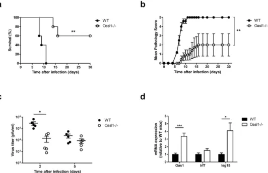
In our previousstudy, we found that OASL1 inhibited the translation of IRF7, a master transcription factor for type I IFN pro-duction15. Thus, OASL1 negatively regulates excessive production of type I IFN to limit hyperinflammatory responses. In this regard, Oasl1−/− mice produce more type I IFN after poly (I:C) treatment and are more resistant to systemic virus infection than WT mice15. To determine whether Oasl1−/− mice are also more resistant to local mucosal virus infection, we infected Oasl1−/− and littermate WT control mice intravaginally with 1000 pfu of WT HSV-2. Only two-fifths of Oasl1−/− mice died after genital HSV-2 infection, while all WT mice died within 11 days of infection (Fig. 1a). Moreover, Oasl1−/− mice showed only mild clinical pathology (Fig. 1b), and viral titers from vaginal washes were markedly lower in Oasl1−/− mice at early time points post-infection as compared to WT mice (Fig. 1c). Interestingly, we found that ISGs such as OAS1 and ISG15 were markedly increased in vaginal tissue of Oasl1−/− mice even in the absence of infection (Fig. 1d). This indicates that the enhanced antiviral state within the vaginal tract limited viral replication early after infection in Oasl1−/− mice and that these mice are more resistant to local mucosal HSV-2 infection than are WT mice.
Oasl1
−/−hematopoietic cells are sufficient for enhanced protection against mucosal HSV-2
infection.
Unlike systemic viral infection, both hematopoietic and stromal compartments take part in innate immune responses after mucosal HSV-2 infection. Mucosal epithelial cells are the first cell types infected with HSV-2 that produce type I IFN, albeit much less than hematopoietic cells. These type I IFNs, primarily IFN-β , initiate a positive feedback loop, thus promoting robust production of more type I IFN by hematopoietic cells recruited to the site of infection. In this regard, Shen and Iwasaki reported that mice lacking IFNα β R expres-sion on hematopoietic cells displayed a more severe phenotype in response to mucosal HSV-2 infection than mice lacking IFNα β R on stromal cells21. To elucidate whether Oasl1−/− hematopoietic cells are sufficient for the enhanced protection against mucosal HSV-2 infection, we generated irradiation-induced BM chimera mice that lack OASL1 expression in hematopoietic cells but have intact OASL1 expression in stromal cells. Compared with Oasl1+/−→ WT mice, Oasl1−/−→ WT mice survived longer and showed milder disease pathology following intravaginal infection with WT HSV-2 (Fig. 2a,b). Interestingly, Oasl1−/−→ WT mice produced a profuse amount of IFN-α at the infection site two days post-infection (Fig. 2c).The recently identified type III IFN, IFN-λ , has been shown to confer antiviral protection to the mucosal epithelia10,11. Type III IFNs bind different receptors than type I IFNs, but induce the same signaling pathways9. Thus, to determine whether OASL1 also regulates type III IFN, we measured the level of IFN-λ in vaginal washes after mucosal HSV-2 infection. Unlike IFN-α , the level of IFN-λ at the infection site in Oasl1−/−→ WT mice was comparable to Oasl1+/−→ WT mice at early time points post-infection. However, high levels of IFN-λ in
www.nature.com/scientificreports/
Figure 1. OASL1 deficiency enhances immune protection against mucosal HSV-2 infection. WT and
Oasl1−/− mice were infected intravaginally with 1000 pfu of WT HSV-2. (a) Survival and (b) disease scores were monitored for one month post-challenge (n = 5 mice per group). Data are representative of four independent experiments. (c) At the indicated days post infection, HSV-2 viral titers from vaginal washes were measured on Vero cells (n = 5 mice). Data are representative of three independent experiments. (d) Expression of ISGs in vaginal tissue of uninfected Oasl1−/− mice relative to that of uninfected Oasl1+/− mice was determined by qRT-PCR (n = 5 mice). Data are representative of two independent experiments. *p < 0.05; **p < 0.01; ***p < 0.001. Error bars: SEM.
Figure 2. Hematopoietic cells contribute to enhanced immune protection against mucosal HSV-2 infection in Oasl1−/− mice. Oasl1+/−→ WT and Oasl1−/−→ WT chimera mice were infected intravaginally with 10000 pfu of WT HSV-2. (a) Survival and (b) disease scores were monitored until all mice succumbed. (c,d) On the indicated days post infection, vaginal washes were collected and the level of (c) IFN-α and (d) IFN-λ was measured by ELISA (Oasl1+/−→ WT, n = 9 mice; Oasl1−/−→ WT, n = 8 mice). *p < 0.05; **p < 0.01; ***p < 0.001. Error bars: SEM.
Oasl1−/−→ WT mice were prolonged until late time points post-infection, while IFN-λ levels in Oasl1+/−→ WT mice gradually decreased (Fig. 2d). Taken together, these data suggest that hematopoietic cells are sufficient for protection against mucosal HSV-2 infection in Oasl1−/− mice through the enhanced production of type I and type III IFNs.
Production of type I IFNs, but not type III IFNs and proinflammatory cytokines, is enhanced in
Oasl1
−/−bone marrow cells.
Based on the above results, we next wanted to examine the role ofhemato-poietic cells in Oasl1−/− mice in response to HSV-2 infection. To this end, we treated bone marrow (BM) cells with TK- HSV-2 at various multiplicities of infection (MOI) in vitro. We found that Oasl1−/− BM cells produced significantly more type I IFN, including IFN-α and IFN-β , when stimulated with HSV-2 (Fig. 3a,b). However, IFN-λ production by Oasl1−/− BM cells was comparable to that of Oasl1+/− BM cells after stimulation with HSV-2 (Fig. 3c). In addition, similar amounts of IL-12p40, a pro-inflammatory cytokine important for the differ-entiation of Th1 cells, were produced by infected control and Oasl1−/− BM cells (Fig. 3d). These data indicate that OASL1 selectively suppresses the production of type I IFN, but not type III IFN or pro-inflammatory cytokines after HSV-2 infection.
Ly6C
highmonocytes are major sources of enhanced production of type I IFNs in Oasl1
−/−mice
in response to HSV-2 infection.
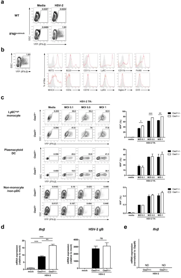
Although most types of cells can produce type I IFN, certain cells such as pDCs robustly produce type I IFN in response to viral infection. In the case of HSV infection, it has been reported that pDCs are indispensable for early antiviral protection due to their ability to produce type I IFN22. However, a recent study using transgenic mice selectively depleting pDCs showed that pDCs are critical for antiviral immu-nity against systemic HSV, but not local HSV infection23. In addition, this study suggested that pDCs are not the only source of IFN-α during systemic HSV infection. To elucidate what is the primary source of type I IFN during HSV-2 infection, we infected BM cells isolated from IFNβ mob/mob mice, which express yellow fluorescent protein (YFP) in an IFN-β -dependent manner, with HSV-2 in vitro. First, we confirmed that YFP was expressed specifi-cally in this reporter mouse after infection with HSV-2 (Fig. 4a). Next, we investigated the surface phenotype of YFP+/IFN-β -producing cells in BM cells stimulated with HSV-2 in order to determine the cell type producing type I IFN following HSV-2 infection. Strikingly, while YFP+/IFN-β -producing cells were positive for surface markers of pDCs such as BST2, B220, and CD11c, some molecules not expressed by pDCs, such as CD11b, were also expressed on YFP+/IFN-β -producing cells. In this regard, we found that YFP+/IFN-β -producing cells also expressed high levels of Ly6C and F4/80, and intermediate levels of Ly6G and not Siglec-F, suggesting that these cells are Ly6Chigh monocytes (Fig. 4b).As shown in Fig. 3, Oasl1−/− cells produced more type I IFN after infection with HSV-2. To determine whether enhanced production of type I IFN in Oasl1−/− cells is cell-type dependent, we examined the expression of YFP in BM cells from Oasl1-heterozygous IFNβ mob/mob mice or Oasl1-deficient IFNβ mob/mob mice after infection with HSV-2. YFP expression was induced by both Ly6Chigh monocytes and pDCs after stimulation with HSV-2 in MOI-dependent manner. We detected greater YFP expression in Ly6Chigh monocytes, but not in pDCs or other cells, in Oasl1−/− mice than in control mice after HSV-2 infection (Fig. 4c and supplementary Fig. 1). In addition, to examine whether Ly6Chigh monocytes became directly infected by HSV-2 and then produced type I IFN or Figure 3. Oasl1−/− BM cells produce more type I IFNs, but not IFN-λ and IL-12p40. BM cells from Oasl1+/− and Oasl1−/− mice were stimulated with TK- HSV-2 at the indicated MOIs for 18 h. Levels of (a) IFN-α , (b) IFN-β , (c) IFN-λ , and (d) IL-12p40 in supernatants were measured by ELISA (n = 2). Data are representative of two to three independent experiments. *p < 0.05; **p < 0.01; ***p < 0.001. Error bars: SEM.
www.nature.com/scientificreports/
Figure 4. Ly6Chigh monocytes are major contributors of the enhanced production of type I IFNs in
Oasl1−/− mice after infection with HSV-2. (a,b) BM cells from WT and IFNβ mob/mob mice were stimulated with TK- HSV-2 (MOI 5) for 18 h. (a) YFP expression was assessed using flow cytometry. Plots were gated on PI- cells. (b) Surface expression of indicated molecules (red histograms) on YFP+ cells from BM of IFNβ mob/ mob mice was analyzed by flow cytometry. Shaded gray histograms indicate isotype control. (c) BM cells from
Oasl1-heterozygous IFNβ mob/mob and Oasl1-deficient IFNβ mob/mob mice were stimulated with TK- HSV-2 at the indicated MOIs for 18 h. left. YFP expression in Ly6Chigh monocytes, pDCs, and non-monocytes/non-pDCs
if Ly6Chigh monocytes produced type I IFN in a paracrine manner, we examined the expression of GFP in BM monocytes after infection with GFP HSV-1. We found that GFP expression in BM monocytes increased in an MOI-dependent manner, and the level of GFP expression was not different between Oasl1+/− and Oasl1−/− BM monocytes (Supplementary Fig. 2).
Next, to investigate whether Ly6Chigh monocytes infiltrated in vaginal tissues contributed to the enhanced production of type I IFN in Oasl1−/− mice in vivo, we compared IFN-β mRNA expression in Ly6Chigh monocytes sorted from infected vaginal tissues with BM monocytes (mock control) (Fig. 4d and supplementary Fig. 3). Interestingly, we detected increased IFN-β mRNA expression in vaginal Ly6Chigh monocytes following HSV-2 infection, whereas IFN-β expression was not detected in vaginal CD4+ T cells (Fig. 4d,e). Further, Oasl1−/− mice showed higher expression of IFN-β in vaginal Ly6Chigh monocytes than did Oasl1+/− mice (Fig. 4d). Furthermore, similar levels of HSV-2 glycoprotein B (gB) were detected in Ly6Chigh monocytes sorted from vaginal tissues of genital HSV-2 infected Oasl1+/− and Oasl1−/− mice (Fig. 4d).
Taken together, our data indicate that the major cell type contributing to the robust production of IFN-β in
Oasl1−/− mice are Ly6Chigh monocytes in vitro and in vivo, although substantial amounts of IFN-β can be pro-duced by both Ly6Chigh monocytes and pDCs in response to HSV-2 infection. Moreover, Ly6Chigh monocytes from Oasl1−/− mice produce higher levels of type I IFNs than those from Oasl1+/− mice, even though these cells are infected by HSV-2 at similar levels.
Recruitment of innate immune cells in Oasl1
−/−and control mice is comparable.
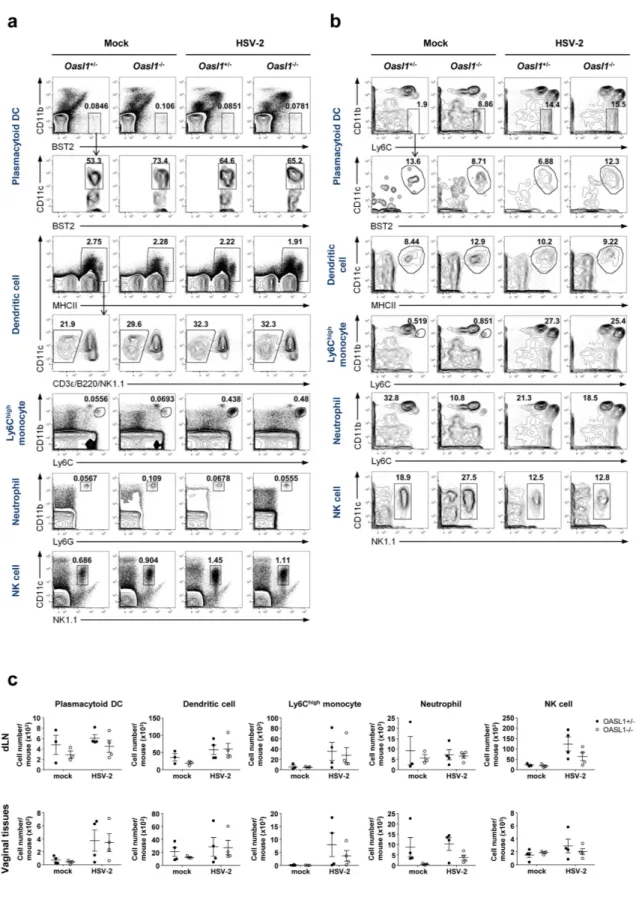
After viralinfection, innate effector cells migrate to the site of infection to defeat the infection through cytokine production and killing of viral-infected cells. To investigate the possibility that OASL1 deficiency affects the trafficking of innate immune cells to the site of infection, we examined the proportion and the number of effector immune cell subsets, including pDCs, DCs, Ly6Chigh monocytes, neutrophils, and NK cells, in draining lymph nodes and vag-inal tissues early after infection (Fig. 5). We found that there was no substantial difference between Oasl1+/− and
Oasl1−/− mice in the proportion or number of innate immune cells in either draining lymph nodes or vaginal tissues. Taken together, these results suggest that migration of innate effector cells is not affected by the absence of OASL1 protein.
Expression of co-stimulatory and MHC molecules on antigen presenting cells is similar between
Oasl1
−/−and control mice.
Type I IFN modulates various immune cell functions including thedevelop-ment, maturation, migration, and antigen presentation of antigen presenting cells (APC), the most important mediators of innate and adaptive immunity6. To investigate whether the increased production of type I IFN in
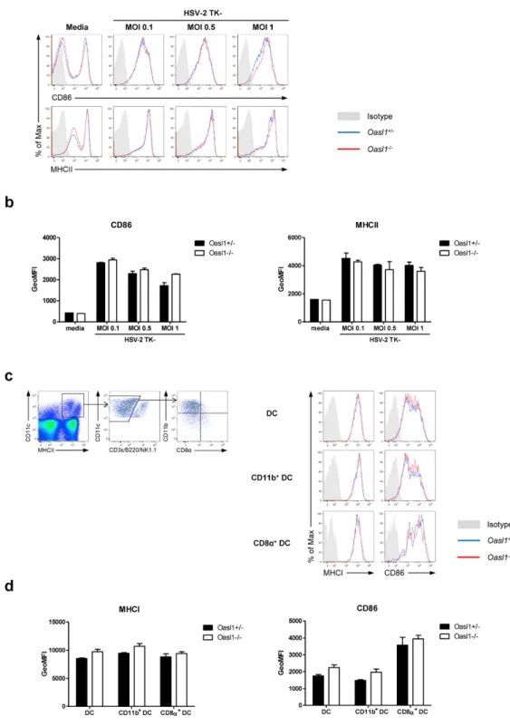
Oasl1−/− mice affects DC maturation, we measured the expression of co-stimulatory and MHC molecules on DCs following HSV-2 infection. To this end, BM-derived DCs from Oasl1−/− mice showed comparable expression of CD86 and MHCII to DCs from control mice after stimulation with HSV-2 in vitro (Fig. 6a,b). Moreover, the level of expression of CD86 and MHCI on various DC subsets including CD11b+ DCs and CD8α + DCs was not significantly different between Oasl1−/− and control mice three days after mucosal HSV-2 infection (Fig. 6c,d). Collectively, DC maturation was not enhanced in Oasl1−/− mice, even though production of type I IFNs was greatly enhanced in Oasl1−/− mice in response to HSV-2 infection. These results suggest that the amount of type I IFN produced by control mice might be sufficient for inducing DC maturation after HSV-2 infection.
Augmented CTL priming in Oasl1
−/−mice following mucosal HSV-2 infection.
There have beenmany studies investigating the role of type I IFNs in antiviral T cell responses6,24. To determine whether the abil-ity of Oasl1−/− mice to induce enhanced type I IFN responses also affects the priming of virus-specific T cells, we measured IFN-γ production by CD4+ and CD8+ T cells from draining lymph nodes after mucosal HSV-2 infection. We found that the level of IFN-γ produced by CD8+ T cells, but not CD4+ T cells, was increased in Oasl1−/− mice compared to WT mice (Fig. 7a). Consistently, the frequency of IFN-γ -producing activated (defined as CD44+) CD8+ T cells was also increased in Oasl1−/− mice in response to mucosal HSV-2 infection (Fig. 7b,c). Collectively, these results suggest that CTL priming, but not Th1 priming, is augmented in Oasl1−/− mice following mucosal HSV-2 infection.
Discussion
In this study, we investigated how OASL1 affects antiviral protection against mucosal HSV-2 infection. We show that Oasl1−/− mice exhibit better survival rates and efficiently suppressed viral replication in spite of recruit-ment comparable to that of WT mice of effector immune cells into the site of infection. Notably, this enhanced protection was attributed to hematopoietic cells. In this regard, we also show that BM cells from Oasl1−/− mice produced markedly higher levels of type I IFN after stimulation with HSV-2, and that this enhanced produc-tion of type I IFN was induced in Ly6Chigh monocytes and not by pDCs or other cell types. However, the level was assessed using flow cytometry. Numbers indicate the percentage of cells with YFP expression. right. Bar graphs show the percentage of YFP expression shown in left panels (n = 2). Data are representative of three independent experiments. (d) Expression of Ifnβ and HSV-2 gB in sorted Ly6Chigh monocytes from vaginal tissue of intravaginal WT HSV-2 infected Oasl1+/− and Oasl1−/− mice relative to sorted Ly6Chigh monocytes from BM cells of uninfected WT mice (mock) was determined by qRT-PCR (n = 6 mice pooled per group). (e) Expression of Ifnβ in sorted CD4+ T cells from vaginal tissue of intravaginal WT HSV-2 infected Oasl1+/− and Oasl1−/− mice was determined by qRT-PCR. Data are representative of two independent experiments. *p < 0.05; **p < 0.01; ***p < 0.001; ns, not significant. Error bars: SEM.
www.nature.com/scientificreports/
Figure 5. Innate immune cells migrate normally into the site of mucosal HSV-2 infection of Oasl1−/− mice.
Oasl1+/− and Oasl1−/− mice were infected intravaginally with 500 pfu of WT HSV-2. At day 3 post-infection, (a) iliac lymph nodes and (b) vaginal tissues were collected, and the frequency of immune cells was analyzed by flow cytometry. Plots were gated on PI- (iliac LN) and PI-CD45.2+ (vaginal) cells. Numbers indicate the percentage of gated cells. Results are representative of two experiments with 3–4 mice per group. (c) Number of innate immune cells in iliac lymph nodes and vaginal tissues was assessed using flow cytometric analysis (n = 3–4 mice per group). Data are a compilation of two experiments. Error bars: SEM.
Figure 6. Expression of co-stimulatory molecules on antigen presenting cells in Oasl1−/− mice is
comparable to that in control mice. (a,b) BM-derived dendritic cells (BM-DCs) generated from Oasl1+/− and
Oasl1−/− mice were stimulated with TK- HSV-2 at the indicated MOIs for 18 h. (a) Surface expression of CD86 and MHCII on CD11c+ cells from BM-DCs of Oasl1+/− (blue histograms) and Oasl1−/− (red histograms) mice was analyzed by flow cytometry. Shaded gray histograms indicate isotype control. (b) Geometric mean fluorescent intensity (GeoMFI) of surface expression of CD86 and MHCII molecules shown in (a) (n = 2). Data are representative of two independent experiments. (c,d) Oasl1+/− and Oasl1−/− mice were infected intravaginally with 107 pfu of TK- HSV-2. At day 3 post-infection, iliac lymph nodes were collected. (c) left. Gating strategies of DCs and DC subsets. right. Surface expression of MHCI and CD86 on the indicated cell types from draining lymph nodes of infected Oasl1+/− (blue histograms) and Oasl1−/− (red histograms) mice were analyzed by flow cytometry. Shaded gray histograms indicate isotype control. DC was defined as MHCII+CD11c+CD3ε -B220-NK1.1- cells, CD11b+ DC as CD11b+CD8α - DC, and CD8α + DC as CD8α +CD11b- DC. (d) GeoMFI of surface expression of MHCI and CD86 molecules shown in (c) (n = 2). Data are representative of two independent experiments. Error bars: SEM.
www.nature.com/scientificreports/
of type III IFN was not increased in Oasl1−/− BM cells compared with Oasl1+/− BM cells after stimulation with HSV-2. Furthermore, after mucosal HSV-2 infection, cytotoxic T cell responses, including IFN-γ production, are augmented in Oasl1−/− mice compared to WT mice although the level of DC maturation is not enhanced in
Oasl1−/− mice.
The higher level of type I IFN and effective protection against viral infection observed in the present study complements our previous study in which we showed that Oasl1−/− mice are more resistant to systemic viral infections due to enhanced production of type I IFN15. Notably, one important difference between these studies is the route of viral entry and, thus, the cell types participating in innate immune responses. Unlike systemic viral infection, both hematopoietic and stromal cells take part in antiviral immunity against local mucosal viral infection. Upon respiratory viral infection, airway epithelial cells rapidly recognize viral pathogens through surface-expressed or cytoplasmic pattern recognition receptors. They, in turn, produce various kinds of antiviral proteins including antimicrobial peptides, IFNs, cytokines and chemokines, promoting recruitment of immune Figure 7. CTL priming is accelerated in Oasl1-deficient mice during mucosal HSV-2 infection. (a) WT and
Oasl1−/− mice were infected intravaginally with 106 pfu of TK- HSV-2. At day 6 post-infection, CD4 and CD8 T cells isolated from draining lymph nodes were restimulated with heat-inactivated HSV-2 or gB peptide for 72 h, and IFN-γ production was measured by ELISA (n = 2 mice). Data are representative of two independent experiments. (b,c) Oasl1+/− and Oasl1−/− mice were infected intravaginally with 5000 pfu of WT HSV-2. (b) At day 6 post-infection, IFN-γ production from activated CD4+ or CD8+ T cells isolated from draining lymph nodes was measured by intracellular cytokine staining after stimulation with PMA and ionomycin. (c) Frequency of CD44+IFN-γ + of CD4+ and CD8+ T cells was assessed (n = 4 mice). Data are representative of three independent experiments. *p < 0.05; ns, not significant. Error bars: SEM.
cells and inducing adaptive immune responses25. Likewise, epithelial cells in the female reproductive tract, as well as hematopoietic cells, function in antiviral immunity26,27. In the case of IFN signaling, because hematopoietic cells were reported to be more important than stromal cells21, we hypothesized that hematopoietic cells from
Oasl1−/− mice contribute more to the enhanced production of type I IFNs and provide better protection against mucosal HSV-2 infection. Using BM chimera mice, we confirmed that hematopoietic cells from Oasl1−/− mice are sufficient for the enhanced antiviral protective immunity (Fig. 2). Of note, how stromal compartments, including epithelial cells, contribute to the enhanced protection against mucosal HSV-2 infection observed in Oasl1−/− mice versus WT mice remains to be investigated.
The type III IFNs (IFN-λ ) are newly identified cytokines important in mucosal antiviral protection7,8. Although type III IFN binds to a different receptor complex than type I IFN, these cytokines share similar sig-naling pathways including the activation of IRF79. In our study, in vitro stimulation with HSV-2 did not induce increased production of IFN-λ despite the induction of much higher levels of IFN-α and IFN-β in Oasl1−/− BM cells compared to Oasl1+/− BM cells (Fig. 3a–c). In the case of mucosal HSV-2 infection, the level of IFN-λ in vaginal washes from Oasl1−/−→ WT mice did not decrease and was maintained until late time points post-infection; however, the amount of IFN-λ at early time points in Oasl1−/−→ WT mice did not differ from that of Oasl1+/−→ WT mice (Fig. 2d). This apparent discrepancy between in vitro and in vivo assays regarding the effect of OASL1 on IFN-λ production might be explained by dynamic IFN production including the effects of positive feedback. Thus, because the type I IFN receptor system also mediates positive feedback of IFN-λ expres-sion13, high levels of type I IFN in the vaginal tract of Oasl1−/−→ WT mice at early times after HSV-2 infection could promote the enhanced production of IFN-λ at late time points post-infection. However, we could not detect any differences in the IFN-λ production of BM cells likely due to the limited time points of stimulation.
While pDCs are known to be an important source of type I IFN in response to HSV-2 infection and antibody-mediated depletion of pDCs abrogates the expression of type I IFN22, recent study using CLEC4C-DTR transgenic mice to specifically deplete pDCs showed that pDCs are not the only source of type I IFNs during HSV infection23. To this end, we identified an additional cellular source of type I IFN after stimulation with HSV-2, Ly6Chigh monocytes (Fig. 4b). Interestingly, Ly6Chigh monocytes, but not pDCs, contributed to the enhanced expression of type I IFNs in Oasl1−/− cells after stimulation with HSV-2 (Fig. 4c). This is in agreement with our previous study that showed Oasl1−/− mouse embryonic fibroblasts and BM-derived pDCs do not express more type I IFN than do WT cells although the amount of IRF7 protein in Oasl1−/− cells was much greater than that in WT cells15. Based upon the observation that pDCs showed constitutively high levels of IRF728, translation of IRF7 in Oasl1+/− pDCs was probably sufficient to produce type I IFN after stimulation with HSV-2, thus leading to no substantial differences in the expression of type I IFN between Oasl1+/− and Oasl1−/− cells. Taken together, in Oasl1−/− mice diverse cell types may differentially contribute to type I IFN production in response to various viruses and routes of infection.
In addition to suppressing early viral replication, enhanced levels of type I IFN in Oasl1−/− mice act both directly and indirectly on CD8+ T cells to provide further protection from viral infection. Although type I IFN was reported to act on CD4+ T cells to promote Th1 differentiation in vivo29,30, CD4+ T cell priming after mucosal HSV-2 infection in Oasl1−/− mice was comparable to that in Oasl1+/− mice in the present study (Fig. 7). Although both IFN-α and IL-12 can activate T cells to produce IFN-γ , a cytokine essential for protection against viral infection, IL-12 induces more efficient production of IFN-γ than does IFN-α 30. Thus, these results are consistent with the observation that after stimulation with HSV-2, Oasl1+/− and Oasl1−/− cells showed comparable levels of IL-12p40 production (Fig. 3d). Type I IFN was also reported to enhance the function of DC by promoting their maturation including upregulation of co-stimulatory and MHC molecules31–33. In the case of HSV-2 infection, expression of co-stimulatory and MHC molecules was not significantly higher in Oasl1−/− mice than in Oasl1+/− mice in vivo and in vitro (Fig. 6). In this regard, it is likely that the level of type I IFNs produced in Oasl1+/− mice was sufficient to induce DC maturation.
Consistent with our previous study, the present study demonstrates that loss of OASL1, a negative regulator of IRF7, promotes antiviral immunity against mucosal HSV-2 infection through robust production of type I IFN. These results emphasize the potential of OASL1 as an antiviral target for boosting the production of type I IFN during HSV-2 infection of the genital mucosa.
Methods
Mice.
Oasl1−/− mice were described in our previous study15. IFNβ mob/mob mice have been reported previ-ously34. Oasl1-deficient IFNβ mob/mob mice were bred in-house. Specific pathogen-free C57BL/6 mice were pur-chased from DBL Co. Ltd, Korea. All mice were housed in a specific pathogen-free facility at KAIST. All animal experiments were performed in accordance with the guidelines and policies for rodent experimentation provided by the Institutional Animal Care and Use Committee (IACUC) of KAIST. The study protocol was approved by the IACUC of KAIST (IACUC-13-140).Virus and intravaginal infection.
HSV-2 WT, HSV-2 TK-, and HSV-1 GFP strains provided by A. Iwasaki (Yale University, New Haven, CT) were used for all experiments. HSV-2 was propagated and titrated by a plaque assay on Vero cells. For intravaginal virus infection, mice were injected subcutaneously with medroxyproges-terone acetate (Tokyo Chemical Industry Co., Ltd.) at 2 mg/mouse in 100 μ L volume 5–7 d prior to infection, swabbed with calcium-alginate, and inoculated intravaginally with 500–10000 pfu of HSV-2 WT, or with 106 or 107 pfu HSV-2 TK- in a 10 μ L volume using a blunt-ended micropipette tip, as previously described35. Upon WT HSV-2 challenge, the severity of disease was scored as follows36: 0, no sign of disease; 1, slight genital erythema and edema; 2, moderate genital inflammation; 3, purulent genital lesions; 4, hind-limb paralysis; 5, pre-moribund. Due to humane concerns, animals were euthanized prior to reaching the moribund state.www.nature.com/scientificreports/
Vaginal viral titration and cytokine measurement.
Vaginal fluids were collected on the indicated days by pipetting a volume of 50 μ L of PBS in and out of the vagina 20 times. Viral titers were measured by titration of vaginal fluids on Vero cells for 72 h in duplicate as described previously37. The levels of IFN-α (eBioscience, San Diego, CA) and IFN-λ (R & D systems, Minneapolis, MN) in vaginal fluids were measured by ELISA according to manufacturer’s instructions.RNA isolation, cDNA preparation, and RT-PCR.
RNA was isolated from sorted cells or total vaginal tis-sue through mechanical homogenization followed by use of the RNeasy Plus Mini Kit (QIAGEN), and cDNA was synthesized using the NobleZyme™
M-MLV reverse transcriptase (Noble Bio). Both were performed according to the manufacturer’s instructions. Quantitative PCR was performed using the CFX96 Real-Time PCR detection system with SYBR Green-based quantification (Bio-Rad) with the following gene-specific forward (F) and reverse (R) primers: Oas1 F:GCCTGATCCCAGAATCTATGC and R:GAGCAACTCTAGGGCGTACTG38; Irf7 F:CAG CAGCAGTCTCGGCTTGTG and R:TGACCCAGGTCCATGAGGAAGTG20; Isg15 F:GGTGTCCGTGACTA ACTCCAT and R: TGGAAAGGGTAA-GACCGTCCT39; Ifnβ F:GCACTGGGTGGAATGAGACTATTG and R:TT CTGAGGCATCAACTGACAGGTC ; HSV-2 gB F:CACCGCTACTCCCAGTTTATG and R: CGGTGGT CTCCATGTTGTT ; Hprt F:GTTGGATACAGGCCAGACTTTGTTG and R:GAGGGT-AGGCTGGCCTAT TGGCT; Gapdh F:CACTCTTCCACCTTCGATGCC and R:CCTTGGAGGCCATGTAGGCC. Genes of interest were normalized to Hprt or Gapdh and results displayed as the fold difference relative to WT control mice.RT-qPCR for sorted CD4
+T cells.
Sorted CD4+ T cells from vaginal tissues were lysed and one-step RT-qPCR was performed using SingleShot™
SYBR Green One-Step Kit (Bio-rad) according to the manufac-turer’s instructions. The RT-qPCR reaction was performed using the CFX96 Real-Time PCR detection system (Bio-Rad) with the primers described above.Generation of bone marrow chimera mice.
BM cells were isolated from the femurs and tibiae of mice, and single cells were prepared by passage through 70 μ m cell strainers (SPL). To generate chimeric mice, recipient mice (C57BL/6) were lethally irradiated two times with 4.75 Gy of γ -irradiation 3 h apart for a total exposure of 9.5 Gy from a 137Cs source (Gamma Cell-1000 Irradiator; Nordion Inc, Kanata, ON, Canada). Each recipient received 5 × 10⁶ BM cells via tail vein injection. Transplanted mice were maintained on oral antibiotic in the drinking water for 3 weeks. Eight weeks post-reconstitution, chimeric mice were used for experiments.Stimulation of total bone marrow cells.
BM cells were isolated from the femurs and tibiae of mice, and single cells were prepared by passage through 70 μ m cell strainers (SPL). 5 × 10⁵ or 1 × 10⁶ BM cells were stimu-lated with GFP HSV-1 or TK- HSV-2 at the indicated MOIs and cultured at 37 °C for 18 h. Levels of IFN-α , IFN-β (eBioscience), IFN-λ (R & D systems), and IL-12p40 (BD Biosciences, San Jose, CA) from supernatants were measured by ELISA. Cells were collected and stained for flow cytometric analysis.Stimulation of BM-derived DCs.
BM-derived DCs (BM-DCs) were generated by incubation of BM cells with 5% granulocyte-macrophage colony-stimulating factor (GM-CSF)-supplemented RPMI 1640 media con-taining 10% FBS (HyClone, Logan, UT) and penicillin/streptomycin (Welgene) for 5 days, as described previ-ously40. 2.5 × 10⁵ BM-DCs were stimulated with TK- HSV-2 at the indicated MOIs and cultured at 37 °C for 18 h. Cells were collected, stained, and analyzed by flow cytometric analysis.Flow Cytometry.
Single-cell suspensions were prepared from the iliac lymph nodes and vaginal tissue based on previously described methods, with modifications41. Briefly, lymph nodes were digested with Collagenase type IV (Worthington) and DNase I (Roche). Vaginas were treated with Dispase II (Roche) for 15 min and then digested with Collagenase type IV (Worthington), DNase I (Roche), and hyaluronidase (MP Biomedicals) for 45 min. Single cells were pretreated with anti-CD16/32 (2.4G2) antibody to block Fc receptors and stained with following antibodies: B220 (RA3-6B2), Ly6C (AL-21), Ly6G (1A8), CD45.2 (104), CD11c (HL3), CD11b (M1/70), MHCII (M5/114.15.2), NK 1.1 (PK136), CD8α (53-6.7), and CD86 (GL1) (BD Biosciences); CD3ε (145-2C11), CD11c (N418), and CD317 (BST2, eBio927) (eBioscience); MHCI (H-2Kb, AF6-88.5) (BioLegend, San Diego, CA). BM cells stimulated with HSV-2 were stained with following antibodies: B220 (RA3-6B2), Ly6C (AL-21), Ly6G (1A8), CD11b (M1/70), and Siglec-F (E50-2440) (BD Biosciences); CD3ε (145-2C11), CD19 (eBio1D3), CD317 (BST2, eBio927), F4/80 (BM8), and CD49b (DX5) (eBioscience); Siglec-H (551), CD11c (N418), and MHCII (M5/114.15.2) (BioLegend). BM-DCs stimulated with HSV-2 were pretreated with anti-CD16/32 (2.4G2) antibody to block Fc receptors and stained with following antibodies: MHCII (M5/114.15.2), CD11b (M1/70), and CD86 (GL1) (BD Biosciences); CD11c (N418) (BioLegend). Cells were gated on the basis of forward and side scatter properties, and live cells were gated on the basis of propidium iodide (PI) or 4’,6-diamidino-2-phenylindole (DAPI) exclusion. All samples were acquired on an LSR Fortessa or LSRII (BD Biosciences). Leukocytes from iliac lymph nodes of infected mice were cultured in the presence of 50 ng/mL phorbol myristate (PMA) (Sigma-Aldrich, St. Louis, MO) and 500 ng/mL ionomycin (Sigma-Aldrich) for 5 h, and 2 μ M GolgiStop (BD Biosciences) was added for the final 2 h. Cells were stained with Fixable Viability Stain 450 (BD Biosciences) in order to exclude dead cells. Cells were then surface stained with CD3ε (145-2C11), CD4 (GK1.5) (eBioscience), CD8α (53-6.7), and CD44 (IM7) (BD Biosciences) and were fixed and permeabilized using a Cytofix/Cytoperm kit (BD Biosciences) according to manufacturer’s instructions. For intracellular cytokine staining, APC-labeled anti-mouse IFN-γ Ab (XMG1.2; BD Biosciences) was used. All data were analyzed with FlowJo (Treestar).Vaginal/BM monocyte and vaginal CD4
+T cell sorting.
Single-cell suspensions were prepared fromthe bone marrow of uninfected WT mice and intravaginal WT HSV-2 infected vaginal tissues of Oasl1+/− or
Oasl1−/− mice. To obtain FACS-sorted Ly6Chigh monocytes and CD4+ T cells, cells were stained with Ly6C (AL-21), Ly6G (1A8), CD11b (M1/70), and CD45.2 (104) (BD Biosciences); CD3ε (145-2C11) and CD4 (GK1.5) (Tonbo biosciences) after incubating for 15 min on ice in the presence of anti-CD16/32 (2.4G2, Tonbo bio-sciences) antibody to block Fc receptors. Live cells were gated on the basis of DAPI exclusion. Stained cells were sorted by FACS Aria (BD Biosciences). Sorted vaginal Ly6Chigh monocytes (CD45.2+Ly6G-Ly6ChighCD11b+) and CD4+ T cells (CD45.2+CD3ε +CD4+) were more than 90% pure as assessed by post-sort analyses.
CD4
+and CD8
+T cell responses.
HSV-specific T cell responses were analyzed as previously described42. At day 6 post-infection, CD4+ and CD8+ T cells were isolated from the draining lymph nodes of mice infected intravaginally with 106 pfu of TK- HSV-2 using anti-CD4- or anti-CD8-conjugated microbeads (Miltenyi Biotech) according to the manufacturer’s instructions. 1 × 10⁵ CD4+ and CD8+ T cells were co-cultured with 2 × 10⁵ splenocytes as antigen presenting cells and restimulated with various amounts of heat-inactivated HSV-2 or HSV glycoprotein B (gB) peptide (SSIEFARL) at 37 °C for 72 h. IFN-γ production in supernatants was meas-ured by ELISA.Statistical analysis.
Data are expressed as mean ± standard error of means (SEM). Differences between groups at individual time points were analyzed using the unpaired, two-tailed Student’s t-test. Disease scores were analyzed by two-way ANOVA test. Differences in survival were evaluated by the Log-Rank test. Statistical analysis was performed using GraphPad Prism 5.01 software (GraphPad Software, San Diego, CA). Differences were considered significant when the P value was < 0.05 and are indicated as follows: *p < 0.05; **p < 0.01; and ***p < 0.001.References
1. Wald, A. & Link, K. Risk of human immunodeficiency virus infection in herpes simplex virus type 2-seropositive persons: a meta-analysis. J. Infect. Dis. 185, 45–52 (2002).
2. Johnston, C., Koelle, D. M. & Wald, A. HSV-2: in pursuit of a vaccine. J. Clin. Invest. 121, 4600–4609 (2011).
3. Gill, N., Deacon, P. M., Lichty, B., Mossman, K. L. & Ashkar, A. A. Induction of innate immunity against herpes simplex virus type 2 infection via local delivery of Toll-like receptor ligands correlates with beta interferon production. J. Virol. 80, 9943–9950 (2006). 4. Stark, G. R., Kerr, I. M., Williams, B. R. G., Silverman, R. H. & Schreiber, R. D. How cells respond to interferons. Annu. Rev. Biochem.
67, 227–264 (1998).
5. Katze, M. G., He, Y. P. & Gale, M. Viruses and interferon: A fight for supremacy. Nat. Rev. Immunol. 2, 675–687 (2002).
6. Crouse, J., Kalinke, U. & Oxenius, A. Regulation of antiviral T cell responses by type I interferons. Nat. Rev. Immunol. 15, 231–242 (2015).
7. Kotenko, S. V. et al. IFN-lambdas mediate antiviral protection through a distinct class II cytokine receptor complex. Nat. Immunol.
4, 69–77 (2003).
8. Sheppard, P. et al. IL-28, IL-29 and their class II cytokine receptor IL-28R. Nat. Immunol. 4, 63–68 (2003).
9. Donnelly, R. P. & Kotenko, S. V. Interferon-Lambda: A New Addition to an Old Family. J. Interferon Cytokine Res. 30, 555–564 (2010).
10. Sommereyns, C., Paul, S., Staeheli, P. & Michiels, T. IFN-lambda (IFN-lambda) is expressed in a tissue-dependent fashion and primarily acts on epithelial cells in vivo. PLoS Path. 4, e1000017 (2008).
11. Pott, J. et al. IFN-lambda determines the intestinal epithelial antiviral host defense. Proc. Natl. Acad. Sci. USA 108, 7944–7949 (2011).
12. Nice, T. J. et al. Interferon-lambda cures persistent murine norovirus infection in the absence of adaptive immunity. Science 347, 269–273 (2015).
13. Ank, N. et al. An important role for type III interferon (IFN-lambda/IL-28) in TLR-induced antiviral activity. J. Immunol. 180, 2474–2485 (2008).
14. Ank, N. et al. Lambda interferon (IFN-lambda), a type III IFN, is induced by viruses and IFNs and displays potent antiviral activity against select virus infections in vivo. J. Virol. 80, 4501–4509 (2006).
15. Lee, M. S., Kim, B., Oh, G. T. & Kim, Y.-J. OASL1 inhibits translation of the type I interferon-regulating transcription factor IRF7.
Nat. Immunol. 14, 346–355 (2013).
16. Honda, K., Takaoka, A. & Taniguchi, T. Type I interferon gene induction by the interferon regulatory factor family of transcription factors. Immunity 25, 349–360 (2006).
17. Akira, S. & Takeda, K. Toll-like receptor signalling. Nat. Rev. Immunol. 4, 499–511 (2004).
18. Sato, M. et al. Positive feedback regulation of type I IFN genes by the IFN-inducible transcription factor IRF-7. FEBS Lett. 441, 106–110 (1998).
19. Honda, K. et al. IRF-7 is the master regulator of type-I interferon-dependent immune responses. Nature 434, 772–777 (2005). 20. Lee, M. S., Park, C. H., Jeong, Y. H., Kim, Y. J. & Ha, S. J. Negative Regulation of Type I IFN Expression by OASL1 Permits Chronic
Viral Infection and CD8(+ ) T-Cell Exhaustion. PLoS Path. 9, e1003478 (2013).
21. Shen, H. & Iwasaki, A. A crucial role for plasmacytoid dendritic cells in antiviral protection by CpG ODN-based vaginal microbicide. J. Clin. Invest. 116, 2237–2243 (2006).
22. Lund, J. M., Linehan, M. M., Iijima, N. & Iwasaki, A. Cutting Edge: Plasmacytoid Dendritic Cells Provide Innate Immune Protection against Mucosal Viral Infection In Situ. J. Immunol. 177, 7510–7514 (2006).
23. Swiecki, M., Wang, Y. M., Gilfillan, S. & Colonna, M. Plasmacytoid Dendritic Cells Contribute to Systemic but Not Local Antiviral Responses to HSV Infections. PLoS Path. 9, e1003728 (2013).
24. Le Bon, A. et al. Cross-priming of CD8(+ ) T cells stimulated by virus-induced type I interferon. Nat. Immunol. 4, 1009–1015 (2003).
25. Vareille, M., Kieninger, E., Edwards, M. R. & Regamey, N. The Airway Epithelium: Soldier in the Fight against Respiratory Viruses.
Clin. Microbiol. Rev. 24, 210–229 (2011).
26. Wira, C. R., Grant-Tschudy, K. S. & Crane-Godreau, M. A. Epithelial cells in the female reproductive tract: A central role as sentinels of immune protection. Am. J. Reprod. Immunol. 53, 65–76 (2005).
27. Sato, A. & Iwasaki, A. Induction of antiviral immunity requires Toll-like receptor signaling in both stromal and dendritic cell compartments. Proc. Natl. Acad. Sci. USA 101, 16274–16279 (2004).
28. Prakash, A., Smith, E., Lee, C. K. & Levy, D. E. Tissue-specific positive feedback requirements for production of type I interferon following virus infection. J. Biol. Chem. 280, 18651–18657 (2005).
www.nature.com/scientificreports/
29. Huber, J. P. & Farrar, J. D. Regulation of effector and memory T-cell functions by type I interferon. Immunology 132, 466–474 (2011). 30. Matikainen, S. et al. IFN-alpha and IL-18 synergistically enhance IFN-gamma production in human NK cells: differential regulation
of Stat4 activation and IFN-gamma gene expression by IFN-alpha and IL-12. Eur. J. Immunol. 31, 2236–2245 (2001).
31. Gallucci, S., Lolkema, M. & Matzinger, P. Natural adjuvants: Endogenous activators of dendritic cells. Nat. Med. 5, 1249–1255 (1999).
32. Luft, T. et al. Type I IFNs enhance the terminal differentiation of dendritic cells. J. Immunol. 161, 1947–1953 (1998).
33. Montoya, M. et al. Type I interferons produced by dendritic cells promote their phenotypic and functional activation. Blood 99, 3263–3271 (2002).
34. Scheu, S., Dresing, P. & Locksley, R. M. Visualization of IFN beta production by plasmacytoid versus conventional dendritic cells under specific stimulation conditions in vivo. Proc. Natl. Acad. Sci. USA 105, 20416–20421 (2008).
35. Lee, H. K. et al. Differential roles of migratory and resident DCs in T cell priming after mucosal or skin HSV-1 infection. J. Exp. Med.
206, 359–370 (2009).
36. Morrison, L. A., Da Costa, X. J. & Knipe, D. M. Influence of mucosal and parenteral immunization with a replication-defective mutant of HSV-2 on immune responses and protection from genital challenge. Virology 243, 178–187 (1998).
37. Zhao, X. et al. Vaginal Submucosal Dendritic Cells, but Not Langerhans Cells, Induce Protective Th1 Responses to Herpes Simplex Virus-2. J. Exp. Med. 197, 153–162 (2003).
38. Wang, R. X. et al. Antiviral Responses in Mouse Embryonic Stem Cells differential development of cellular mechanisms in type i interferon production and response. J. Biol. Chem. 289, 25186–25198 (2014).
39. Pan, Y. et al. Smurf2 negatively modulates RIG-I-dependent antiviral response by targeting VISA/MAVS for ubiquitination and degradation. J. Immunol. 192, 4758–4764 (2014).
40. Ichinohe, T., Lee, H. K., Ogura, Y., Flavell, R. & Iwasaki, A. Inflammasome recognition of influenza virus is essential for adaptive immune responses. J. Exp. Med. 206, 79–87 (2009).
41. Iijima, N., Mattei, L. M. & Iwasaki, A. Recruited inflammatory monocytes stimulate antiviral Th1 immunity in infected tissue. Proc.
Natl. Acad. Sci. USA 108, 284–289 (2011).
42. Lee, H. K. et al. In Vivo Requirement for Atg5 in Antigen Presentation by Dendritic Cells. Immunity 32, 227–239 (2010).
Acknowledgements
The authors would like to thank the members of the Laboratory of Host Defenses for helpful advice on experiments and data discussions. The authors also thank SJ Kang for MOB mice. This work was supported by the National Research Foundation (NRF-2015R1A4A1042416, NRF-2014M3A9A5044964, NRF-2012R1A1A2046001, and NRF-2012M3A9B4028274), the Converging Research Center Program (2014M3C1A8048778), and the KAIST Future Systems Healthcare project funded by the Ministry of Science, ICT, and Future Planning of Korea. This study was also supported by the Korean Health Technology R & D Project (A100920), which is funded by the Ministry of Health & Welfare, Republic of Korea. YJ Kim was supported by the Bio & Medical Technology Development Program of the National Research Foundation funded by the Ministry of Science, ICT & Future Planning (NRF-2012M3A9B4028272).
Author Contributions
J.E.O. and H.K.L. designed and performed the research, M.S.L. and Y.-J.K. generated Oasl1 deficient mice, J.E.O. and H.K.L. conceived the study, analyzed the data, and wrote the manuscript.
Additional Information
Supplementary information accompanies this paper at http://www.nature.com/srep Competing financial interests: The authors declare no competing financial interests.
How to cite this article: Oh, J. E. et al. OASL1 deficiency promotes antiviral protection against genital herpes simplex virus type 2 infection by enhancing type I interferon production. Sci. Rep. 6, 19089; doi: 10.1038/ srep19089 (2016).
This work is licensed under a Creative Commons Attribution 4.0 International License. The images or other third party material in this article are included in the article’s Creative Commons license, unless indicated otherwise in the credit line; if the material is not included under the Creative Commons license, users will need to obtain permission from the license holder to reproduce the material. To view a copy of this license, visit http://creativecommons.org/licenses/by/4.0/