299
Molecular Characterization of a
β
-1,4-Endoglucanase Gene
from
Bacillus subtilis
H12
Jin-Hwan Oh, Jeong-Ah Cha, and Min-Ho Yoon*
Department of Bio-Environmental Chemistry, College of Agriculture and Life Science, Chungnam National University, Daejeon 305-764, Republic of Korea
Received August 8, 2008; Accepted September 4, 2008
A β-1,4-endoglucanase gene from Bacillus subtilis H12 was cloned into Escherichia coli JM109
(pBC8) and sequenced. The endoglucanase gene with an insert DNA of 2.5 kb possessed an open reading frame of 1,500 bp encoding a mature protein of 499 amino acids with a calculated molecular mass of 55 kDa. The deduced amino acid sequence showed similarity to those of the known neutral cellulase genes of B. subtilis PAP115 (99.2%) and BSE616 (97.8%), as well as
the alkaline gene of Bacillus sp. N4 (55.1%). The endoglucanase activity expressed by E. coli
(pBC8) was localized in the periplasmic fraction (80%) and the cytoplasmic fraction (20%). An endoglucanase was purified from the periplasmic fraction by performing gel filtration and anion exchange chromatography. The molecular weight of the purified enzyme was estimated to be 31 kDa by SDS-PAGE, and the maximum activity occurred at pH 7 and 40oC. The enzyme easily
hydrolyzed soluble substrates such as carboxymethyl cellulose and barely β-glucan, whereas the
sigmacell and xylan, the known insoluble substrates, were not entirely hydrolyzed. Key words: Bacillus subtilis H12, cloning, β-1,4-endoglucanase, purification
Cellulose, which consists of long chains of β-1,4-linked
glucose units, is one of the most abundant renewable biomass on the earth. Thus, numerous studies have been performed on cellulases during the last few decades, because this carbohydrate can be converted by enzymatic degradation into useful products such as soluble sugars, bio-ethanol, and industrial chemicals [Lynd et al., 2002]. The cellulose is hydrolyzed into glucose by the synergistic action of endo-β-1,4-glucanase (EC 3.2.1.4), exo-β
-1,4-glucanase (EC 3.2.1.91), and β-glucosidase (EC3.2.1.21)
[Woodward, 1991], among which, the action mechanism of endoglucanase, as well as the substrate-binding site and the hydrolysis site, has been extensively studied [Murasima et al., 2002; Yoon et al., 2007]. A number of microbial endoglucanase genes have been cloned and sequenced from most bacteria and fungi [Ozaki et al., 1991; Wymelenberg et al., 2002]. The encoded enzymes
were found to differ markedly in their substrate specificities. Furthermore, the sequence analysis revealed important information relating to the evolutionary relationship of the genes as well as the primary structure and the enzymatic function [Yoda et al., 2005].
The genus Bacillus is one of the representative cellulolytic bacteria. To date, cloning and characterization of several endo- or exo-glucanase genes from the genus Bacillus have been extensively reported from B. subtilis [MaCkay et al., 1986], B. latus [Hansen et al., 1992],and B. polymyxa [Baird et al., 1990]. Recently, several cellulase genes of the Bacillus strain have been cloned into E. coli to investigate the catalytic domain and the cellulose-binding domain of the genes [Lima et al., 2005; Bischoff et al., 2007].
The present study was undertaken to determine the neucleotide sequence of the gene coding the endoglucanase gene of B. subtilis H12 isolated from soil. In addition, the properties of the endoglucanase expressed in E.coli were investigated.
Materials and Methods
Bacterial strains, plasmids, and culture condition.B. subtilis H12 isolated from a green house soil was used as
*Corresponding author
Phone: +82-42-821-6733; Fax: +82-42-823-9241 E-mail: [email protected]
Abbreviations: CMC, carboxymethyl cellulose; LB, Luria-Ber-tani; ORF, open reading frame
a source of DNA for the endoglucanase gene. B. subtilis
H12 was grown at 30oC in Dubos’ broth (0.1% NaNO 3,
0.05% MgSO4, 0.05% NaCl, 0.001% FeSO4, pH 7)
containing 1% bacto soyton, 1% CMC, and 0.05% yeast extract. E. coli JM109, used as the host organism for
transformations, was routinely cultured in LB medium at 37oC. The pBluescript II SK (+) and pGEM3Zf (-) were
used as vectors for cloning.
Construction of endoglucanase gene library. The gene encoding endoglucanase from B. subtilis H12 was
isolated from a shotgun genomic library prepared in the pBluscript vector. Chromosomal DNA from B. subtilis
H12 was prepared by the method of Saito and Miura [1963], digested with PstI, and electrophoresed on 0.8%
agarose gel. The fragments of 2-5 kb were ligated with pBluscript II SK(+), previously digested with PstI, and
dephosphorylated with the calf alkaline phosphatase. The ligated DNA was used to transform the CaCl2-treated
competent E. coli JM109 [Cohen et al., 1973]. E. coli
transformants were spread on the LB agar containing ampicillin (50µg/mL), X-gal, and IPTG, and the selection
of the positive clones expressing endoglucanase activity was performed by using the Congo red test [Teather and Wood, 1982]. The recombinant clones were tooth-picked onto the LB agar containing ampicillin and incubated at 37oC overnight. The colonies were overlaid with 0.6%
top agar supplemented with 0.5% CMC in 0.1 M sodium acetate buffer (pH 6.4), 0.0002% Congo red, and 0.02% triton X-100, and incubated at 45oC for up to 12 h. The
positive clone developing a clear zone on LB agar was selected into the transformant with endoglucanase gene.
Nucleotide sequence analysis. The insert DNA fragment of 2.5 kb (pBC8), which was a real gene expressing endoglucanase from the selected recombinant plasmid (pBC5) with a 5.4-kb insert gene, was deleted in both orientation by using exonuclease III and SI nuclease (Promega, Madison, WI) to sequence the middle region of the insert DNA. The ends of the fragments were subcloned into the pGEM3Zf(-) vector after blunting with the DNA polymerase (Klenow fragment). The nucleotide sequence of the pBC8 clone was determined by sequencing the entire 2.5-kb insert of the recombinant plasmid using a Big Dye Terminator cycle sequencing kit (Perkin Elmer Applied-Biosystems, Forster city, CA) and an automated DNA sequencer (Model 377, Perkin Elmer Applied-Biosystems).
Preparation of cell extract enzymes. The extraction of periplasmic and cytoplasmic enzymes was performed by using the cold osmotic shock procedure of Nossal et al. [1966] to examine the cellular localization of
endoglucanase in E. coli carrying pBC8. The E. coli
transformant cells were suspended in 30 mM Tris-HCl
buffer (pH 8) containing 20% sucrose and 1 mM EDTA and centrifuged at 4oC for 10 min at 12,000×g. The cell
pellet was suspended in ice-cold water and slowly shaken for 10 min for osmotic shock. After centrifugation, the supernatant was used as the periplasmic enzyme. To prepare the cytoplasmic fraction, the osmotically shocked cells were sonicated on ice for 2 min, followed by centrifugation at 4oC for 10 min at 12,000×g. The
supernatant was used as the cytoplasmic enzyme.
Enzyme purification. The periplasmic enzyme (30 mL) was concentrated by ultrafiltration using a PM10 membrane (Millipore, Beverly, MA) and a 2-mL aliquot of the concentrated solution was separated on a Superdex R-200 column (1.6×100 cm; bed volume, 115 mL; Amersham Bioscience, Uppsala, Sweden) equilibrated with 20 mM phosphate buffer, pH 7.0. The active fractions containing endoglucanases were pooled and concentrated by ultrafiltration using a PM10 membrane (Millipore). The enzymatically active filtrate was applied onto a DEAE-Sephadex A50 column (2.3×30 cm; bed volume, 130 mL) equilibrated with 50 mM Tris buffer, pH 7.5, and the elution was performed at a linear gradient of NaCl from 0 to 0.5 M in the same buffer. One active fraction was separated, and the purified enzyme was identified as homogeneous by SDS-PAGE.
Enzyme assay. β-1,4-Endoglucanase activity was
determined by measuring the amount of reducing sugars released from CMC as described previously [Yoon et al.,
2007] using the method of Nelson and Somogyi [1956]. A unit of enzyme activity was defined as the amount of enzyme that liberates 1µmol of glucose per min under
the assay condition.
Results and Discussion
Isolation of B. subtilis H12. A strain of genus Bacillus,
which produces high level of endoglucanase and amylase, was isolated from green house soil with a high salt content. The isolate was classified as a novel species belonging to B. subtilis based on chemotaxanomic and
phylogenetic analyses (data not shown).
Cloning of an endoglucanase gene from B. subtilis
H12. DNA libraries of B. subtilis H12 were constructed
in pBluescript SK(+), and their recombinant DNAs were transformed into E. coli JM109. The E. coli transformants
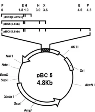
were screened by Congo red test, in which the positive clones developed a clear zone on the LB agar plate containing CMC. Only one clone among 2,500 clones showed halo, indicating the activity of CMC hydrolysis, and the transformant was named as pBC5 (Fig. 1). Plasmid analysis revealed that the insert gene of pBC5 had restriction sites of EcoRI, HincII, AvaI, XbaI, and
Sau3AI within 4.8 kb of the PstI DNA insert (Fig. 2). Subcloning. The insert DNA of pBC5 was separately
restricted with EcoRI and XbaI, and each fragment was
subcloned into the pGEMZf vector to determine the location of the gene expressing endoglucanase at 4.8 kb. The subclones, pBC6 and pBC8, harboring the foreign DNAs of 3.6 and 2.47 kb, respectively, were finally obtained by estimating the endoglucanase activity (Fig. 2). The endoglucanase activities of both pBC6 and pBC8 appeared to be similar to that of pBC5, suggesting the
2.47 kb fragment of pBC8 is the gene encoding endoglucanase.
Nucleotide sequence. The insert DNA of the
recombinant plasmid (pBC8) consisting of 2,472 bp was sequenced, and a single ORF of 1,500 bp encoding for a protein of 499 amino acid residues with a calculated molecular mass of 55 kDa was found in the inserted DNA. Shine-Dalgarno sequence, presumptive as a possible ribosome-binding site (5'-AGGAGG-3'), was found in 12 bases upstream of the putative ATG initiation codon. In addition, a putative promotor consensus was predicted using the Neural Network Promoter Prediction v. 2.2 software [Reese, 2001] with 5'-TAGACA-3' as the potential -35 region and 5'-TCCAAT-3' as the potential -10 region in the upstream region of ORF (Fig. 3). An inverted repeat sequence (5'-CGGACATC-3'), which could act as the transcriptional termination signal for the gene was followed by downstream of the stop codon (TAG). The 499-bp amino acid sequence of the ORF protein was compared with those of several bacterial endoglucanases by using the FASTDB alignment program. Comparison
Fig. 1. Comparison of endoglucanase activity encoded by E. coli transformants harboring pBC5. The cells
grown on LB agar were overlaid with 8 mL of top agar containing 0.5% CM-cellulose at 37oC for 12 h, followed
by Congo red staining. A: Bacillus sp. H12; B: E. coli
JM109/pBC5; C: E. coli JM109/pBluescript II SK(+).
Fig. 2. Restriction map of the recombinant plasmid pBC5 and subclones. Restriction endonuclease: X, XbaI;
E, EcoRI; H, HincII; P, PstI.
Fig. 3. Nucleotide sequence of the endo-β-1,4-glucanase
gene and deduced amino acid sequence. The deduced amino acid sequence is shown below the nucleotide sequence. Possible -35 and -10 regions of the promoter and a possible Shine-Dalgarno sequence are indicated. A putative signal sequence of the enzyme is underlined. Long arrows indicate the inverted repeats with the potential to form a rho-independent transcription termination structure.
of the deduced amino acid sequence showed significant similarities with the neutral cellulase genes of B. subtilis
PAP115 (99.2%) [MacKay et al., 1986], B. subtilis
BSE616 (97.8%) [Park et al., 1991], and Bacillus sp. N4
(55.1%) [Fukumori et al., 1986] known as the alkaline
cellulase gene (Fig. 4).
Localization of endoglucanase activity expressed in E. coli. The cellular localization of endoglucanase in E. coli carrying pBC8 was examined in extracellular,
periplasmic, and cytoplasmic fractions by measuring the endoglucanase activity. The endoglucanase enzymes produced by the E. coli transformant (pBC8) were
localized in the periplasmic (80%) and the cytoplasmic (20%) fractions. In contrast, no enzyme activity was observed in the extracellular fraction. (Table 1). In general, extracellular enzymes are known to contain a probable leader signal peptide for membrane transport
into the periplasmic space or the outer membrane [Wickner, 1979]. However, the leader signal sequence was not found in the N-terminal region of ORF, supporting the confinement of the endoglucanase activity of pBC8 in the cytoplasmic space without further excretion into the culture medium through the inner membrane.
Purification of endoglucanase. An endoglucanase
from the periplasmic fraction of the E. coli transformant
(pBC 8) was purified to homogeneity by means of gel filtration and anion exchange column chromatography. The molecular weight of the purified enzyme was estimated to be 31 kDa on SDS-PAGE (Fig. 5), and the optimal pH and temperature were 7.0 and 40oC,
respectively (data not shown). The apparent molecular weight of 31 kDa was somewhat low compared with the putative enzyme of 55 kDa encoded from the ORF, presumably because the carboxyl-terminus of the soluble enzyme was hydrolyzed by the post-translocation modification or proteolysis of the mature protein. The proteolysis of the recombinant proteins expressed in E. coli is not unusual, and similar proteolytic events have
been observed in other recombinant cellulases expressed
Fig. 4. Amino acid sequence homology between deduced sequences of the endo-β-1,4-glucanases from:
1) Bacillus sp. H12; 2) B. subtilis PAP115; 3) B. subtilis BSE616; 4) Bacillus sp. N4.
Table 1. Localization of endoglucanase in E. coli transformant (pBC8)
Strains Total cell extracts Endoglucanase activity (unit/min/mg of protein)Extracellular fraction Periplasmic fraction Cytoplasmic fraction
E. coli (pBC8) 1.5 - 1.2(80)a 0.3(20)a
B. subtilis H12 1.06 1.06 N.Db N.Db
aNumbers in parentheses indicate the percent distribution of activity between the cytoplasmic and the periplasmic
compartments.
bN.D: not determined.
Fig. 5. SDS-PAGE of each protein samples during purification step of endoglucanase from E. coli (pBC8).
Lane 1. Protein markers. Lane 2. Cytoplasmic fraction of
E. coli (pBC8). Lane 3. Periplasmic fraction of E. coli
(pBC8). Lane 4. Active fraction from gel filtration. Lane 5. Enzyme purified by anion chromatography.
in E. coli [Reverbel-Leroy et al., 1996].
Substrate specificities of endoglucanase. Substrate
specificity of the enzyme purified from the E. coli
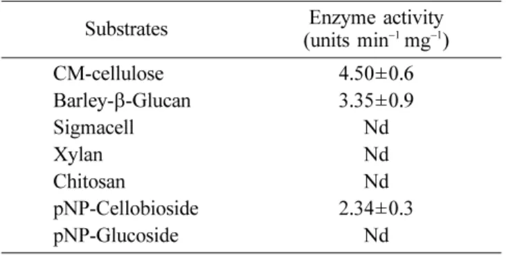
transformants harboring pBC8 was investigated by assessing its ability to hydrolyze various cellulosic substrates (Table 2). Specific activity of the enzyme was very high toward CMC and barely-β-glucan, whereas the
enzyme was completely inactive toward the microcrystalline celluloses such as xylan and sigmacell. These results suggest that endoglucanase encoded from pBC8 prefers the endo-type to the exo-type glycosides as the substrate, based on the report that CMC and barely-β-glucan are
typical substrates for endoglucanase [Yoon et al., 2007]. References
Baird SD, Johnson DA, and Seligy VL (1990) Molecular cloning, expression, and characterization of endo-β
-1,4-glucanase genes from Bacillus polymyxa and Bacillus circulans. J Bacteriol172, 1576-1586.
Bischoff K, Liu S, and Hughes SR (2007) Cloning and characterization of a recombinant family 5 endogluca-nase from Bacillus licheniformis strain B-41361. Pro-cess Biochem42, 1150-1154.
Cohen SN, Chang ACY, and Hso L (1973) Nonchromo-somal antibiotic resistance in bacteria: Genetic transfor-mation of Escherchia coli by R-factor DNA. Proc Natl Acad Sci USA70,3240-3244.
Fukumori F, Sashihara N, Kudo T, and Horikoshi K (1986) Nucleotide sequences of two cellulase genes from alkalophilic Bacillus sp. strain N-4 and their strong homology. J Bacteriol168, 479-485.
Hansen C, Diderichsen B, and Jorgensen PL (1992) celA from Bacillus lautus PL236 encodes a novel cellulose-binding endo-β-1,4-glucanase. J Bacteriol 174,
3522-3531.
Lima AO, Quecine MC, Fungaro M, Andreote FD, Maccheroni Jr M, Araújo WM, Silva-Filho M, Pizzirani-Kleiner A, and Azevedo JL (2005) Molecular
character-ization of a β-1,4-endoglucanase from an endophytic
Bacillus pumilus. Appl Microb Biotech 68, 57-65. Lynd LR, Weimer PJ, van Zyl WH, and Pretorius IS (2002)
Microbial cellulose utilization: fundamentals and biotech-nology. Microbiol Mol Biol Rev66, 506-577.
Mackay RM, Lo A, Willick G, Zuker M, Baird S, Dove M, Moranelli F, and Seligy V (1986) Structure of a Bacil-lus subtilis endo-β-1,4-glucanase gene. Nucleic Acid Res
14, 9159-9170.
Murasima K, Nisimura T, Nakamura Y, Koga J, Moriya T, and Sumida N (2002) Purification and characterization of new endo1,4-beta-glucanases from Rhizopus oryzae.
Enzyme Microb Tech30, 319-326.
Nossal NG, and Heppel LA (1966) The release of enzymes by osmotic shock from Escherichia coli in exponential phase. J Biol Chem241, 3055-3062. Ozaki K, Sumitmoto N, and Ito S (1991) Molecular
clon-ing and nucleotide sequence of a gene encodclon-ing an endo-β-1,4-glucanase gene from Bacillus sp. KSM-330.
J Gen Microbiol137, 2299-2305.
Park SH, Kim HK, and Pack MY (1991) Characterization and structure of the cellulase gene of Bacillus subtilis
BSE616. Agr Biol Chem55, 441-448.
Reese MG (2001) Application of a time-delay neural net-work to promoter annotation in Drosophila melano-gaster genome. Comput Chem 26, 51-56.
Reverbel-Leroy C, Belaich A, Bernadac A, Gaudin C, Bela-ich JP, and Tardif C (1996) Molecular study and overex-pression of the Clostridium cellulolyticum celF cellulose gene in Escherichia coli. Microbiology142, 1013-1023. Saito H and Miura K (1963) Preparation of transforming
DNA by phenol treatment. Biochim Biophys Acta 72, 619-629.
Sashihara N, Kudo T, and Horikoshi K (1984) Molecular cloning and expression of cellulase genes of alkalophilic
Bacillus sp. strain N-4 in Escherichia coli. J Bacteriol
158, 503-506.
Somogyi M (1952) Notes on sugar determination. J Biol Chem195, 19-23.
Teather RM & Wood PJ (1982) Use of Congo red-polysac-charide interactions in enumeration and characterization of cellulolytic bacteria from the bovine rumen. Appl Environ Microb43, 777-780.
Wickner W (1979) The Assembly of proteins into biologi-cal membranes: the membrane trigger hypothesis. Ann Rev Biochem48, 23-45.
Woodward J (1991) Synergism in cellulase systems. In
Enzymatic Hydrolysis of Cellulose, Walker LP and Wil-son DB (ed), pp. 67-75. Elsevier Applied Science, New York.
Wymelenberg AV, Denman S, Dietrich D, Bassett J, Yu X, Atalla A, Predki P, Rudsander U, Teeri T, and Cullen D (2002) Transcript analysis of genes encoding a family 61endoglucanase and a putative membrane-anchored family 9 glycosyl hydrolase from Phanerochaete chry-sosporium. Appl Environ Microb68, 5765-5768.
Table 2. Substrate specificities of endoglucanase purified from E. coli (pBC8)
Substrates (units minEnzyme activity−1 mg−1)
CM-cellulose 4.50±0.6 Barley-β-Glucan 3.35±0.9 Sigmacell Nd Xylan Nd Chitosan Nd pNP-Cellobioside 2.34±0.3 pNP-Glucoside Nd Nd: not detected
Yoda K, Toyoda A, Mukoyama Y, Nakamura Y, and Minato H (2005) Cloning, sequencing, and expression of a
Eubacterium cellulosolvens 5 gene encoding an endoglu-canase (Cel5A) with novel carbohydrate-binding mod-ules, and properties of Cel5A. Appl Environ Microb71,
5787-5793.
Yoon MH and Choi WY (2007) Characterization and action patterns of two β-1,4-glucanases purified from