142
INTRODUCTION
Esophageal carcinoma frequently metastasizes to the lungs and liver, but carcinoma of the esophagus has been considered an uncommon source of metastatic brain tumor [1,2]. Three studies from Japan reported the incidence of brain metastasis in patients diagnosed with esophageal cancer in the 1980s to early 2000s as ranging from 1.4–1.5% [3,4]. Several other stud-ies reported that the incidence of brain metastasis in a con-temporary series of patients with carcinoma of the esophagus ranged from 0.5% to 4.8% [3-5]. Although some studies have mentioned esophageal cancer with solitary brain metastasis [6-8] or with meningocerebral metastasis [1,2,9] or with skull metastasis [1,10], multiple meningocerebral metastasis and extensive skull metastasis from squamous cell carcinoma of esophagus has not been reported in the literature. We report a patient with an extensive osteolytic change of the skull and multiple meningocerebral metastases from esophageal
carci-Multiple Meningocerebral Metastasis and Extensive Skull
Metastasis from Squamous Cell Carcinoma of Esophagus:
A Case Report and Review of Literature
Min Kyun Na, Choong Hyun Kim, Jae Min Kim, Jin Whan Cheong, Je Il Ryu, Hyun Woo Kim
Department of Neurosurgery, Hanyang University Guri Hospital, Guri, Korea
Received December 16, 2015 Revised September 7, 2016 Accepted September 19, 2016 Correspondence
Choong Hyun Kim Department of Neurosurgery, Hanyang University Guri Hospital, 153 Gyeongchun-ro,
Guri 11923, Korea Tel: +82-31-560-2322 Fax: +82-31-560-2327
E-mail: [email protected]
Esophageal carcinoma rarely metastasizes to the brain. Although some studies have mentioned esoph-ageal cancer with solitary brain metastasis or with meningocerebral metastasis or with skull metastasis, multiple meningocerebral metastasis and extensive skull metastasis from squamous cell carcinoma of esophagus has not been reported in the literature. We encountered a case of an extensive osteolytic change of the skull and multiple meningocerebral metastases from esophageal carcinoma.
Key Words Brain metastasis; Esophageal carcinoma.
noma. In this report, we emphasized the clinical and radiologi-cal findings of the patient with skull and multiple meningocer-ebral metastases from an esophageal carcinoma, and reviewed the pertinent literatures.
CASE REPORT
A 71-year-old man presented with a 3-month history of headache and enlarging painless scalp mass on both high pa-rietal convexities. He was undertaken esophagectomy, Ivor-Lewis operation, for esophageal cancer and confirmed as a sq-uamous cell carcinoma of the esophagus 3 years ago. He had received chemotherapy and radiotherapy for esophageal car-cinoma. The physical examination showed a subcutaneous mass over both parietal convexities. There were several small and non-tender lymph nodes in the right supraclavicular re-gion, but no axillary lymphadenopathy. There were no sig-nificant deficits on the neurological examination. The brain computed tomography (CT) scans demonstrated brain me-tastasis with peritumoral edema in the right temporal lobe and the left parietal lobe. In addition, expansile osteolytic change was observed in the skull vault and both parietal convexities (Fig. 1). The soft tissue tumor was measured about 8.9× CASE REPORT Brain Tumor Res Treat 2016;4(2):142-144 / pISSN 2288-2405 / eISSN 2288-2413http://dx.doi.org/10.14791/btrt.2016.4.2.142
This is an Open Access article distributed under the terms of the Creative Commons Attribution Non-Commercial License (http://creativecommons.org/licenses/by-nc/3.0) which permits unrestricted non-commercial use, distribution, and reproduction in any medium, provided the original work is properly cited.
Copyright © 2016 The Korean Brain Tumor Society, The Korean Society for Neuro-Oncology, and The Korean Society for Pediatric Neuro-Oncology
MK Na et al.
143
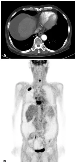
5.5×4.3 cm in size with intra- and extra-cranial compression of the brain. Brain magnetic resonance imaging (MRI) scans showed two brain metastatic lesions with peritumoral edema in the right temporal lobe and the left parietal lobe. Diffuse meningeal thickening and enhancement was also noted in the fronto-parietal area (Fig. 2). A CT scan of his chest showed diffuse wall thickening of the lower esophagus from subcari-na to epiphrenic portion (Fig. 3A). Positron emission tomog-raphy showed multiple metastatic foci in the brain, bones, right lung, liver, anastomotic site of esophagus, right adrenal gland, and mesenteric lymph nodes (Fig. 3B). The patient underwent operation by a bicoronal scalp incision with bi-frontal craniectomy, removal of the bony tumor, and cranio-plasty. Pathological findings revealed the metastatic squamous cell carcinoma. The final diagnosis was squamous cell carci-noma of the esophagus with skull metastasis. The patient was referred for concurrent chemotherapy and radiation therapy. Two month later, the patient complained chest discomfort and underwent fine needle aspiration biopsy, diagnosed as a small cell lung cancer. Then, he could not take chemotherapy due to his age and poor general condition.
DISCUSSION
The incidence of esophageal cancer has been increasing over the past thirty years [11], with an expected 16,470 cases and 14,530 deaths in the United States in 2009 [12]. Nevertheless,
esophageal carcinoma is among the most challenging onco-logic problems. It is well known that esophageal cancer may cause distant metastasis, most frequently in the lungs, pleura, liver, peritoneum, and the adrenal gland. However, metastasis to the bones is infrequent, and skull metastasis from esopha-geal cancer may be extremely rare [1,2]. Brain metastasis sec-ondary to esophageal carcinoma is considered to be a rare event, with a reported incidence of between 0.5 and 4.8% [3-5]. Several studies in the United States also reported low inci-dence of brain metastasis in patients with esophageal cancer [5,11,13]. Although some studies have reported esophageal cancer with brain metastasis [6-8], dural metastasis has been reported only 3 case in the past literature [1,2,9]. Akhavan and Navabii [1] reported a case of leptomeningeal carcino-matosis from squamous cell carcinoma of esophagus pre-sented by hoarseness. Chen and Huang [2] reported the case of esophageal squamous cell carcinoma with dural and bone marrow metastases. This report also discussed the pathogen-esis of unusual metastatic diseases and differential diagnosis of pachymeningeal thickening. Irie et al. [9] reported the case of solitary meningocerebral metastasis from squamous cell car-cinoma of the esophagus. Stark et al. [14] reported the charac-teristic clinical features of metastatic skull tumor, which are distinctive from those of primary skull tumor. Metastatic skull tumor typically appear as expansile, osteolytic, hypervascular lesions and MRI scans showed iso- or hypointensity on T1 and T2 weighted images with moderate enhancement. And
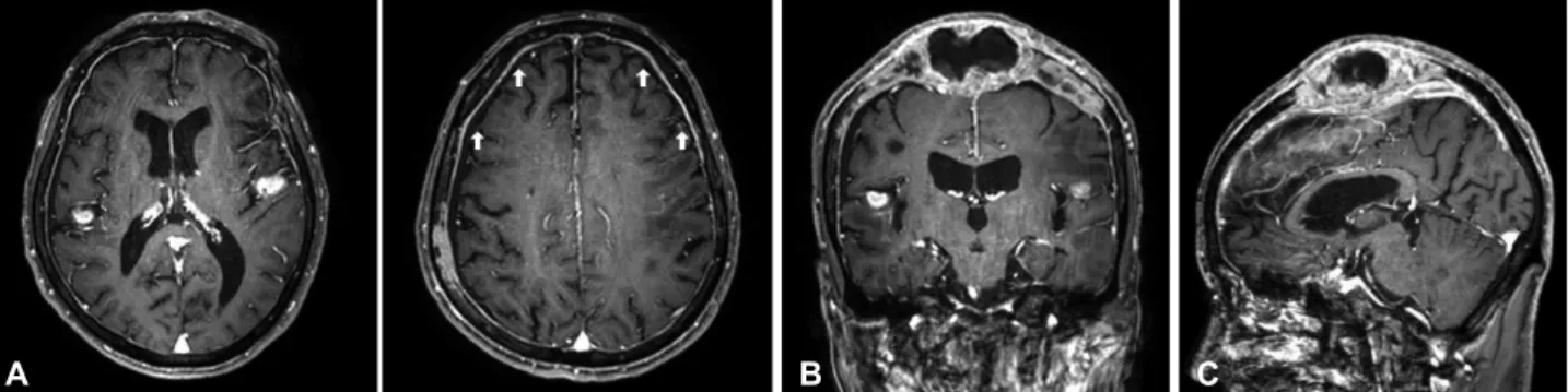
Fig. 2. Magnetic resonance images with gadolinium enhancement of the patient. A: Axial image shows well-enhanced mass the in right
temporal lobe and left parietal lobe. Diffuse dural thickening and enhancement was also noted in bilateral pachymeninges (arrows). Coronal (B) and sagittal (C) images represent the extensive osteolytic lesion in the vault of the skull and enhanced tumor in the left temporal lobe.
A B C
144 Brain Tumor Res Treat 2016;4(2):142-144 Meningocerebral Metastasis of Esophageal Carcinoma
can cause local swelling that is usually painless, but they rarely lead to neurologic dysfunction [14]. However, Ellis and Mc-Donald [15] reported a case of acute epidural hematoma sec-ondary to skull metastasis from esophageal carcinoma, which proved that skull metastasis can also lead to severe neurolog-ic dysfunction. For the metastatneurolog-ic skull tumor, surgneurolog-ical treat-ment should be considered in patients who have neurologic consequences, for example, high risk of extradural or
intra-tumoral hemorrhage or brain compression. Surgical resection might not cure the underlying disease, but it is a relatively safe and satisfactory treatment for relief of neurologic symptoms in patients with cranial metastasis.
This is an uncommon case of squamous cell carcinoma of the esophagus including leptomenigeal dissemination, exten-sive osteolytic skull lesions, and multiple brain metastases. Conflicts of Interest
The authors have no financial conflicts of interest. REFERENCES
1. Akhavan A, Navabii H. Leptomeningeal metastasis from squamous cell carcinoma of oesophagus with unusual presentation. BMJ Case Rep 2012 Nov 21 [Epub]. http://dx.doi.org/10.1136/bcr-02-2012-5846. 2. Chen YH, Huang CH. Esophageal squamous cell carcinoma with dural and bone marrow metastases. World J Gastroenterol 2014;20:12691-5. 3. Ogawa K, Toita T, Sueyama H, et al. Brain metastases from esophageal
carcinoma: natural history, prognostic factors, and outcome. Cancer 2002;94:759-64.
4. Yoshida S. Brain metastasis in patients with esophageal carcinoma. Surg Neurol 2007;67:288-90.
5. Weinberg JS, Suki D, Hanbali F, Cohen ZR, Lenzi R, Sawaya R. Metas-tasis of esophageal carcinoma to the brain. Cancer 2003;98:1925-33. 6. Nwankwo N, Mirrakhimov AE, Wind KP, Bucher N. Brain metastasis
from oesophageal adenocarcinoma. BMJ Case Rep 2013 Apr 19 [Epub]. http://dx.doi.org/10.1136/bcr-2013-009395.
7. Agrawal R, Shukla P, Shukla V, Chauhan A. Brain metastasis from esophageal carcinoma. J Cancer Res Ther 2009;5:137-9.
8. Averbuch SD, Blinder MA. Brain metastasis from esophageal carcino-ma. CA Cancer J Clin 1983;33:164-6.
9. Irie K, Austin E, Morgenstern L. Solitary meningocerebral metastasis from squamous cell carcinoma of the esophagus: a case report. Cancer 1978;42:2461-5.
10. Kao CH, Shang CT, Lin YC, Li YF, Cheng YL. Esophageal carcinoma presented with a skull tumour. Can J Surg 2009;52:E215-6.
11. Pohl H, Welch HG. The role of overdiagnosis and reclassification in the marked increase of esophageal adenocarcinoma incidence. J Natl Cancer Inst 2005;97:142-6.
12. Jemal A, Siegel R, Ward E, Hao Y, Xu J, Thun MJ. Cancer statistics, 2009. CA Cancer J Clin 2009;59:225-49.
13. Brown LM, Devesa SS, Chow WH. Incidence of adenocarcinoma of the esophagus among white Americans by sex, stage, and age. J Natl Cancer Inst 2008;100:1184-7.
14. Stark AM, Eichmann T, Mehdorn HM. Skull metastases: clinical fea-tures, differential diagnosis, and review of the literature. Surg Neurol 2003;60:219-25; discussion 225-6.
15. Ellis MJ, McDonald PJ. Acute epidural hematoma secondary to skull metastasis from esophageal carcinoma. Can J Neurol Sci 2007;34:491-3.
A
B
Fig. 3. CT and Positron emission tomography (PET) scans show
esophageal cancer and multiple metastatic lesions. A: Contrast en-hanced computed tomography scan of the chest showing diffuse wall thickening of the lower portion of esophagus (arrow). B: PET scans showing multiple metastatic foci in bones, right lung, anasto-motic site of esophagus, right, and mesenteric lymph nodes.