Received May 20, 2010; Accepted October 11, 2010 Corresponding author: Jung Hwa Choi
Department of Rehabilitation Medicine, Yonsei University College of Medicine, Sinchon-dong, Seodaemun-gu, Seoul 120-752, Korea Tel: 82-2-2227-3131, Fax: 82-2-2019-3499, E-mail: [email protected]
This study was supported by a faculty research grant of Yonsei University College of Medicine for 2008 (6-2008-0177). Copyright © 2011 by Korean Academy of Rehabilitation Medicine
Myogenic Differentiation of Human
Adipose-Derived Stem Cells
Yoon Ghil Park, M.D.
1, Ah Mi Baek
2, Byung Rok Do, Ph.D.
3, Jung Hwa Choi, M.D.
4, Sun
Do Kim, M.D.
41
Department of Rehabilitation Medicine and 2Rehabilitation Institute of Muscular Disease, Yonsei University College of Medicine, Seoul 135-720, 3Hurim Biocell, Seoul 157-810, 4Department of Rehabilitation Medicine, Yonsei University College of Medicine, Seoul 120-752, Korea
Objective Cell therapy has been extensively studied as a gene complementation approach in muscular dystrophy
including Duchenne muscular dystrophy (DMD), and adipose tissue has recently been identified as a uniquely abun-dant and adequately accessible source of pluripotent cells. In the present work, we investigated myogenic potentials of adipose-derived stem cells (ADSCs) depending on culture media and isolation with using surface markers.
Method Human ADSCs were obtained by liposuction and cultured in two different media; control and myogenic
media. In addition we attempted to isolate ADSCs by utilizing surface markers: CD45 and CD133. The following obser-vations were made to evaluate myogenic differentiation as the expression of myogenic regulatory factors (MyoD, Myf-5 and Myf-6) and desmin by RT-PCR and immunoflurescence study.
Results Conversion of ADSCs to myogenic phenotype was observed by indirect immunoflurescence study of MyoD and
Myf-5 in regardless of media type and isolation method. In addition mRNA of MyoD and Myf-5 were positive in both culture media, and there were no differences of MyoD and Myf-5 responses between CD45− and CD45−CD133− ADSCs. However, secondary myogenic regulatory factor (Myf-6) was not expressed constantly, and desmin were negative in all cultural condition.
Conclusion Our findings suggest that human ADSCs might have myogenic potentials. However, further studies are
needed to express the secondary myogenic regulatory factors and proteins in myoblasts.
Key Words Muscular dystrophy, Stem cell, Differentiation, Myogenic regulatory factor, Desmin
INTRODUCTION
Duchenne muscular dystrophy is recessively inherited through sex chromosomes. It is known to be caused by the genetic variation of dystrophin located in the short arm (Xp21) of the X chromosome, and occurs in 1 out
of 3,500 baby boys.1-3 There is currently no cure for
DMD, although comprehensive rehabilitation inducing physiotherapy, kinesitherapy and pulmonary rehabilita-tion have been adhibited to minimize progress and complications.2 But in recent times, intensive research is underway. One of those experimental treatments is
based on myoblast transfer. The myoblast transfer has been developed to fuse normal cells with the patients’ myocytes and thus to induce the production of dystro-phin protein. However, this approach has a number of limitations including a low rate of spread, the poor survival of the injected myoblasts, and an insufficiency of myoblasts, so there has been still controversy as to its clinical efficacy.3-6
In particular, immunorejection is a principal problem in myoblast graft. To solve the problem, an attempt has been made to apply stem cells obtained from cord blood, bone marrow, muscles and fat. Adipose tissue eases donors’ burden as it can be collected through lip-osuction, as well as tissue can be collected in quantity.7-12
However, the problem is that adipose-derived stem cells (ADSCs) may be differentiated into various tissues such as bone, cartilage and muscle as most of them are mesenchymal tissues. Thus, it is imperative to control them to be differentiated only into myocytes in the process of isolation and culture.13
Embryologically, it is known that various factors are concerned in the process of myogenic differentiation, but the whole process has not been unraveled yet. Meanwhile, the basic helix-loop-helix (bHLH) tran-scription factor, i.e., a myogenic regulatory factor (MRF), has been reported to take an important role. MyoD and Myf-5, primary MRFs, lead ADSCs to myoblasts. Myo-genin and Myf-6, secondary MRFs, differentiate myo-blasts into myotubes,13-15 which can be proved by mor-phologic changes, caused by stem cells’ being differ-entiated into myocytes, and MRFs’ being expressed in the process of differentiation.
Since it is difficult to control the differentiation of stem cells, various differentiations have been attempted under various conditions. The diversification of media is one of such methods.12,13,16,17 But according to previous
studies, the diversification of media has limitations to the perfect myogenic differentiation. To overcome such limitations, it may be effective to separately culture myogenic stem cells. In this connection, surface mark-ers, such as CD45, CD133, CD34, Sca-1 and c-kit, can be used to sort cells effective for myogenic differentia-tion.18,19
The Purpose of this study was to identify proper conditions for myogenic differentiation, by extension, to ascertain whether surface markers are useful to sort cells. In line with these objectives, an observation was made of whether myogenic differentiation would be in-fluenced by different media of which composition was
adjusted by stem cells, for which the expression of MRFs was checked. In addition, an observation was made of the differentiation of adipose-derives stem cells sorted by surface markers.
MATERIALS AND METHODS
Cell sorting and primary culture
HURIM BIOCELL, a biotech company located in Seoul, Korea, primarily cultured ADSCs by using adipose tissue collected through liposuction, and delivered them to re-searchers as in the following.
The collected adipose tissue was rinsed with phos-phate buffered saline (PBS, GIBCO BRL, Grand Island, USA) 2 times, which removed blood and foreign sub-stances. Then, it was suspended in 0.075% collagenase (Sigma, St. Louis, USA) which dissolved in PBS at 37oC
for 30 minutes, whereby extracellular matrix was lysed. For reference, the mixture was the same amount as the adipose tissue. After that, it was mixed with Dulbeco’s modified Eagles medium (DMEM, GIBCO) and 10% fetal bovine serum (FBS, GIBCO) in order to decelerate lysis. Next, it was centrifuged at 250 g for 10 minutes, which precipitated cells. The precipitated cells were treated with culture medium (10% FBS-DMEM) and 0.16 M NH4Cl, waited for 10 minutes in room temper-ature, and then were centrifuged at 250 g for 10 minutes. The precipitate was soaked in culture medium (10% FBS-DMEM), and was divided onto 75-square-cen-timeter culture flasks (NUNC, Rocklde, Denmark) by 1×10⁶/mL. It was cultured in the 5% CO
2 incubator at
37oC, and the culture medium was replaced at intervals of 1 week. When the confluence of cells reached 70% and over, cells were separated from the culture dish by the use of 0.05% trypsin-0.53 mM EDTA. After being centrifuged, it was subcultured. Since then, the culture medium was replaced at intervals of 3 days.
Induction of myogenic differentiation
Media-based differentiation: After being primarily cul-tured, cells were divided into two groups. One group was cultured in a medium composed of 10% FBS- DMEM, 1% penicillin and streptomycin (GIBCO). The medium was defined as the control medium, and the group cultured in the control medium was defined as the control group. The other group was cultured in a medium composed of the control medium, 5% horse serum (GIBCO) and 50 μM hydrocortisone (Sigma). The
medium was used in previous studies.12,20 The medium was defined as the myogenic medium, and the group cultured in the myogenic medium was defined as the myogenic group. The two groups were cultured in the 5% CO2 incubator at 37oC for 21 days, whereat the
ex-pression of MRFs and myofascial protein, i.e., desmin, was observed through immunofluorescene stain and Western blot. The expression of mRNA was observed through RT-PCR.
Surface markers-based differentiation: CD45 and CD133, markers of myogenic stem cells, were applied to mag-netic-activated cell sorting (MACS) as in the following: After being primarily cultured, ADSCs were treated with the CD45 antibody (Miltenyi Biotec GmbH, Bergisch Gladbach, Germany) in the proportion of 1:100 at room temperature for 30 minutes. Cells, which re-sponded to the primary antibody, were cleansed with 1×PBS (original concentration) 2 times. Then, they were made to respond to 20 μL of goat anti-mouse IgG mi-crobeads (Miltenyi Biotec GmbH) at 6 to 12oC for 15
minutes after being soaked in 80 μL of PBS. Next, the supernatant was removed by centrifugation after dilu-tion with decuple PBS. By the use of MiniMACS separa-tion system (Miltenyi Biotec GmbH), the cells were div-ided into ones sorted by the magnetic field (CD45+) and ones were not sorted (CD45−), only CD45− cells of which were cultured. Likewise, CD133 cells were sorted by the use of CD45− cells in the same way. A group composed of cells that responded negatively to CD45− cells and another group of cells that responded neg-atively not even to the CD133 antibody (Miltenyi Biotec GmbH) (CD45−CD133−) were cultured in the control medium and the myogenic medium respectively. An analysis was made of differentiation, like in primarily cultured cells.
Analysis of differentiation
RT-PCR (reverse transcription-polymerase chain reac-tion): The differentiated cells were washed with 1×PBS 2 times. Total RNAs were extracted with the Trizol Reagent as per the manufacturer’s protocol (Invitrogen, Carlsbad, USA). Three microgram amounts of total RNA were reversetranscribed using M-MuLV reverse tran-scriptase (Promega, Madison, USA) at 37oC for 1 h.
Gene expression of human cells was evaluated using primers specific for human cDNAs. These were im-plemented by the use of PTC-500 thermocycler (MJ Research, Waltham, USA). The followings are primer pairs used for amplification.
MyoD forward: 5'-GTCGAGCCTAGACTGCCTGT-3', reverse: 5'-GGTATATCGGGTTGGGGTTC-3'
Myf-5 forward: 5'-GCCTGAAGAAGGTCAACCAG-3', reverse: 5'-CCATCAGAGCAGTTGGAGGT-3'
Myf-6 forward: 5'-GCCAAGTGTTTCCGATCATT-3', reverse: 5'-CTTCTCCACCACTTCCTCCA-3'
Desmin forward: 5'-CCTACTCTGCCCTCAACTTC-3', re-verse: 5'-AGTATCCCAACACCCTGCTC-3'
ImmunoFluorescence stain: The cultured cells were washed with 1×PBS after being fixed with a mixture of cold acetone and methanol (1:1). After this, the sam-ples were blocked with PBS containing 2% bovine-se-rum albumin (BSA, Sigma) and 5% goat sebovine-se-rum (Sigma) for 30 minutes at room temperature, and incubated with the primary antibody (Santa Cruz Biotechnology, Santa Cruz, USA) of MyoD, Myf-5, Myf-6 and desmin overnight at 4oC. The cells were washed with 1×PBS 3 times and then they were kept at room temperature for 30 minutes with secondary species-specific Abs (Vector Laboratories, Inc., USA). They were washed with 1×PBS 3 times, incubated with avidin-fluorescein iso-thiocyanate conjugated (FITC) for 30 minutes at room temperature, and again washed with 1×PBS. The ensu-ing color development was observed through a fluo-rescence microscope (Nikon Eclipse 80i, Nikon, Tokyo, Japan). MyoD, Myf-5 and Myf-6 were regarded as pos-itive only when their nuclei were stained. In the case of desmin, the positive reaction was confined to cases where cytoplasm was stained.
RESULTS
Differentiation in primarily cultured cells
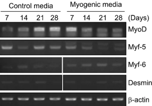
Expression of MRFs (MyoD, Myf-5 and Myf-6): A 28- day culture was performed with the control medium and the myogenic medium, during which the mRNA levels of MRFs (MyoD, Myf-5 and Myf-6) were meas-ured by RT-PCR. MyoD was expressed in both groups. In the case of the control group, the expression of MyoD increased in proportion to time. In the myogenic group, expression increased immediately after replace-ment of medium but gradually decreased with time. Myf-5 was similar to MyoD in the pattern of expres-sion, but its expression more increased in the control group than in the myogenic group. The expression of Myf-6 was more increased in the myogenic group than in the control group, but its increase was gradual, un-like MyoD and Myf-5 (Fig. 1).
Fig. 1. Myogenic differentiation of primary cultured ADSCs
(adipose-derived stem cells), as determined by RT-PCR for expression of MyoD, Myf-5, Myf-6 and Desmin mRNAs. Expression of β-actin mRNA was used as a loading control. ADSCs were cultured in each control and myogenic media.
Fig. 2. Myogenic regulatory
fac-tors and desmin expression by ADSCs (adipose-derived stem cells) cultured in control and myo-genic media. ADSCs were stained with avidin-FITC(fluorescein iso-thiocyanate conjugated) and vi-sualized by a fluorescent micro-scope on days 7 and 21. By day 7 MyoD and Myf-5 were ex-tensively expressed in the nu-clei of most cells (×200). How-ever, Myf-6 was not expressed in the nuclei, and desmin was totally negative through day 21.
To examine the protein expression changes of MRFs under different conditions, primary cells were cultured for 7 and 21 days respectively using control or myo-genic medium. In the case of MyoD and Myf-5, intra-nuclear expression increased 7 days later in both the control medium and the myogenic medium, but inter-group significant differences were not observed. Myf-6 did not show intranuclear staining even 21 days later (Fig. 2).
Desmin: During the period, any significant expressions were not observed not only in protein but in mRNA regardless of the conditions of media (Fig. 1 and 2). Differentiation in surface markers
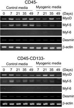
Expression of MRFs (MyoD, Myf-5 and Myf-6): ADSCs, divided into CD45− and CD45−CD133−, were cultured in the control medium and the myogenic medium re-spectively for 49 days, during which the mRNA ex-pressions of MyoD, Myf-5 and Myf-6 were measured by RT-PCR at intervals of 2 weeks. MyoD was expressed throughout the period and seemed to increase with time, but did not show a meaningful tendency. Myf-5 was more expressed in CD45− and CD45−CD133− con-trol media than in the myogenic medium. The ex-pression of Myf-6 was not observed throughout the pe-riod (Fig. 3).
Cells, divided into CD45− and CD45−CD133−, were cultured in the control medium and the myogenic me-dium for 21 days. On the immunofluorescence stain test, MyoD and Myf-5 were observed in both groups, but intergroup significant differences were not observed. Myf-6 did not show intranuclear staining in both groups.
Desmin: During the study, protein was not expressed not only on the immunofluorescence stain test but on
Fig. 3. Myogenic differentiation of isolated ADSCs with
us-ing CD45/CD133, as determined by RT-PCR for expression of MyoD, Myf-5, Myf-6 and Desmin mRNAs. Expression of β-actin mRNA was used as a loading control. ADSCs were cultured for 42 days in each control and myogenic media.
the Western blot regardless of surface markers (not shown), and besides, significant changes were not ob-served even in the mRNA level (Fig. 3).
DISCUSSION
Mammals’ adipose tissue consists of adipocytes and collagenous fiber in addition to the stromal vascular fraction. Thus, it has been reported that adipose tissue can be variously differentiated according to culture con-ditions, in common with bone marrow.12,21,22 Asakura et
al.23 and Csete et al.24 reported that myoblasts could be
differentiated into osteocytes and adipocytes, which shows the possibility that musculoskeletal cells, osteo-cytes, chondrocytes and adipocytes have the same mes-enchymal origin and they share regulatory process and factors when they are differentiated into each organ. Mesenchymal stem cells, extracted from such organs, are easily differentiated into related cells.25
So far, mesenchymal stem cells have been extracted from bone marrow in many cases. But it causes incon-venience to donors and also they cannot be produced
in quantity. As the solution, interest has been con-centrated on adipose tissue extracted through liposuc-tion.
Several studies reported that ADSCs could be differ-entiated to hoped-for cells by changing the composi-tions of media.12,17,22 In this study, the expression of
primary MRFs showed a positive reaction with time, regardless of the conditions of media. But when the differentiation of primarily cultured cells was induced the myogenic medium, MyoD was early expressed and decreased as compared to the control medium, which is assumed that the myogenic medium would accelerate the myogenic differentiation of ADSCs. It is because in the process of myogenic differentiation, secondary MRFs are expressed after primary MRFs are activated, and then primary MRFs tend to be deactivated. But in the process where myogenic differentiation, MyoD and Myf- 5, primary MRFs, showed positive reactions on the im-munofluorescence stain test early in culture. On the other hand, Myf-6, a secondary MRF, showed a negative reaction even after culture. In the case of desimin pro-tein in myoblasts, a negative reaction was continuously maintained and as a result was judged to be unsuitable for myogenic differentiation. The results imply that a perfect differentiation cannot be induced by myogenic medium used in this study and the applied condition. Accordingly, there is a need to conduct a further study with factors to promote differentiation and various analyses.
ADSCs, cultured after being sorted by CD45 and CD133, were not much different from primarily cul-tured cells in differentiation. It means that cells, sorted by surface markers, are not effective to induce differ-entiation. Thus, it may be effective to apply other methods to the induction of differentiation without ad-ditional cell sorting.
CONCLUSION
In this study, a myogenic differentiation was partially induced by the use of ADSCs. These findings suggest that human ADSCs might have myogenic potentials. So the results of this study are expected to be a basic step for the differentiation of ADSDs into myoblasts. However, further studies are needed to express the sec-ondary myogenic regulatory factors and proteins in myoblasts.
REFERENCES
1) Hoffman EP, Brown RH Jr, Kunkel LM. Dystrophin: the protein product of the Duchenne muscular dystrophy locus. Cell 1987; 51: 919-928
2) Kilmer DD. Myopathy. In: DeLisa JA, editor. Physical med-icine and Rehabilitation: principles and practice, 4th ed, Philadelphia: Lippincott Williams & Wilkins, 2005, 913- 929
3) Huard J, Labrecque C, Dansereau G, Robitaille L, Tremblay JP. Dystrophin expression in myotubes formed by the fu-sion of normal and dystrophic myoblasts. Muscle Nerve 1991; 14: 178-182
4) Morgan JE, Hoffman EP, Partridge TA. Normal myogenic cells from newborn mice restore normal histology to de-generating muscles of the mdx mouse. J Cell Biol 1990; 111: 2437-2449
5) Partridge TA, Morgan JE, Coulton GR, Hoffman EP, Kunkel LM. Conversion of mdx myofibres from dystrophin-neg-ative to -positive by injection of normal myoblasts. Nature 1989; 337: 176-179
6) Kang SW, Choi YC. Rehabilitation of Neuromuscular disease. In: Park CI, Moon JH, editors. Rehabilitation med-icine, 1st ed. Seoul: Hanmi, 2007, 649-677
7) Kogler G, Callejas J, Hakenberg P, Enczmann J, Adams O, Daubener W, Krempe C, Gobel U, Somville T, Wernet P. Hematopoietic transplant potential of unrelated cord blood: critical issues. J Hematother 1996; 5: 105-116 8) Querol S, Capmany G, Azqueta C, Gabarro M, Fornas O,
Martin-Henao GA, Garcia J. Direct immunomagnetic meth-od for CD34+ cell selection from cryopreserved cord blood grafts for ex vivo expansion protocols. Transfusion 2000; 40: 625-631
9) Ferrari G, Cusella-De Angelis G, Coletta M, Paolucci E, Stornaiuolo A, Cossu G, Mavilio F. Muscle regeneration by bone marrow-derived myogenic progenitors. Science 1998; 279: 1528-1530
10) Rubinstein P, Dobrila L, Rosenfield RE, Adamson JW, Migliaccio G, Migliaccio AR, Taylor PE, Stevens CE. Pro-cessing and cryopreservation of placental/umbilical cord blood for unrelated bone marrow reconstitution. Proc Natl Acad Sci USA 1995; 92: 10119-10122
11) Lequerica JL, Mirabet V, Montero JA, Hurtado C, Piquer S, Carbonell F. In vitro proliferation, differentiation and im-muno-magnetic bead purification of human myoblasts. Ann Transplant 1999; 4: 103-108
12) Zuk PA, Zhu M, Mizuno H, Huang J, Futrell JW, Katz AJ, Benhaim P, Lorenz HP, Hedrick MH. Multilineage cells from human adipose tissue: implications for cell-based therapies. Tissue Eng 2001; 7: 211-228
13) Park YG, Moon JH, Lee EY, Do BR, Kim JH, Kim KT, Kim DS. Myogenic potential of human adipose-tissue-derived cells. J Korean EMG Electrodiagn Med 2007; 9: 69-74 14) Rudnicki MA, Jaenisch R. The MyoD family of
tran-scription factors and skeletal myogenesis. Bioessays 1995; 17: 203-209
15) Sabourin LA, Rudnicki MA. The molecular regulation of myogenesis. Clin Genet 2000; 57: 16-25
16) Mizuno H, Hyakusoku H. Mesengenic potential and future clinical perspective of human processed lipoaspirate cells. J Nippon Med Sch 2003; 70: 300-306
17) Kim M, Choi YS, Yang SH, Hong HN, Cho SW, Cha SM, Pak JH, Kim CW, Kwon SW, Park CJ. Muscle regeneration by adipose tissue-derived adult stem cells attached to in-jectable PLGA spheres. Biochem Biophys Res Commun 2006; 348: 386-392
18) Montarras D, Morgan J, Collins C, Relaix F, Zaffran S, Cumano A, Partridge T, Buckingham M. Direct isolation of satellite cells for skeletal muscle regeneration. Science 2005; 309: 2064-2067
19) Jankowski RJ, Haluszczak C, Trucco M, Huard J. Flow cy-tometric characterization of myogenic cell populations ob-tained via the preplate technique: potential for rapid iso-lation of muscle-derived stem cells. Hum Gene Ther 2001; 12: 619-628
20) Di Rocco G, Iachininoto MG, Tritarelli A, Straino S, Zacheo A, Germani A, Crea F, Capogrossi MC. Myogenic potential of adipose-tissue-derived cells. J Cell Sci 2006; 119: 2945- 2952
21) Bacou F, el Andalousi RB, Daussin PA, Micallef JP, Levin JM, Chammas M, Casteilla L, Reyne Y, Nougues J. Trans-plantation of adipose tissue-derived stromal cells increases mass and functional capacity of damaged skeletal muscle. Cell Transplant 2004; 13: 103-111
22) Safford KM, Hicok KC, Safford SD, Halvorsen YD, Wilkison WO, Gimble JM, Rice HE. Neurogenic differentiation of murine and human adipose-derived stromal cells. Biochem Biophys Res Commun 2002; 294: 371-379
23) Asakura A, Komaki M, Rudnicki M. Muscle satellite cells are multipotential stem cells that exhibit myogenic, osteo-genic, and adipogenic differentiation. Differentiation 2001; 68: 245-253
24) Csete M, Walikonis J, Slawny N, Wei Y, Korsnes S, Doyle JC, Wold B. Oxygen-mediated regulation of skeletal muscle satellite cell proliferation and adipogenesis in culture. J Cell Physiol 2001; 189: 189-196
25) Sordella R, Jiang W, Chen GC, Curto M, Settleman J. Modulation of Rho GTPase signaling regulates a switch between adipogenesis and myogenesis. Cell 2003; 113: 147-158