Extremely Low Frequency Magnetic Fields Do Not Elicit Oxidative
Stress in MCF10A Cells
Mi-Na HONG
1,2†, Na-Kyung HAN
1,2†, Hyung-Chul LEE
1,2, Young-Kyu KO
2, Sung-Gil CHI
2,
Yun-Sil LEE
3, Yoon-Myung GIMM
4, Sung-Ho MYUNG
5and Jae-Seon LEE
1*
ELF-MF/Oxidative stress/MCF10A cells.
The aim of this study was to determine whether extremely low frequency magnetic fields (ELF-MF) could affect intracellular reactive oxygen species (ROS) levels and antioxidant enzyme activity. After MCF10A human breast epithelial cells were exposed to 1 mT of 60 Hz ELF-MF for 4 hours, intracellular ROS level, superoxide dismutase (SOD) activity, and reduced to oxidized glutathione (GSH/GSSG) ratio were measured. The cells exposed to ELF-MF did not evidence statistically significant changes in the above-mentioned biological parameters as compared to either the incubator controls or sham-exposed cells. By way of contrast, the IR-exposed cells exhibited marked changes in ROS level, SOD activity, and GSH/GSSG ratio. When we assessed morphological changes and senescence-associated beta-galactosidase (SA-β-Gal) activity, only the IR-exposed cells were positive. According to our results, it could be con-cluded that ELF-MF has no effect on intracellular ROS level, SOD activity, and GSH/GSSG ratio under our exposure condition.
INTRODUCTION
Extremely low frequency magnetic fields (ELF-MF), which are conventionally defined as those produced by alter-nating current at between 30 and 300 Hz, are generated by power distribution networks, industrial machinery, and elec-tric appliances. General public is exposed continuously to ELF-MF on a daily basis in industrialized nations.1)
There-fore, the biological effects of ELF-MF have been a subject of investigation for a long period of time. A great deal of research has been focused on the possible relationship between ELF-MF and variety of biological processes.2–5)
ELF-MF exposure has been previously suggested its associ-ation with increased incidence of certain tumor types, par-ticularly leukemia and brain cancer.6–8) Those epidemiological
data have been explained by the radical pair mechanism.9)
It has been suggested that 50 Hz magnetic fields may affect the formation of superoxide radicals.10) It has been
also reported that ELF-MF exposure can modulate redox related protein expression or activity, finally leading to ROS generation.11,12) Excessive ROS generation induces DNA
damage, which may lead to carcinogenesis, implicating that ELF-MF could act as a cancer initiator in different cell types and under different experimental conditions.13,14) By way of
contrast, antioxidant enzyme activities and protein expres-sions which were involved in regulation of cellular redox state have been appeared not to be affected by ELF-MF.15,16)
Absence of genotoxicity in cells which were exposed to ELF-MF proved that excessive oxidative stress may not be induced due to ELF-MF exposure.17) Since the relationship
between exposure to ELF-MF and formation of free radicals in the living cells remains ambiguous as mentioned above, further elucidation is still needed.
ROS is a product of normal aerobic metabolism and are generally produced in the mitochondria. The ROS genera-tion processes also play a decisive role in important biolog-ical processes, such as inflammation, cell division, differen-tiation, apoptosis, and cellular senescence. Currently, it has been demonstrated that ROS and ROS-mediated biochemical modifications perform important functions not only in the carcinogenic process, but also in the physiological aging progression which is strictly associated with radical-mediated oxidative damage.18) ROS is a known causative factor in the
oxidative damage of cellular structures and macromolecules such as lipids, proteins, and nucleic acids.19) Cells harbor
*Corresponding author: Phone: +82-2-970-1388, Fax: +82-2-970-2402, E-mail: [email protected]
1Division of Radiation Cancer Research, Korea Institute of Radiological &
Medical Sciences, Seoul 139-706, Korea; 2School of Life Sciences and
Biotechnology, Korea University, Seoul 136-713, Korea; 3College of
Pharmacy & Division of Life Science and Pharmaceuticals, Ewha Womans University, Seoul 120-750, Korea; 4EMF Safety, Dankook University,
Yongin-si 448-701, Korea; 5Smart Grid Research Division, Korea
Electrotechnology Research Institute, Changwon-si 642-120, Korea.
†MN HONG and NK HAN are contributed equally to this work.
large quantities of antioxidants to prevent or repair the dam-age caused by ROS, and to regulate redox-sensitive signal-ing pathways. Intracellular ROS concentrations are under the control of antioxidant enzymes. The intrinsic antioxidant enzyme system has been identified as a defense mechanism that protects cells against oxidative injury.20) Three primary
antioxidant enzymes which are necessary for life in all oxygen-metabolizing cells are superoxide dismutase (SOD), catalase (CAT) and the substrate-specific peroxidase, glu-tathione peroxidase (GPx).21)
Free radical theory was recently proposed as one of the most
important and possible biological mechanisms of ELF-MF activity. The World Health Organization (WHO) Research Agenda (2007) classified the in vitro studies regarding the evaluation of reactive oxygen species into a high-priority line of inquiry in the research field of biological effect of EMF. The principal objective of this study was to elucidate whether ELF-MF exposure could elicit oxidative stress in cultured human breast epithelial MCF10A cells. We deter-mined intracellular ROS level, antioxidant enzyme activity, GSH/GSSG ratio, and cellular senescence in ELF-MF exposed cells.
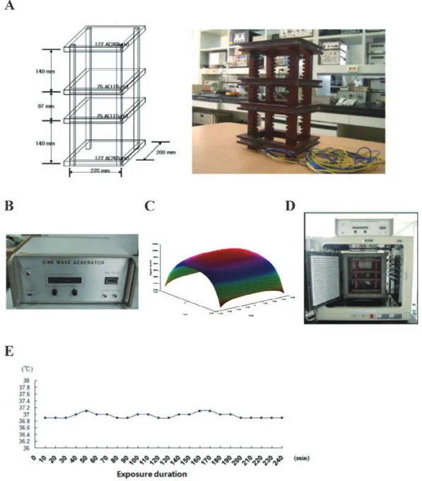
Fig. 1. ELF-MF exposure system. A: Diagram and photo of exposure system. B: Power generator. C: The voltage fluc-tuation rate and harmonic rate of power quality. D: Total exposure system including incubator. E: Temperature variation during experimental period in exposure system.
MATERIALS AND METHODS
Cell cultures
MCF10A human breast epithelial cells was purchased from the American Type Culture Collection (ATCC, Manassas, USA) and grown in DMEM/F12 medium with L-glutamine (Invitrogen, Carlsbad, USA) supplemented with 10% heat-inactivated horse serum (Invitrogen, Carlsbad, USA), 21 ng/mL of EGF (Peprotech, NJ, USA), 0.525 μg/mL of Hydrocorti-zone (Sigma, St. Louis, USA), 0.05 μg/mL of CholeraToxin (Sigma, St. Louis, USA), 11.2 μg/mL of Insulin (Sigma, St. Louis, USA), 100 mg/mL of streptomycin (WelGENE, Daegu, Korea), and 100 U/mL of penicillin (WelGENE, Daegu, Korea) in a 5 % CO2 incubator at 37°C.
Extremely low frequency magnetic fields (ELF-MF)
exposure system
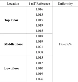
ELF-MF generation equipment was designed and con-structed by Korea Electrotechnology Research Institute as shown in Fig. 1A. ELF-MF monitoring was conducted under observation of the current injected to exposure system, since ELF-MF is proportional to injected current. The field genera-tor consists of four square-shaped coils and one cage with three testing floor (top, middle, and bottom floor). Power sup-ply is shown in Fig. 1B. The voltage fluctuation rate and har-monic rate of power quality using power amplifier was under 1%. Figure 1C shows calculated magnetic field distribution at the middle floor in exposure system. Fixing the magnetic field at the center of the middle floor at 1 mT. The fields at various points were measured under the condition of uniformity of ELF-MF exposure system is shown in Table 1. The spatial variation of magnetic field was under 3%. This strongly
dem-onstrates that the field generator is good for a small-sized in
vitro study. Total ELF-MF exposure system including
incuba-tor is demonstrated in Fig. 1D. The temperature of incubaincuba-tor was maintained 37 ± 0.3°C during exposure period using water-jet cooling system as shown in Fig. 1E. Magnetic field shielding system using ferrite material was adopted to shield strong magnetic field in outer of ELF-MF exposure system.
Exposure to ELF-MF and
γ
-radiation
ELF-MF exposure system was warmed up for 15 min to equilibrate it prior to ELF-MF exposure. And then, cells in 100 mm Petri dish were exposed to 60 Hz at 1 mT for 4 h. Maximum MF of our exposure system (1 mT, ~ two folds of the occupational reference level of 60 Hz magnetic field which was guided by International Commission on Non-ionizing Radiation Proteiction (ICNIRP)) was chosen for this study.22) After exposure, the cells were immediately
transferred to a cell culture incubator and kept for the post-exposure time (10, 24, or 48 h) before harvesting the cells. For the sham exposure, the cells were kept in the ELF-MF exposure device, but were not exposed to ELF-MF. The tem-perature of the exposure device was monitored throughout the exposure period in the sham-exposed and the ELF-MF exposed groups. For the positive control group, cells were exposed to 0.5, 2, or 4 Gy of γ-radiation from a 137Cs gamma
ray source (Atomic Energy of Canada, Mississauga, Canada) at a dose rate of 3.81 Gy/min.
Measurement of SOD activity
SOD activities in the MCF10A cells were measured using a SOD assay kit-WST (Dojindo Molecular Technologies, Kumamoto, Japan), according to the manufacturer’s instruc-tions. Briefly, Cells were harvested and suspensed in lysis buffer (100 μL of ice-cold 50 mmol/L of D-mannitol, 2 mmol/L of Tris-HCl, pH 7.4, 0.1% of Triton X-100) and sonicated 30 seconds for 8–10 times until the solution was cleared. After 15 minutes, the sonicated sample at 1,000 × g for 15 min at 4°C, the supernatant was transferred to a new tube and diluted with dilution buffer to prepare a sample solution. The SOD activity was expressed as inhibition rate percent per the manufacturer’s instructions.
Measurement of reduced and oxidized glutathione level
A Biooxytech GSH/GSSG-412 kit (Oxis International, Portland, USA) was used to measure reduced and oxidized glutathione (GSH/GSSG). To measure GSH/GSSG ratio, cells were harvested and suspensed with lysis buffer which were described above. Lysates mixed gently with or without 33 mmol/L 1-methyl-2-vinyl-pyridinium trifluoromethane sulfonate (M2VP), a scavenger of reduced glutathione (GSH). And then, six volumes of 5% metaphosphoric acid added to the sample solutions. The sample solutions were centrifuged at 1,000 × g for 10 min. For GSH estimation, 20 μL supernatant was mixed with 1,220 mL assay buffer (100Table 1. Uniformity of MF exposure system. Location 1 mT Reference Uniformity
Top Floor 1.016 1%–2.6% 1.013 1.015 1.019 1.015 Middle Floor 1.018 1.019 1.021 1.008 Low Floor 1.013 1.012 1.010 1.019 1.026
mmol/L NaPO4 and 5 mmol/L EDTA, pH 7.5) For GSSG
estimation, 50 μL supernatant was mixed with 700 μL
GSSG buffer (100 mmol/L NaPO4 and 5 mmol/L EDTA, pH
10.05). The samples were mixed with 1.262 mmol/L 5,5-dithiobis-(2-nitrobenzoic acid) and 15 units/mL GSH reduc-tase. The mixture was incubated at room temperature for 5 minutes, and the absorbance was recorded at 412 nm for 3 minutes after the addition of NADPH.
Flow cytometric analysis of reactive oxygen species
The intracellular ROS level was assessed using the 2’, 7’-dichloro-fluorescein diacetated compound (DA). DCF-DA (Sigma, St. Louis, USA) was added directly to the cul-ture medium at a final concentration 10 μM. Cells were then incubated at 37°C for 20 minutes, washed once in PBS (WelGENE, Daegu, Korea) and immediately analyzed using a FACScan flow cytometer (Becton Dickinson, NJ, USA) with excitation and emission at 490 and 530 nm.Statistical analysis
All data were expressed as mean ± standard deviation (M ± SD) from three or five independent experiments in duplicate. Differences between control and exposed groups were ana-lyzed using a Student’s t-test. Values of p < 0.05 were con-sidered statistically significant.
RESULTS
Effect of ELF-MF exposure on intracellular ROS level
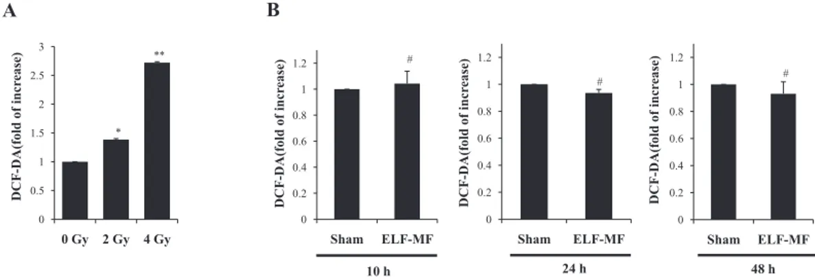
We evaluated the effects of ELF-MF on intracellular ROS levels using DCF-DA. Exposure to 60 Hz ELF-MF at 1 mT for 4 hours did not affect intracellular ROS levels relative to the sham-exposed group (Fig. 2B). On the contrary, IR expo-sure increased intracellular ROS levels in a dose-dependent manner compared to the controls in MCF10A cells at each time point (Fig. 2A). These results were analyzed statisticallyand values of p < 0.05 were considered statistically significant.
Effect of ELF-MF exposure on SOD activity
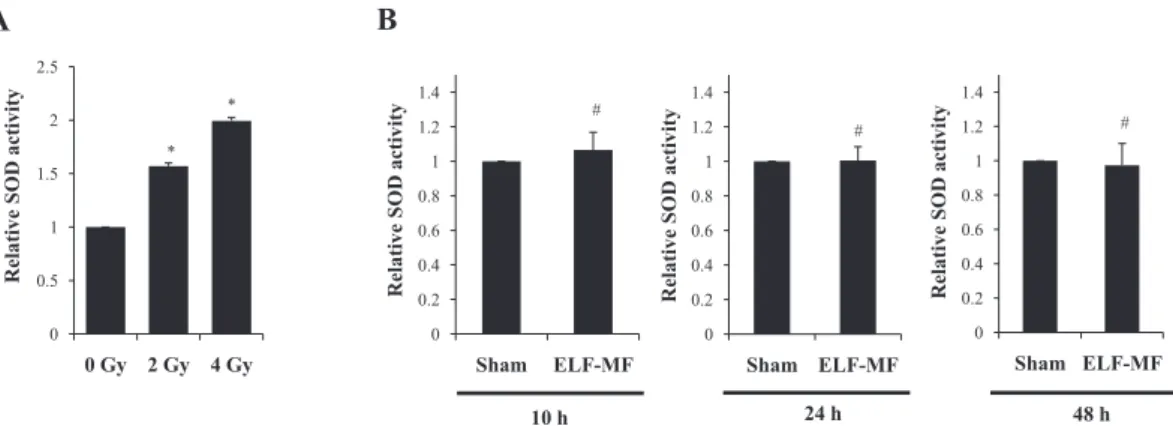
Next, we conducted SOD activity assays, as SOD has been recognized as an antioxidant enzyme that elicits a pri-mary defense response when cells are challenged by oxida-tive stress. As shown in Fig. 3B, exposure to 60 Hz ELF-MF at 1 mT did not significantly alter SOD activity in a time- or dose-dependent manner, relative to the sham-exposed group. However, SOD activity was significantly increased in the IR-exposed groups (Fig. 3A).
Effect of ELF-MF exposure on reduced/oxidized
glu-tathione levels
Reduced glutathione (GSH) level is noted in all cellular compartments and GSH functions as a cell protectant, spe-cifically as a radical scavenger via a cycling redox process. We measured reduced to oxidized glutathione ratio (GSH/ GSSG), because oxidized glutathione accumulates in cells under oxidative stress challenge conditions.20) When we
measured GSH/GSSG ratio following exposure of the cells to ELF-MF, the ratio was reduced in the IR-exposed groups as shown in Fig. 4A. By way of contrast, GSH/GSSG ratio evidenced no statistical difference between the ELF-MF-exposed and sham-ELF-MF-exposed groups (Fig. 4B).
Effect of ELF-MF on cellular morphology and SA-
β
-Gal activity
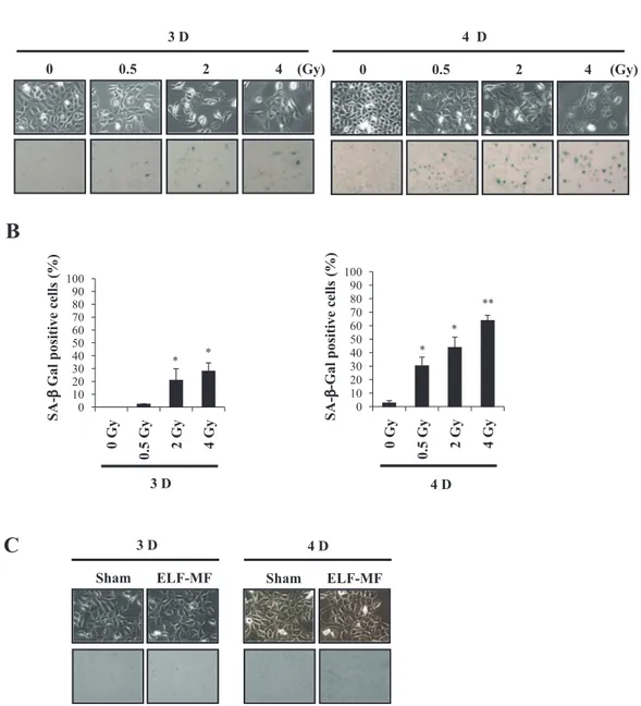
Finally, we evaluated the effects of ELF-MF on senescence-associated phenotypic changes. IR-exposed cells exhibited senescence-specific large and flat cellular morphology and stained positively for SA-β-Gal staining. Such senescence-associated phenotypic changes were detected in dose- and time-dependent manners in the IR-exposed groups (Fig. 5A). Quantitative analysis of SA-β-Gal positivity is shown in Fig. 5B. However, 60 Hz ELF-MF at 1 mT affected neither cellular
Fig. 2. Effect of ELF-MF on intracellular ROS levels in MCF10A cells. A: Effect of IR on intracellular ROS level in MCF10A cells exposed to 0, 2, and 4 Gy doses of IR. ROS levels were evaluated 48 h after IR exposure. B: Effect of ELF-MF on intracellular ROS level in MCF10A cells. Cells were exposed continuously to 1 mT for 4 h and ROS level was analyzed at 10, 24, and 48 h after recovery times via flow cytometry. Bars show the mean values and standard deviations (M ± SD) of five independent experiments in duplicate. Significantly different: *p < 0.05 versus 0 Gy, **p < 0.01 versus 0 Gy, insignificantly different: #p > 0.05 versus sham.
morphology nor SA-β-Gal activity (Fig. 5C). When SA-β-Gal positivity was assessed in sham and ELF-MF exposed groups, there was no statistical difference (data not shown).
DISCUSSION
ROS including oxygen free radicals and other activated oxygen species have been previously implicated in a variety of physiological and pathological conditions: namely, the aging process, inflammatory injury, atherosclerosis, and arthritis.23) Intracellular ROS concentration was determined
according to the balance between the ROS production via mitochondrial or nonmitochondrial pathways and clearance rates by various antioxidant compounds or antioxidant enzymes. As increased levels of intracellular ROS could be associated directly with DNA damage and carcinogenesis, it is important to assess and elucidate the effects of ELF-MF on oxidative stress.
Several studies have previously demonstrated that ELF-MF could affect ROS production, and may function as an oxidative stress. It has linked the 50 Hz ELF-MF capacity for the activation of immune-relevant cells to the free radi-cal-based physiological changes.24,25) Moreover, it was
reviewed that ELF-MF exposure resulted in excessive free radical production through immune cell activation, in turn leading to genotoxic event.26) On the other hand, Frahm et
al.27) reported that 50 Hz ELF-MF elevated phagocytic activity
and intracellular ROS level in the absence of any genotoxic effects after 24 h exposure at 1 mT. It has been also reported that 50 Hz ELF-MF exposure reduced cell tolerance towards oxidative attack without any genotoxic and cytotosic effect in neuroblastoma cells.28) Fiorani et al.29) proved that 50 Hz
MF (0.2–0.5 mT) could be able to potentiate the cellular damage induced by oxidizing agents in rabbit red blood cells. Other study showed that ELF-MF did not cause oxi-dative damage in cellular DNA in human placental
cotyle-Fig. 3. Effect of ELF-MF on SOD activity in MCF10A cells. A: SOD activity in MCF10A cells exposed to 0, 2, and 4 Gy doses of IR. B: Effect of ELF-MF on SOD activity in MCF10A cells. Cells were exposed continuously to 1 mT for 4 h and SOD activity was measured at 10, 24, and 48 h after recovery times. Bars show the mean values and standard deviations (M ± SD) of five indepen-dent experiments in duplicate. Significantly different: *p < 0.05 versus 0 Gy, insignificantly different: #p > 0.05 versus sham.
Fig. 4. Effect of ELF-MF on GSH/GSSG level in MCF10A cells. A: Analysis of GSH/GSSG levels in MCF10A cells exposed to 0, 2, and 4 Gy doses of IR. B: Analysis of GSH/GSSG levels in MCF10A cells which were exposed to 60 Hz ELF-MF at 1 mT for 4 h. GSH/GSSG levels was assesssed at 10, 24, and 48 h after recovery times. Bars show the mean values and standard deviations (M ± SD) of five independent experiments in duplicate. Significantly different: *p < 0.05 versus 0 Gy, insignificantly different: #p >
dons in vitro after exposure to low dose 50 Hz ELF-MF (2 and 5 mT for 3 h).30) Luukkonen et al.31) recently found that
pre-exposure to 50 Hz MF enhanced menadione-induced DNA damage, DNA repair rate, and micronucleus formation in human SH-SY5Y neuroblastoma cells. However, they found no effect of ELF-MF alone exposure on DNA damage and repair rate. As menadione is a radical-inducing agent, authors described that MF might affect free radical processes, as suggested by the radical pair mechanism. Brocklehurst and McLauchlan9) have been suggested, although the radical
pair mechanism has been hypothsized as a possible route whereby MF of environmental strength might affect a biolog-ical system, freely diffusing pairs of radbiolog-icals are unlikely to produce significant effects in biological system. In the present study, we assessed ELF-MF influence on the ROS level and found that exposure to 60 Hz ELF-MF at 1 mT did not affect intracellular concentration of ROS in MCF10A cells.
Cellular macromolecules, i.e, DNA, protein, and lipid are the most susceptible molecules to free radical attack. There-fore, living organisms harbor a variety of defense systems
Fig. 5. Effect of ELF-MF on cellular morphology and SA-β-Gal activity. A: Cellular morphology and SA-β-Gal activity in MCF10A cells exposed to 0.5, 2 and 4 Gy doses of IR. B: Quantitative analysis of SA-β-Gal positivity in IR-exposed cells. C: Cellular morphology and SA-β-Gal activity in MCF10A cells exposed to 60 Hz ELF-MF at 1 mT for 4 h. Cellular morphology and SA-β-Gal activity were assessed at 3 and 4 days after exposure, via phase-contrast microscopy. Bars show the mean values and stan-dard deviations (M ± SD) of three independent experiments. Significantly different: *p < 0.05 versus 0 Gy, **p < 0.01 versus 0 Gy.
including antioxidant vitamins, SOD, CAT, GSH, and glu-tathione peroxidase (GSH-Px) to protect cells against oxida-tive stress. Among them, GSH/GSSG, one of the best-char-acterized antioxidant defense mechanisms, is the most rapid and abundant weapon against ROS. Its protective activity is predicated on the oxidation of the thiol group of its cysteine residue with the formation of GSSG, which is, in turn, catalytically reduced back to the thiol form (GSH) by glu-tathione reductase (GSSG-Rd).32) Decrease of intracellular
GSH level was reported as the result of 50 Hz ELF-MF simultaneous exposure at 0.5 mT for 90 min with the Fe(II)/ ascorbate system in rabbit erythrocytes.28) It has been
sug-gested that GSH could play an important role in the antiox-idant defense mechanism, due to finding of a positive corre-lation between reduced glutathione (GSH) and ROS levels after 50 Hz ELF-MF exposure at 1 mT for 7 days in rat cortical neurons.33) Another antioxidant enzyme, SOD,
per-forms a crucial role in the system, protecting cells against destructive free radical activity. This antioxidant defense system could be deteriorated by a 50 Hz ELF-MF at 8.8–84 μT MF strength and 20–133 V/m electric field strength, which might lead to oxidative stress in human red blood cells.34) Zwirska-Korczala et al. demonstrated that 180–195
Hz ELF-MF influences enzyme activities of SOD, catalase, GSH-Px, and GSSG-Rd in 3T3-L1 preadipocytes after ELF-MF exposure (120 μT, 36 min daily for 2 days).35) It was also
demonstrated that SOD activity was increased as result of 60 Hz ELF-MF exposure at 1.2 mT for 3 h in mouse brain, sug-gesting that such increase may be reflective of a compensa-tory reaction against oxidative stress.36) Our study employed
assessments of the GSH/GSSG ratio and SOD activity, since both of which reflect the strength of the antioxidant defense system and are also indicatives of oxidative stress. We found that exposure to 60 Hz ELF-MF at 1 mT affected no signif-icant changes in GSH/GSSG and SOD activity. Intracellular ROS (superoxide and hydrogen peroxide) are produced from the mitochondrial electron transport chain and enzymatic reactions. Superoxide is converted to hydrogen peroxide by SOD, and catalase neutralizes hydrogen peroxide to water.37)
Since we measured intracellular ROS level using DCF-DA and SOD activity, it would be interesting if intracellular superoxide level was further assessed using dihydroethidium (DHE) probe. In addition, since we monitored ELF-MF alone effect on oxidative stress, it would be also valuable to elucidate whether EMF-MF could potentiate oxidative stress which is induced oxidative stimuli in various cell lines. Therefore, more extensive studies to elucidate whether MF might induce genetic damage and/or affect a biological sys-tem via generation of oxidative stress are necessary.
Cellular senescence is irreversible cell cycle arrest, which is activated in response to variety of cellular stresses, specif-ically to the oxidative stress.38,39) Actually, the radical theory
is concerned in the field of cellular senescence by a growing body of evidence that damaged cellular function due to the
increase of free radical could be associated with a number of senescence-related phenotypes.22,38) Senescent cells
undergo morphological changes with flat and large size and most commonly used senescence biomarker is SA-β-Gal positivity.40,41) Therefore, we evaluated effect of ELF-MF on
cellular senescence via the examination of cellular morphol-ogy and SA-β-Gal activity. We could not observe any detect-able changes in cellular morphology and SA-β-Gal activity after ELF-MF exposure.
Collectively, the results of this study demonstrated that 1 mT ELF-MF exposure did not affect intracellular ROS level, SOD enzyme activity, GSH/GSSG ratio, and phenotypical changes of cellular senescence in MCF10A cells under our exposure condition. The publications regarding the biological impact of ELF-MF have been accumulated, however, contra-dictory and ambiguoucity are still existed due to the variations of biological systems and endpoints. In addition, some papers have not been considered temperature variation in in vitro exposure system during the ELF-MF exposure. We suggest that future experiments have to be performed under the care-fully controlled exposure conditions and with proper positive and negative controls to get closer to final conclusion regard-ing concerns about harmful effect of ELF-MF exposure.
ACKNOWLEDGEMENTS
This work was supported by the Power Generation & Electricity Delivery of the Korea Institute of Energy Tech-nology Evaluation and Planning (KETEP) grant funded by the Korea government Ministry of Knowledge Economy (No. 2009101030003E).
REFERENCES
1. WHO (2007) Extremely low frequency fields. Environmental Health Criteria, World Health Organization, Vol. 238, Geneva. 2. Santini MT, Rainaldi G and Indovina PL (2009) Cellular
effects of extremely low frequency (ELF) electromagnetic fields. Int J Radiat Biol 85: 294–313.
3. Nordenson I, et al (1994). Chromosomal aberrations in human amniotic cells after intermittent exposure to fifty hertz mag-netic fields. Bioelectromagmag-netics 15: 293–301.
4. Santora N, et al (1997) Effect of extremely low frequency magnetic field exposure on morphological and biophysical properties of human lymphoid cell line Raji. Biochim Biophys Acta 1357: 281–290.
5. Tonini R, et al (2001) Calcium protects differentiating neuro-blastoma cells during 50 Hz electromagnetic radiation. Bio-phys J 81: 2580–2589.
6. Wertheimer N, Savitz DA and Leeper E (1995) Childhood cancer in relation to indicators of magnetic fields from ground current sources. Bioelectromagnetics 16: 86–96.
7. Loomis DP and Savitz DA (1990) Mortality from brain cancer and leukemia among electrical workers. Br J Ind Med 47: 633–638.
risk and electromagnetic fields (EMFs): assessing the geo-magnetic component. Arch Environ Health 56: 314–319. 9. Brocklehurst B and McLauchlan KA (1996) Free radical
mechanism for the effects of environmental electromagnetic fields on biological systems. Int J Radiat Biol 69: 3–24. 10. Mannerling AC, et al (2010) Effects of 50-Hz magnetic field
exposure on superoxide radical anion formation and HSP70 induction in human K562 cells. Radiat Environ Biophys 49: 731–741.
11. Frahm J, Mattsson MO and Simkó M (2010) Exposure to ELF magnetic fields modulate redox related protein expression in mouse macrophages. Toxicol Lett 192: 330–336.
12. Zwirska-Korczala K, et al (2004) Influence of extremely-low-frequency magnetic field on antioxidative melatonin proper-ties in AT478 murine squamous cell carcinoma culture. Biol Trace Elem Res 102: 227–243.
13. Wolf FI, et al (2005) 50 Hz extremely low-frequency electro-magnetic fields enhance cell proliferation and DNA damage: Possible involvement of a redox mechanism. Biochim Bio-phys Acta 1743: 120–129.
14. Lai H and Singh NP (2004) Magnetic-field-induced DNA strand breaks in brain cells of the rat. Environ Health Perspect 112: 687–694.
15. Benfante R, et al (2008) The expression of PHOX2A, PHOX2B and of their target gene dopamine-beta-hydroxylase (DbetaH) is not modified by exposure to extremely-low-fre-quency electromagnetic field (ELF-EMF) in a human neu-ronal model. Toxicol In Vitro 22: 1489–1495.
16. Stronati L, et al (2004) Absence of genotoxicity in human blood cells exposed to 50 Hz magnetic fields as assessed by comet assay, chromosome aberration, micronucleus, and sister chromatid exchange analyses. Bioelectromagnetics 25: 41–48. 17. Amara S, et al (2007) Influence of a static magnetic field (250
mT) on the antioxidant response and DNA integrity in THP1 cells. Phys Med Biol 52: 889–898.
18. Kregel KC and Zhang HJ (2007) An integrated view of oxi-dative stress in ageing: basic mechanisms, functional effects, and pathological considerations. Am J Physiol Regul Integr Comp Physiol 292: 18–36.
19. Valko M, et al (2007) Free radicals and antioxidants in normal physiological functions and human disease. Int J Biochem Cell Biol 39: 44–84.
20. Shull S, et al (1991) Differential regulation of antioxidant enzymes in response to oxidants. J Biol Chem 266: 24398–403. 21. Droge W (2002) Free radicals in the physiological control of
cell function. Physiol Rev 82: 47–95.
22. ICNIRP (1998) Guidelines for limiting exposure to time-vary-ing electric, magnetic, and electromagnetic fields (up to 300 GHz). Health Phys 74: 494–522.
23. Stadtman ER (1992) Protein oxidation and aging. Science 257: 1220–1224.
24. Lupke M, et al (2006) Gene expression analysis of ELF-MF exposed human monocytes indicating the involvement of the alternative activation pathway. Biochim Biophys Acta 1763: 402–412.
25. Zmyslony M, et al (2004) The effect of weak 50 Hz magnetic fields on the number of free oxygen radicals in rat
lympho-cytes in vitro. Bioelectromagnetics 25: 607–612.
26. Simkó M and Mattsson MO (2004) Extremely low frequency electromagnetic fields as effectors of cellular responses in vitro: possible immune cell activation. J Cell Biochem 93: 83–92. 27. Frahm J, et al (2006) Alteration in cellular functions in mouse
macrophages after exposure to 50 Hz magnetic fields. J Cell Biochem 99: 168–177.
28. Falone S, et al (2007) Fifty hertz extremely low-frequency electromagnetic field causes changes in redox and differenti-ative status in neuroblastoma cells. Int J Biochem Cell Biol 39: 2093–2106.
29. Fiorani M, et al (1997) In vitro effects of 50 Hz magnetic fields on oxidatively damaged rabbit red blood cells. Bioelec-tromagnetics 18: 125–131.
30. Lopucki M, et al (2005) Low dose magnetic fields do not cause oxidative DNA damage in human placental cotyledons in vitro. Virchows Arch 446: 634–639.
31. Luukkonen J, et al (2011) Pre-exposure to 50 Hz magnetic fields modifies menadione-induced genotoxic effects in human SH-SY5Y neuroblastoma cells. PLoS One 6:e18021. 32. Meister A and Anderson ME (1983) Glutathione. Annu Rev
Biochem 52: 711–760.
33. Di Loreto S, et al (2009) Fifty hertz extremely low-frequency magnetic field exposure elicits redox and trophic response in rat-cortical neurons. J Cell Physiol 219: 334–343.
34. Sharifian A, et al (2009) Effect of extremely low frequency magnetic field on antioxidant activity in plasma and red blood cells in spot welders. Int Arch Occup Environ Health 82: 259– 266.
35. Zwirska-Korczala K, et al (2005) Effect of extremely low fre-quency of electromagnetic fields on cell proliferation, antiox-idative enzyme activities and lipid peroxidation in 3T3-L1 preadipocytes--an in vitro study. J Physiol Pharmacol 56 Suppl 6: 101–108.
36. Lee BC, et al (2004) Effects of extremely low frequency mag-netic field on the antioxidant defense system in mouse brain: a chemiluminescence study. J Photochem Photobiol B 73: 43–48. 37. Chaudhuri L, Sarsour EH, and Goswami PC (2010) 2-(4-Chlorophenyl) benzo-1,4-quinone induced ROS-signaling inhibits proliferation in human non-malignant prostate epithe-lial cells. Environ Int 36: 924–930.
38. Lee JJ, et al (2010) Prevention of premature senescence requires JNK regulation of Bcl-2 and reactive oxygen species. Oncogene 29: 561–575.
39. Lee JJ, et al (2011) PTEN status switches cell fate between premature senescence and apoptosis in glioma exposed to ion-izing radiation. Cell Death Differ 18: 666–677.
40. Dimri GP, et al (1995) A biomarker that identifies senescent human cells in culture and in aging skin in vivo. Proc Natl Acad Sci USA 92: 9363–9367.
41. Byun HO, et al (2009) Cathepsin D and eukaryotic translation elongation factor 1 as promising markers of cellular senes-cence. Cancer Res 69: 4638–4637.
Received on March 25, 2011 Revision received on August 26, 2011 Accepted on August 30, 2011