ISSN 2234-3806 • eISSN 2234-3814 Ann Lab Med 2015;35:416-422
http://dx.doi.org/10.3343/alm.2015.35.4.416
Direct Identification of Urinary Tract Pathogens From
Urine Samples Using the Vitek MS System Based on
Matrix-Assisted Laser Desorption Ionization–Time of
Flight Mass Spectrometry
Yeongsic Kim, M.D.1, Kang Gyun Park, M.T.1, Kyungwon Lee, M.D.2, and Yeon-Joon Park, M.D.1
Department of Laboratory Medicine1, College of Medicine, The Catholic University of Korea, Seoul; Department of Laboratory Medicine2, Yonsei University
College of Medicine, Seoul, Korea
Background: We evaluated the coincidence rate between Vitek MS system (bioMérieux, France) and Vitek 2 in identifying uropathogens directly from urine specimens.
Methods: Urine specimens submitted to our microbiology laboratory between July and September 2013 for Gram staining and bacterial culture were analyzed. Bacterial identifi-cation was performed by using the conventional method. Urine specimens showing a sin-gle morphotype by Gram staining were processed by culturing and matrix-assisted laser desorption ionization–time of flight mass spectrometry (MALDI-TOF MS). Of 2,370 urine specimens, 251 showed a single morphotype on Gram staining, and among them, 202 were available for MALDI-TOF MS.
Results: In these 202 specimens, colony growth was observed in 189 specimens, and 145 specimens had significant growth of single-colony morphotype in culture. One hun-dred and ten (75.9%) of them had colony counts of ≥105 colony-forming units (CFU)/mL and included 71 enteric gram-negative bacteria (GNB), 5 glucose-non-fermenting GNB, 9 gram-positive cocci (GPC), and 25 yeasts. Furthermore, 70 (98.6%), 3 (60.0%), 4 (44.4%), and 5 (20.0%), respectively, of these were correctly identified by Vitek MS. Thirty-one specimens (21.4%; 11 GNB, 7 GPC, 12 yeasts, and 1 gram-positive bacillus) had colony counts of 104-105 CFU/mL. Four specimens (2.8%) yielded colony counts of 103-104 CFU/mL.
Conclusions: Vitek MS showed high rate of accuracy for the identification of GNB in urine specimens (≥105 CFU/mL). This could become a rapid and accurate diagnostic method for urinary tract infection caused by GNB. However, for the identification of GPC and yeasts, further studies on appropriate pre-treatment are warranted.
Key Words: Urinary tract pathogens, MALDI-TOF MS, Vitek MS, UTI
Received: October 6, 2014
Revision received: November 13, 2014 Accepted: May 11, 2015
Corresponding author: Yeon-Joon Park
Department of Laboratory Medicine, College of Medicine, The Catholic University of Korea, 222 Banpo-daero, Seocho-gu, Seoul 137-701, Korea Tel: +82-2-2258-1640
Fax: +82-2-2258-1719 E-mail: [email protected]
© The Korean Society for Laboratory Medicine
This is an Open Access article distributed under the terms of the Creative Commons Attribution Non-Commercial License (http://creativecom-mons.org/licenses/by-nc/3.0) which permits unrestricted non-commercial use, distribution, and reproduction in any medium, provided the original work is properly cited.
INTRODUCTION
Urinary tract infection (UTI) is the most commonly encountered nosocomial infection, and the major risk factor is urinary cathe-terization [1]. Although Escherichia coli is the most common
uropathogen for UTI, other gram-negative bacteria (GNB; en-terobacteria other than E. coli, Pseudomonas spp., and Acineto-bacter spp.), gram-positive cocci (GPC), and Candida spp. are common uropathogens. In patients positive for the same organism in both urine and blood culture, the five leading
micro-organisms found in blood cultures are E. coli, Candida spp., Klebsiella spp., Acinetobacter spp., and Staphylococcus aureus [2]. Therefore, early identification of uropathogens is necessary, especially in nosocomial UTI, and it could help select appropri-ate antibiotics for treatment. However, urine culture (quantitative culture of urine specimens on solid medium followed by bio-chemical characterization of isolates), which is the gold standard for diagnosis, takes 24-72 hr before results are available [3]. Matrix-assisted laser desorption ionization time-of-flight mass spectrometry (MALDI-TOF MS) becomes a reliable tool for the identification of bacteria and yeast [4, 5]. The Vitek MS system (bioMérieux, Marcy I’Etoile, France) got US Food and Drug Ad-ministration (FDA) clearance on August 2013 for the identifica-tion of bacteria (except Mycobacterium) and yeasts grown on solid media. It was recently reported that MALDI-TOF MS could directly provide rapid and accurate bacterial identification for the majority of urine specimens with a bacterial colony count of ≥105 colony-forming units (CFU)/mL, where an automated de-vice based on flow cytometry was used for the screening test [6, 7]. However, in most clinical laboratories, such an automated device is not used for screening UTIs. For urine specimens with ≥104 CFU/mL of a single uropathogen, the identification and susceptibility test for the uropathogen is recommended because colony counts of <105 CFU/mL in voided specimens in the presence of dysuria and symptoms of UTI should not be ignored [8]. In this study, we evaluated the coincidence rate between Vi-tek MS system (bioMérieux, France) and ViVi-tek 2 in identifying urinary tract pathogens directly from urine specimens with ≥105 CFU/mL, as well as those with 104-105 CFU/mL.
METHODS
1. Study specimens
Of the urine specimens submitted to our microbiology laboratory between July and September 2013 for Gram staining and bac-terial culture, specimens showing a single morphotype by Gram staining were processed both by conventional culture and MALDI-TOF MS. Leukocyte numbers were examined at a low-power field (LPF) and were graded as negative, 1+:<1/LPF, 2+: 1-9/LPF, 3+:10-25/LPF, or 4+:>25/LPF. Bacteria were counted per oil immersion field (OIF) and graded as negative, 1+: <1/ OIF, 2+:1-5/OIF, 3+:6-30/OIF, or 4+:>30/OIF [8].
2. Urine culture and conventional identification
Bacterial identification was performed by the conventional method using the Vitek 2 system. For the conventional culture,
1 µL of well-mixed urine was inoculated and spread onto blood agar plates and MacConkey agar plates using a sterile plastic disposable loop (SPL Lifesciences, Pocheon, Korea). Plates were incubated in an aerobic atmosphere at 37°C for 18-24 hr. When bacterial growth was observed, the colonies on blood agar were counted, and colonies from both types of plates were iden-tified by using the Vitek 2 system.
3. MALDI-TOF MS identification
For MALDI-TOF MS analysis, the preparation method was an adaptation of the protocol described by Ferreira et al. [7] and Lavergne et al. [9]. Briefly, urine (3 mL) was centrifuged at 2,000 g for 30 sec to remove leukocytes. The supernatant was centrifuged at 14,100 g for 10 min to collect bacteria. The pellet was washed once with de-ionized water (DW). To the pellet, 1.5 mL of basic solution of sodium dodecyl sulfate (SDS 0.1%, NaHCO3 0.015M) was added and incubated at 37°C for 10 min. After further centrifugation at 14,100 g for 10 min and washing with DW, the supernatant was removed. The pellet it-self (8-10 μL) was applied to the MALDI plate in duplicate and was dried on a slide warmer (56°C) for 10 min. Then, 1 μL of matrix solution (3.1% [w/v] α-cyano-4-hydroxycinnamic acid, product no. 411071; bioMérieux) was added to each well. For yeasts, 1 μL of formic acid (product no. 411072, bioMérieux) was applied before the addition of matrix solution. No repeat testing was performed.
Following preparation, specimens were analyzed with the Vi-tek MS in linear, positive-ion mode at a laser frequency of 200 Hz, across the mass-to-charge (m/z) ratio of 2,000 to 20,000 Da. External mass calibration was performed by using a TOF mix (bioMérieux). Target plates were calibrated and quality con-trolled using E. coli ATCC8739. After the acquisition of spectra, data were transferred from the Vitek MS acquisition station to the Vitek MS analysis server, and identification results were dis-played by using Myla v3.2 middleware. The Vitek MS identifica-tion system is based on comparison of the characteristics of the obtained spectra with those of the Vitek MS v2.0 database. This database was built by using spectra of known strains for each claimed species. On the basis of this representative data collec-tion, a weight is assigned to each peak for each species accord-ing to its specificity.
A confidence value of ≥60 with the unique spectrum of a sin-gle organism indicated good species-level identification. If no unique identification pattern was found, a list of possible organ-isms was given as “low discrimination” (confidence value of
the database (“no ID”) [4]. If one of the duplicates showed a confidence value of ≥60%, it was considered an acceptable identification. No identification result was given when the dupli-cate had poor confidence levels or did not have the same spe-cies-level identification. The warning messages “bad spectrum” or “insufficient peak” appeared in case of a human error or a poor-quality deposit.
4. Statistical analysis
Statistical analyses were performed by using the SPSS statistical analysis package version 13.0 (SPSS Inc., Chicago, IL, USA). Comparisons of proportions were tested by using chi-square analysis and Fisher’s exact test, as appropriate, and the criterion for statistical significance was set at P <0.05.
RESULTS
1. Urine culture results of specimens available for
MALDI-TOF MS
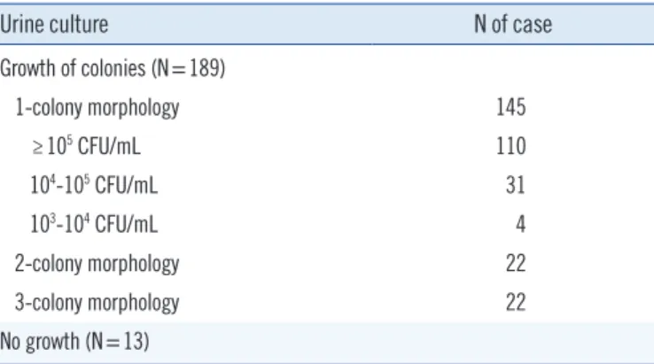
Of the 202 urine specimens, the growth of colonies was ob-served in 189 specimens: 145 specimens with single-colony morphology, 22 specimens with two-colony morphology, and the remaining 22 specimens with morphologies of more than two colonies. Thirteen specimens did not grow in culture, and Vitek MS did not identify any significant protein profile in any of these cases (Table 1).
2. Comparison of the results between the conventional
method and MALDI-TOF
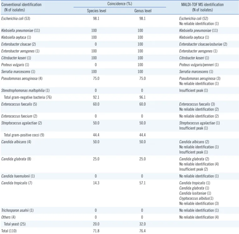
Of the 145 specimens with significant growth of single-colony morphology on agar, 110 (75.9%) of them had colony counts of ≥105 CFU/mL. They included 71 enteric GNB, five
glucose-non-fermenting GNB, nine GPC, and 25 yeasts. Vitek MS cor-rectly identified 70 (98.6%) enteric GNB, three (60.0%) glu-cose-non-fermenting GNB, four (44.4%) GPC, and five (20.0%) yeast isolates (Table 2). Overall, among the 110 specimens with colony counts of ≥105 CFU/mL, microorganism identification coincided at the species level in 79 cases (71.8%) and at the genus level in 84 cases (76.4%). In 26 cases, Vitek MS did not give a reliable identification. We found no major discrepancies at the genus level.
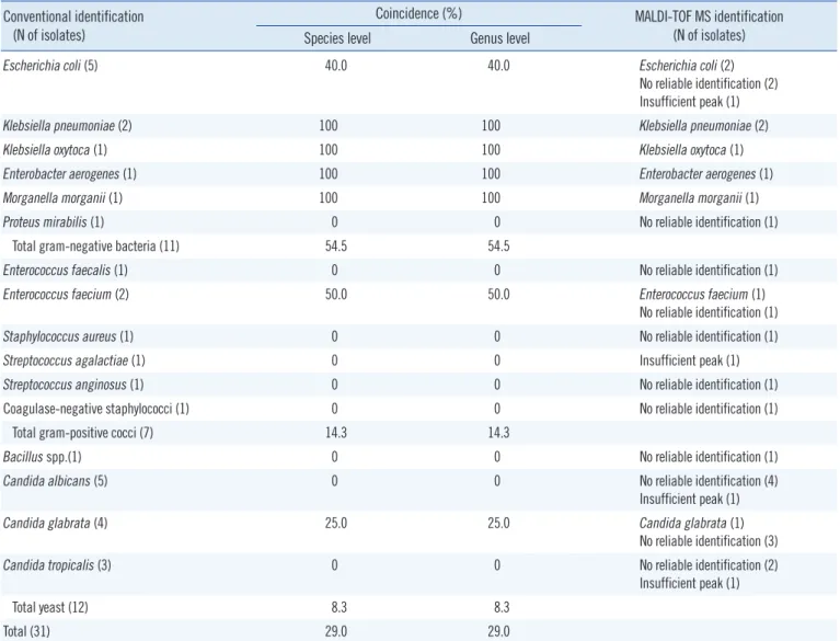
Thirty-one specimens (31/145, 21.4%) showed growth of bacterial colonies between 104 and 105 CFU/mL, and they were 11 GNB, seven GPC, 12 yeasts, and one gram-positive bacillus (GPB) (Table 3). Among them, seven (63.6%) GNB, one (14.3%) GPC, and one (8.3%) yeast were correctly identified by Vitek MS. Overall, the accuracy of identification at the genus level was 29.0% (9/31). The correct identification rate was sig-nificantly higher in urine specimens with ≥105 CFU/mL than in those with 104-105 CFU/mL (P =0.000).
Four specimens (2.8%) yielded colony counts between 103 and 104 CFU/mL, and none of them were correctly identified. According to Gram staining of the microorganisms isolated from the above 141 specimens from which more than 104 CFU/ mL of an microorganism was isolated, 87 were GNB, 16 were GPC, 37 were yeasts, and one was GPB. Of the 87 GNB cases, E. coli was isolated in 58 (66.7%), Klebsiella spp. in 15 (17.2%), chromosomal AmpC-producing Enterobacteriaceae (Enterobacter spp., Citrobacter spp., Serratia spp., Morganella spp.) in seven (8.0%), glucose non-fermenting isolates in five (5.7%), and Proteus spp. in two cases. Overall, the coincidence rate between Vitek 2 and Vitek MS was 92.0% (80/87) for GNB (54/58 E. coli, 15/15 Klebsiella spp., 7/7 chromosomal AmpC-producing Enterobacteriaceae, 3/5 glucose non-fermenting GNB, and 1/2 Proteus spp.), 31.3% (5/16) for GPC (3/6 Entero-coccus faecalis, 1/4 EnteroEntero-coccus faecium, 1/3 StreptoEntero-coccus agalactiae, 0/1 Staphylococcus aureus, 0/1 Streptococcus angi-nosus group, 0/1 coagulase-negative Staphylococcus), 16.2% (6/37) for yeasts, and 0% for GPB (0/1 Bacillus spp.). Among the 50 specimens not identified by Vitek MS, eight had “insuffi-cient peaks” caused by poor quality of the microbial film on the plate, and five of them were yeasts. The number of specimens with high or moderate leukocytes were 28 among the 91 cases correctly identified by Vitek MS, and six among the 50 cases not identified by Vitek MS (chi-square test, P =0.003).
For 22 specimens with significant (≥104 CFU/mL) growth of two-colony morphology in culture, Vitek MS correctly identified one of the mixed microorganisms in seven cases (two E. coli,
Table 1. Urine culture results of 202 specimens available for
MAL-DI-TOF MS
Urine culture N of case
Growth of colonies (N=189) 1-colony morphology 145 ≥105 CFU/mL 110 104-105 CFU/mL 31 103-104 CFU/mL 4 2-colony morphology 22 3-colony morphology 22 No growth (N=13)
Abbreviations: MALDI-TOF MS, matrix-assisted laser desorption ionization time-of-flight mass spectrometry; CFU, colony-forming unit.
two Acinetobacter baumannii complex, one Klebsiella pneu-moniae, one Morganella morganii, and one Streptococcus aga-lactiae).
DISCUSSION
Previous studies have shown excellent correlation between Vitek
MS identification and conventional microbiological identification in clinical bacterial and yeast isolates. In these studies, when there were discrepancies between Vitek MS and conventional identification, sequencing results frequently corresponded with the Vitek MS [4, 5, 10]. However, few studies on direct identifi-cation of urinary tract pathogens from urine specimens using MALDI-TOF MS have been performed [6, 7], and to our
knowl-Table 2. Comparison of the results between the conventional method and MALDI-TOF in 110 specimens with colony counts of ≥105 CFU/mL
Conventional identification (N of isolates)
Coincidence (%) MALDI-TOF MS identification (N of isolates) Species level Genus level
Escherichia coli (53) 98.1 98.1 Escherichia coli (52)
No reliable identification (1)
Klebsiella pneumoniae (11) 100 100 Klebsiella pneumoniae (11)
Klebsiella oxytoca (1) 100 100 Klebsiella oxytoca (1)
Enterobacter cloacae (2) 0 100 Enterobacter cloacae/asburiae (2)
Enterobacter aerogenes (1) 100 100 Enterobacter aerogenes (1)
Citrobacter koseri (1) 100 100 Citrobacter koseri (1)
Proteus vulgaris (1) 0 100 Proteus vulgaris/penneri (1)
Serratia marcescens (1) 100 100 Serratia marcescens (1)
Pseudomonas aeruginosa (4) 75.0 75.0 Pseudomonas aeruginosa (3)
No reliable identification (1)
Stenotrophomonas maltophilia (1) 0 0 Insufficient peak (1)
Total gram-negative bacteria (76) 92.1 96.1
Enterococcus faecalis (5) 60.0 60.0 Enterococcus faecalis (3)
No reliable identification (2)
Enterococcus faecium (2) 0 0 No reliable identification (2)
Streptococcus agalactiae (2) 50.0 50.0 Streptococcus agalactiae (1)
Insufficient peak (1)
Total gram-positive cocci (9) 44.4 44.4
Candida albicans (4) 50.0 50.0 Candida albicans (2)
No reliable identification (1) Insufficient peak (1)
Candida glabrata (8) 25.0 25.0 Candida glabrata (2)
No reliable identification (4) Insufficient peak (2)
Candida haemulonii (1) 0 0 No reliable identification (1)
Candida tropicalis (7) 14.3 57.1 Candida tropicalis (1)
Candida glabrata (1) Candida lusitaniae (1) Cryptococcus albidus(1) No reliable identification (3)
Trichosporon asahii (1) 0 0 No reliable identification (1)
Others (4) 0 0 No reliable identification (4)
Total yeast (25) 20.0 32.0
Total (110) 71.8 76.4
edge, this is the first such study that used Vitek MS and in-cluded specimens with yeasts.
Among urine specimens containing ≥104 CFU/mL, GNB accounted for 61.7% of the uropathogens, and yeast was the second most common (26.2%). Moreover, among the GNB, E. coli comprised only about two thirds, which is in line with a pre-vious study from China where E. coli was found to comprise 43.5% of the uropathogens. Considering this diversity of uro-pathogens, early identification of uropathogens is necessary for appropriate therapy [6].
Based on our results, the overall identification rate of Vitek MS was much higher (76.4%) in urine specimens containing ≥105 CFU/mL than that in the specimens containing 104-105 CFU/mL (29.0%). For GPC and yeasts, the identification rate in
specimens with a lower microbial burden was too low (14.3% and 8.3%, respectively). However, for GNB, the identification rate was 63.6% even for specimens with 104 or 105 CFU/mL of bacteria, which is much higher than that reported in an earlier study where only one of the five GNB were correctly identified [7]. For urine specimens containing bacteria of less than 104 CFU/mL, Vitek MS could not identify bacteria. This indicates that sufficient bacterial count is essential for identification by MALDI-TOF MS and supports the previous report where bacte-rial count needed for a good discrimination score by MALDI-TOF MS was higher for GPC (2.5-5×10⁵ CFU/mL) than for GNB (about 6 ×10⁴ CFU/mL) [6]. Therefore, additional studies are needed to improve the collection quantity and purity of collected bacteria.
Table 3. Comparison of the results between the conventional method and MALDI-TOF in 31 specimens with colony counts of 104-105 CFU/
mL
Conventional identification (N of isolates)
Coincidence (%) MALDI-TOF MS identification (N of isolates) Species level Genus level
Escherichia coli (5) 40.0 40.0 Escherichia coli (2)
No reliable identification (2) Insufficient peak (1)
Klebsiella pneumoniae (2) 100 100 Klebsiella pneumoniae (2)
Klebsiella oxytoca (1) 100 100 Klebsiella oxytoca (1)
Enterobacter aerogenes (1) 100 100 Enterobacter aerogenes (1)
Morganella morganii (1) 100 100 Morganella morganii (1)
Proteus mirabilis (1) 0 0 No reliable identification (1)
Total gram-negative bacteria (11) 54.5 54.5
Enterococcus faecalis (1) 0 0 No reliable identification (1)
Enterococcus faecium (2) 50.0 50.0 Enterococcus faecium (1)
No reliable identification (1)
Staphylococcus aureus (1) 0 0 No reliable identification (1)
Streptococcus agalactiae (1) 0 0 Insufficient peak (1)
Streptococcus anginosus (1) 0 0 No reliable identification (1)
Coagulase-negative staphylococci (1) 0 0 No reliable identification (1)
Total gram-positive cocci (7) 14.3 14.3
Bacillus spp.(1) 0 0 No reliable identification (1)
Candida albicans (5) 0 0 No reliable identification (4)
Insufficient peak (1)
Candida glabrata (4) 25.0 25.0 Candida glabrata (1)
No reliable identification (3)
Candida tropicalis (3) 0 0 No reliable identification (2)
Insufficient peak (1)
Total yeast (12) 8.3 8.3
Total (31) 29.0 29.0
For urine specimens with ≥105 CFU/mL, the identification rate differed according to the microorganisms involved. For en-teric GNB, Vitek MS demonstrated excellent identification (98.6%), and this finding is in line with previous studies [6, 7]. For GPC, however, Vitek MS yielded correct identification only in a small portion (44.4%) of urine specimens with GPC. For E. faecalis, which is the most common uropathogenic GPC, al-though the number of specimens was small, the detection rate was 60%, which corresponds with a study where the coinci-dence with conventional culture method was lower (66.7%) in E. faecalis than in E. coli (97.6%) [7]. However, in the study from China, the coincidence was very high in Enterococcus spp. (81.5-90.0%) [6]. This difference might be attributed to the dif-ference in specimen preparation, because those authors used an extraction procedure using formic acid and acetonitrile [6], while we did not (to avoid the use of a fume hood). Perhaps the formic acid and acetonitrile extraction procedure would have a positive effect on the identification of GPC by MALDI-TOF MS. For specimens containing yeasts, only five (20%) out of the 25 specimens were correctly identified. As Vitek MS showed supe-rior ability of identification of yeasts grown on agar (86.7-96.6%) using the simple formic acid pre-treatment [5, 8, 11, 12], the low identification rate for yeast in this study can be ascribed to the matrix effect of urine. A better pre-treatment step that will allow for high identification rate for yeasts directly from urine specimens needs to be developed.
We also compared leukocyte counts between specimens that were identified or not identified by Vitek MS, because in a previ-ous study [6], a large number of leukocytes in urine specimens was frequently observed in specimens not reliably identified by MALDI-TOF MS. However, the proportion of urine specimens containing a large number of leukocytes was much higher among the specimens correctly identified by Vitek MS, suggest-ing that a high leukocyte count does not interfere with bacterial protein peaks. High concentrates of many endogenous sub-stances such as α-defensins in urine, which can suppress or en-hance the ionization of certain protein spectra of microorgan-isms, might result in incorrect or no identification [13, 14]. In our study, the identification of bacteria by MALDI-TOF MS took about 1 hr, which is similar to that reported in several stud-ies [15, 16]. Identifying bacterial specstud-ies will help clinicians choose appropriate antibiotics for empiric therapy. In addition, when the bacterial species is known, the antimicrobial suscepti-bility test can be performed as soon as the colony is formed on the agar, using the appropriate Vitek card (AST-N224 for Entero-bacteriaceae vs. N225 for glucose non-fermenters,
AST-P600 for Enterococcus spp.).
In conclusion, with a simple pre-treatment using SDS, Vitek MS showed high rate of accuracy for the identification of GNB in urine specimens with ≥104 CFU/mL, without any major error. This could become a rapid and accurate diagnostic method useful in the diagnosis of UTI caused by a single GNB. How-ever, for the identification of GPC and yeasts, further studies are required to improve pre-treatment steps. In addition, Vitek MS could not identify bacterial species in urine specimens with mixed bacterial infections; therefore, for improvements in the Vitek MS software, this shortcoming should be considered and rectified.
Authors’ Disclosures of Potential Conflicts of
Interest
No potential conflicts of interest relevant to this article were re-ported.
Acknowledgments
This work was supported by bioMérieux Korea, but they were not involved in the data collection or preparation of the manu-script.
REFERENCES
1. Trautner BW. Management of catheter-associated urinary tract infec-tion. Curr Opin Infect Dis 2010;23:76-82.
2. Isikgoz Tasbakan M, Durusoy R, Pullukcu H, Sipahi OR, Ulusoy S; 2011 Turkish Nosocomial Urinary Tract Infection Study Group. Hospital-acquired urinary tract infection point prevalence in Turkey: differences in risk factors among patient groups. Ann Clin Microbiol Antimicrob 2013;12:31.
3. Burd EM and Kehl KS. A critical appraisal of the role of the clinical mi-crobiology laboratory in the diagnosis of urinary tract infections. J Clin Microbiol 2011;49(S9):S34-8.
4. Dubois D, Grare M, Prere MF, Segonds C, Marty N, Oswald E. Perfor-mances of the Vitek MS matrix-assisted laser desorption ionization-time of flight mass spectrometry system for rapid identification of bacteria in routine clinical microbiology. J Clin Microbiol 2012;50:2568-76. 5. Won EJ, Shin JH, Lee K, Kim MN, Lee HS, Park YJ, et al. Accuracy of
species-level identification of yeast isolates from blood cultures from 10 university hospitals in South Korea by use of the matrix-assisted laser desorption ionization-time of flight mass spectrometry-based Vitek MS system. J Clin Microbiol 2013;51:3063-5.
6. Wang XH, Zhang G, Fan YY, Yang X, Sui WJ, Lu XX. Direct identification of bacteria causing urinary tract infections by combining matrix-assisted laser desorption ionization-time of flight mass spectrometry with UF-1000i urine flow cytometry. J Microbiol Methods 2013;92:231-5. 7. Ferreira L, Sánchez-Juanes F, González-Avila M, Cembrero-Fuciños D,
of urinary tract pathogens from urine samples by matrix-assisted laser desorption ionization-time of flight mass spectrometry. J Clin Microbiol 2010;48:2110-5.
8. Garcia LS, ed. Clinical microbiology procedure handbook. 3rd ed. Wash-ington DC: ASM press, 2010:3.2.16.
9. Lavergne RA, Chauvin P, Valentin A, Fillaux J, Roques-Malecaze C, Ar-naud S, et al. An extraction method of positive blood cultures for direct identification of Candida species by Vitek MS matrix-assisted laser de-sorption ionization time of flight mass spectrometry. Med Mycol 2013; 51:652-6.
10. Moon HW, Lee SH, Chung HS, Lee M, Lee K. Performance of the Vitek MS matrix-assisted laser desorption ionization time-of-flight mass spec-trometry system for identification of Gram-positive cocci routinely isolated in clinical microbiology laboratories. J Med Microbiol 2013;62:1301-6. 11. Jamal WY, Ahmad S, Khan ZU, Rotimi VO. Comparative evaluation of
two matrix-assisted laser desorption/ionization time-of-flight mass spec-trometry (MALDI-TOF MS) systems for the identification of clinically sig-nificant yeasts. Int J Infect Dis 2014;26:167-70.
12. Westblade LF, Jennemann R, Branda JA, Bythrow M, Ferraro MJ, Gar-ner OB, et al. Multicenter study evaluating the Vitek MS system for identification of medically important yeasts. J Clin Microbiol 2013;51: 2267-72.
13. Tudela E, Muñoz G, Muñoz-Guerra JA. Matrix effect marker for multian-alyte analysis by LC-MS/MS in biological samples. J Chromatogr B Ana-lyt Technol Biomed Life Sci 2012;901:98-106.
14. Köhling HL, Bittner A, Müller KD, Buer J, Becker M, Rübben H, et al. Direct identification of bacteria in urine samples by matrix-assisted laser desorption/ionization time-of-flight mass spectrometry and relevance of defensins as interfering factors. J Med Microbiol 2012;61:339-44. 15. Drancourt M. Detection of microorganisms in blood specimens using
matrix-assisted laser desorption ionization time-of-flight mass spectrom-etry: a review. Clin Microbiol Infect 2010;16:1620-5.
16. Guo L, Ye L, Zhao Q, Ma Y, Yang J, Luo Y. Comparative study of MALDI-TOF MS and VITEK 2 in bacteria identification. J Thorac Dis 2014;6: 534-8.