저작자표시-비영리-변경금지 2.0 대한민국 이용자는 아래의 조건을 따르는 경우에 한하여 자유롭게 l 이 저작물을 복제, 배포, 전송, 전시, 공연 및 방송할 수 있습니다. 다음과 같은 조건을 따라야 합니다: l 귀하는, 이 저작물의 재이용이나 배포의 경우, 이 저작물에 적용된 이용허락조건 을 명확하게 나타내어야 합니다. l 저작권자로부터 별도의 허가를 받으면 이러한 조건들은 적용되지 않습니다. 저작권법에 따른 이용자의 권리는 위의 내용에 의하여 영향을 받지 않습니다. 이것은 이용허락규약(Legal Code)을 이해하기 쉽게 요약한 것입니다. Disclaimer 저작자표시. 귀하는 원저작자를 표시하여야 합니다. 비영리. 귀하는 이 저작물을 영리 목적으로 이용할 수 없습니다. 변경금지. 귀하는 이 저작물을 개작, 변형 또는 가공할 수 없습니다.
MASTER’S THESIS
Purification and Characterization of
Tumorsphere Formation Inhibitor derived
from Saururus chinensis
Sreyhak Sruong
Department of Biotechnology
Graduate School
Jeju National University
Purification and Characterization of
Tumorsphere Formation Inhibitor derived
from Saururus chinensis
Sreyhak Sruong
Supervised by
Professor Dong-Sun Lee
Department of Biotechnology
Graduate School
Jeju National University
Purification and Characterization of
Tumorsphere Formation Inhibitor derived
from Saururus chinensis
Sreyhak Sruong
(Supervised by Professor Dong-Sun Lee)
A thesis submitted in partial fulfillment of the requirement for the degree of Master of Biotechnology
August, 2016
This thesis has been examined and approved by
...
Chairperson of the supervising committees
Dongkee Jeong, Ph.D., College of Applied Life Sciences, Jeju National University
...
Dong-Sun Lee, Ph.D., College of Applied Life Sciences, Jeju National University
…...
Somi Kim Cho, Ph.D., College of Applied Life Sciences, Jeju National University
Department of Biotechnology
Graduate School
i
CONTENTS
CONTENTS ... i
LIST OF ABBREVIATIONS ... iii
LIST OF FIGURES ... iv
LIST OF TABLES ... v
ABSTRACT ... 1
INTRODUCTION ... 2
MATERIALS AND METHODS ... 5
1. General Experimental Procedures ... 5
2. Plant Material ... 6
3. Extraction and Characterization ofHP-1from S. chinensis ... 6
3.1. Preparation of mehanolic crude extracts ... 8
3.2. Fractionation of the methanolic extracts by means of liquid-liquid partitioning ... 8
3.3. Isolation and purification of active substances in the bioactive ethyl acetate fraction by bioassay-guided fractionation ... 9
3.4. Identification of active components by P. TLC using UV254 nm fluorescence detection ... 10
3.5. HPLC analysis ... 11
4. Mammosphere formation assay ... 12
RESULTS ... 14
ii
1.1. The anti-CSCs activity of solvent-partitioning fractions ... 14
1.2. The anti-CSCs activity of fractions obtained from Ethyl acetate aqueous ... 16
1.3. Open ODS column chromatography of the anti-CSCs activity compound from F6 ... 19
1.4. Separation of the anti-CSCs activity compound from 50% of acetonitrile elutes using Sephadex LH-20 column ... 21
1.5. Isolation of the anti-CSCs activity compound from compounds separted using Preparative TLC ... 23
1.6 Purification of active substance in SCE using Preparative HPLC ... 26
2. Identification of active substance in SCE ... 29
DISCUSSION ... 32
CONCLUSION ... 34
REFERENCES ... 35
ACKNOWLEDGEMENTS ... 41
iii
LIST OF ABBREVIATIONS
CHCl3 Chloroform
MeCN Acetonitrile
EtOAc Ethyl Acetate
HPLC High Performance Liquid Chromatography
mAU milli (absorbance unit)
MeOH Methanol
Min minute
MS mass spectrometry
NMR Nuclear Magnetic Resonance
ODS Octadecyl-silica
P. TLC Preparative Thin Layer Chromatography
Rpm The number of Rotations per Minute
RP-HPLC Reversed-Phase High Performance Liquid Chromatography
SiO2 Silicon Dioxide
TLC Thin layer chromatography
TR Retention time
iv
LIST OF FIGURES
Figure 1. The photograph of aerial parts on Saururus chinensis ... 4 Figure 2. Overall of purification procedure of cancer stem cell inhibitor derived
from Saururus chinensis (lizard’s tail) ... 6
Figure 3. A flowchart depicting the purification of methanol extracts from the
aerial parts of Saururus chinensis (SCE) ... 7
Figure 4. Mammospherers derived from MCF-7 human breast cancer cell ... 13 Figure 5. Thin layer chromatography analysis of ethyl acetate extracts of Saururus chinensis ... 15 Figure 6. The purification procedure of CSCs inhibitor derived from Saururus Chinensis using SiO2 gel chromatorgraphy ... 17
Figure 7. The purification procedure of CSCs inhibitor in Saururus chinensis using
Reversed-Phase C18 (ODS) gel chromatography ... 20
Figure 8. Purification procedure of CSCs inhibitor in Saururus chinensis using
Sephadex LH-20 gel chromatography ... 22
Figure 9. Purification procedure of CSCs inhibitor in Saururus chinensis using a
preparative thin layer chromatography ... 25
Figure 10. Fractionation of the preparative TLC fraction using HPLC ... 27 Figure 11. The main peak was fractionated and collected by using High
Performance Liquid Chromatography ... 30
v
LIST OF TABLES
Table 1. Mammospohere formation assay using breast cancer stem cell derived
from MCF-7 cells and completed Mammocult medium for 6-wells plate or 24- wells plate ... 13
1
ABSTRACT
Many studies have supported the existence of cancer stem cells (CSCs) as a subpopulation of cancer cells. CSCs are drug-resistant and radiation-resistant cancer cells to be responsible for tumor progress, maintenance and recurrence of cancer, and metastasis. The breast CSCs inhibitor that inhibits cancer stem cell derived from MCF-7 cells was isolated from the aerial parts of Saururus chinensis. The ethyl acetate fraction obtained from methanol extracts exhibited the outstanding inhibitory effect on mammospohere formation. From the ethyl acetate fraction, a new compound named HP-1 was purified through Silica gel column chromatography, ODS open column chromatography, Sephadex LH-20 column chromatography, preparative thin-layer chromatography, and reversed-phase high performance liquid chromatography. The UV spectra of the HP-1 revealed characteristic absorption peaks at 202 and 261 nm. For the first time, we purified CSCs inhibitor from Saururus chinensis. Thus, the isolated compounds exhibited a strong anti-CSCs activity, which may contribute to the interpretation of the pre-pharmacological and clinical effects of S.chinensis.
Keywords: Saururus chinensis; Saururaceae; inhibitor, Mammospheres formation
2
INTRODUCTION
Saururus chinensis (Lour.), a perennial herb commonly called Chinese lizard’s
tail in Chinese or Sam-baekcho in Korean, a member of the Saururaceae family that is endemic to eastern Asia, is widely distributed in wet soils, swampy areas, pond edges, stream margins and river banks in Japan, Taiwan, the Ryukyu Islands of southwestern Japan, the Philippines, and much of southern and eastern China. Similarly, it has been found in some states of the America. Both the American and the Asian species have historically been used in local traditional medicine, especially it has been popularly used in Chinese and Korean society as an oriental folk medicine to remedy various diseases such as edema, gonorrhea, jaundice, fever, and inflammatory diseases in the past decades1-2). Previous pharmaceutical studies on S. chinensis have been reported that its extracts have many biological activities and medicinal properties such as anti-inflammatory3-7), anti-angiogenic8), anti-asthmatic9-10), anti-oxidant11-13), anti-atopic14), cytotoxic activities, and anti-cancer activities15-16). Phytochemical and pharmacological explorations on this plant have demonstrated that it consists of several types of secondary metabolites, such as flavonoids, aristolactams and lignans (neolignans and other lignans), and their biological properties17-20) which are associated with anit-inflammatory21-24), cytotoxicity25), antioxidant26-27), anit-human immunodeficiency virus (HIV) potential28-20), anti-enterovirus 71 (EV71) activity30), anti-Acyl-CoA:cholesterol acyltransferase (ACAT)-1, 231), and anti-cancer activities32-34). A number of studies have indicated that the compounds derived from medicinal herbs play a crucial role in the anti-cancer activity and have been developed as a new anti-cancer drugs35).
3
Furthermore, recently review studies on natural products have been reported compound Sauchinone derived from S. chinensis presenting the activity in both in vitro and in vivo in the chemoprevention of breast cancer as obtaining anti-tumorigenic activity in MCF-7 human breast cancer cell36). Interestingly, anti-tumorigenesis activities of S. chinensis have not been fully understood in many studies in vitro and in vivo, although few studies have revealed on the reaction mechanism of this plant on antitumor activities37), yet the S. chinensis studies on cancer stem cell have not been explored. Moreover, due to many researches on cancer stem cells (CSCs) have been identified as rare cell populations in many cancers, including leukemia and solid tumors. Thus, the accumulating evidence has suggested that CSCs are capable of self-renewal and differentiation into various types of cancer cells. Hence, CSCs are thought to be responsible for cancer initiation, progression, metastasis, recurrence and drug resistance38). Therefore, in this study, we investigate anti-CSCs activity derived from MCF-7 human breast cancer stem cells (BCSCs) as a tool to isolate the active compound of natural product from S. chinensis.
In our going studies for discovering new anti-CSCs drugs using traditional
herbal medicine plant, a 100% MeOH extracts from the aerial parts of S. chinensis showed a potent anti-CSCs activity88-99% attained in silica gel purified fraction against human breast cancer stem cells. In this study, a detailed chemical and biological investigation of the 100% MeOH extracts has led to the isolation of a new single compound by activity-guided chromatographic separation. As a result, these findings support the use of HP-1 for chemoprevention of breast cancer stem cells.
4
(A) (B)
Figure 1. The photography of aerial parts on Saururus chinensis. (A) Saururus chinensis plants. (B) Saururus chinensis flowers.
5
MATERIALS AND METHODS
1. General Experiment Procedures
Evaporated sample was obtained using rotary evaporator (Hei-VAP Value
Digital, Heidolph, Germany). For collecting and measuring the purity of HP-1, HPLC analysis was carried out using Shimadzu ODS column, LC-20A series, Fraction collector F-10, Shim-pack GIS PREP-ODS, 10 x 250mm, 10 m C18
particle size (Tokyo, Japan). Silica gel preparative TLC (PTLC) glass plates (Kiesel gel 60 F254, 20 x 20cm, 1000 microns, Merck, Germany) before HPLC
were used to separate the active band. Thin layer chromatography (TLC) analysis was performed on Silica gel 60 F254 (20 x 20cm, Merck, Germany) plates. Silica
gel (Kiesel gel 60, 0.063-0.200mm, 70-230 mesh ASTM, Merck, Germany) were used for column chromatography (25 x 340mm, 100g) as a stationary phase, and reverse-phase ODS gel (YMC, ODS-A, pore size 120Å , particle size 10µm) were used for open column chromatography. Sephadex LH-20 gel (LH20 100-100G, Sigma-Aldrich, co., Sweden) was used for gel filtration in column chromatography (20 x 400mm). The spots were detected and captured under UV radiation (UV254nm
and UV365nm). All other chemicals and solvents were of analytical grade, and used
without further purification. MCF-7 human breast cancer cell line was used in the mammosphere formation assay as a bioassay-guided chromatographic fractionation and isolation in each step for this whole experiment.
6 2. Plant Material
The aerial parts of S. chinensis were obtained from cultured S. chinensis .
3. Extraction and Characterization of HP-1from S. chinensis
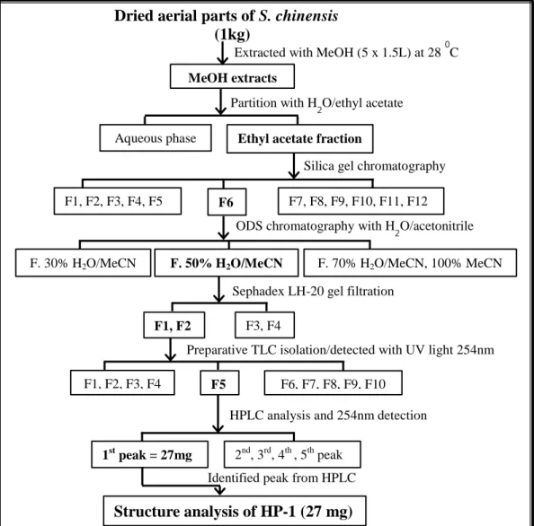
Figure 2. Overall of purification procedure of cancer stem cell inhibitor derived from Saururus chinensis (lizard’s tail).
①
• Extracted with MeOH (Saururus chinensis 1kg + methanol 7.5L)
②
• Extracted with EtOAc : H2O (1:1)
③
• SiO2 column chromatography eluted with CHCl3 : MeOH (30:1)
④
• Reversed-Phase C18 column (ODS) eluted with CH3CN : H2O (30%, 50%, 70% and 100% Acetonitrile)
⑤ • Sephadex LH-20 column chromatography eluted with methanol
⑥
• Prep. Thin layer chromatography
⑦
7
Figure 3. A flowchart depicting the purification of methanol extracts from the aerial parts of Saururus chinensis (SCE). The SCE-suspended in distilled water
was partitioned with ethyl acetate successively 1:1 (v/v) in a separating funnel. The ethyl acetate fraction was further sequentially subjected onto silica gel column chromatography. The silica gel fraction #6 was subsequently fractionated by reversed-phase ODS open column chromatography with solvent gradient 50% of acetonitrile in distilled water. The 50% eluted-sample from ODS was further subjected onto gel filtration Sephadex LH-20, and possibly lead to achieve fourth subfractions, respectively. The Sephadex LH-20 gel filtration fraction #1 & #2 were further separated in Prep TLC, which gave a dark band through the detection of UV254nm. A target band (active band) was collected and injected in HPLC.
Finally, the identified peak was collected by using UV254nm light detection.
MeOH extracts
Aqueous phase
Partition with H2O/ethyl acetate
Ethyl acetate fraction
Silica gel chromatography
Dried aerial parts of S. chinensis (1kg)
Extracted with MeOH (5 x 1.5L) at 28 0C
F1, F2, F3, F4, F5 F6 F7, F8, F9, F10, F11, F12
ODS chromatography with H2O/acetonitrile
F. 50% H2O/MeCN
Sephadex LH-20 gel filtration
F1, F2 F3, F4
Identified peak from HPLC
Preparative TLC isolation/detected with UV light 254nm
HPLC analysis and 254nm detection
F. 30% H2O/MeCN F. 70% H2O/MeCN, 100% MeCN
F1, F2, F3, F4 F5 F6, F7, F8, F9, F10
1st peak = 27mg 2nd, 3rd, 4th , 5th peak
8
3.1. Preparation of methanolic crude extracts
The dried and ground plant material (1kg) of S. chinensis was extracted with
100% methanol (MeOH) (1.5L x 5) in 2800 mL Erlenmeyer flasks at 28oC, 200 rpm for 22 hours in the shaking incubator. After shaking overnight, whole sample were divided into 500 mL plastic bottle and centrifuged at 3500 rpm for 20 min at 4 oC. The methanol-extracted supernatantwas decanted and filtered through a filter paper (Advantec, 100 circles, No. 2, 185mm), and all the solvent extracts were dried under vacuum rotary evaporator to get dried crude S. chinensis extract at 40
o
C to 50 oC under reduced pressure, which was subjected to further fractionation and bioassay.
3.2. Fractionation of the methanolic extracts by means of liquid-liquid partitioning
Methanol-extracted sample of S. chinensis with distilled water (100 mL) was concentrated to remove all methanol and resuspended in 500 mL of distilled water and sequentially partitioned successively with equal volumes of ethyl acetate (EtOAc) in a separating funnel (1:1 v/v) ratio. The lower aqueous phase was discarded and the top ethyl acetate phase was retained and screened for possible anti-CSCs activity. All the solvent phase extracts were dried under vacuum rotary evaporator to get dried crude S. chinensis extract at 40 oC to 50 oC under reduced pressure. The ethyl acetate-soluble fraction was suspended in 120 mL of methanol for a stock solution and subjected for further fractionation and bioassay.
9
3.3. Isolation and purification of active substances in the bioactive ethyl acetate fraction by bioassay-guided fractionation
Silica gel (SiO2) column chromatography: 1.5 mL (from stock 120 mL) of
the bioactive in the ethyl acetate fraction was subjected to normal silica gel column chromatography on a column (silica gel 60, 70-230 mesh ASTM, Merck Co., 100g gel/column). The column (25 x 340mm) was eluted with a solvent system of chloroform: methanol (CHCl3: MeOH = 30:1, v/v) to yield 12 subfractions.
Individual fraction was evaporated to dryness and stocked with MeOH 1.5 mL for further bioassay and fractionation. On the other hand, in each fraction were observed by TLC plates (10 cm x 10 cm, covered with silica gel, Merck 60 F254nm).
Individual fraction was spotted on the TLC plate, and developed in the solvent system (CHCl3: MeOH = 30:1, v/v) to investigate the active fraction.
Reversed-Phase C18 (ODS) gel columns chromatography fractionation:
The fraction #F6 from silica gel was further purified by open column chromatography with Reversed-Phase C18 gel. The sample was reconstituted in methanol and stored for overnight; then centrifuged at 15,000 rpm, 4 oC for 20 min. The aqueous solution parts were dried under vacuum rotary evaporator; then dissolved and eluted in a solvent system of 30% of acetonitrile in water. Then, the following solvent system was used to remove the sample by washing ODS gel with a solvent system 50% of acetonitrile in water, 70% of acetonitrile in water and 100% of acetonitrile. All of the eluents compounds were individually concentrated in vacuum and resuspended in 1.5 mL of methanol. Finally, individual fraction was
10
tested in the mammospheres cell assay to examine the active compounds in order
to confirm the target fraction.
Purification by gel filtration Sephadex LH-20: The active fraction of 50%
acetonitrile sample that reconstituted with methanolwas centrifuged at 15,000 rpm, 4 oC for 20 min, and further purified by column chromatography (20 x 400mm) with gel filtration Sephadex LH-20. The column was eluted with 100% methanol to yield four subfractions of the impure mixed compounds. All of the four subfractions were independently concentrated in vacuum and resuspened in 1 mL of methanol. The single fraction was tested in the mammospheres cell assay to examine the active ingredient. Therefore, F1 and F2 were obtained. Finally, F1 and F2 were combined and dried to remain only 1 mL for further purification.
3.4. Identification of active components by Preparative TLC using UV254nm
fluorescence detection
Preparative TLC was used to separate the gel filtrated sample. The 500 L sample (from 1 mL stocked) was loaded onto the preparative thin layer chromatography (P. TLC) glass plates (20 x 20 cm) covered with Silica gel (60, F254; 1 mm thick) and developed in the solvent gradient of CHCl3: MeOH (30:1,
v/v) using a TLC glass tank pre-saturated with the mobile phase; and each plate was developed to a height of about 26 x 30 cm chamber. After development, plates were removed and dried; the positions of varied compounds were visualized by fluorescence under UV radiation (UV254nm and UV365nm). Individual band was
11
separately removed by scraping off the silica gel from the glass using surgery knife and collected in the 15 mL conical tube. The 5 mL of methanol (twice) was added to purify each compound from the gel. Then, the mixture of gel and solvent was vortexed, filtered and recovered by centrifugation. Hence, the eluents were concentrated in vacuum to give individual band and stocked in 1 mL of methanol for further bioassay and HPLC fractionation.
3.5. HPLC analysis
Liquid chromatography analysis was operated using Shimadzu ODS column, LC-30A/20A/10Avp/10A-series Solvent Delivery Module (Pump), Detector, and Fraction Collector (Tokyo, Japan); and it is also possible to connect and control the SPD-M20A/M30A Photodiode Array Detector from the system controller as a 4-wavelength detector via the network. Chromatographic separations were conducted using Shim-pack GIS PREP-ODS, 10 x 250 mm, 10 m C18 particle size column.
The mobile phase was composed of two solvents: H2O (A) and MeCN (B) as a
linear gradient elution program was applied as follows: 45% B (0-10 min), 45%-60% B (10-30 min), 60%-100% B (30-40 min), 100-100% B (40-50 min), 100-45% B (50-55 min). The flow rate was maintained at a 3 mL min-1, and temperature of the column was set at room temperature.
Briefly, samples from preparative TLC of F5 was performed by HPLC. The sample reconstituted in methanol was filtered through a 0.45-m Millipore filter. Then, the sample injection volume was set up in 500 L, respectively, and the UV
12
detection was performed at 220 and 254nm. However, the elutes were on-line monitored with UV detector (254 nm) and peak fractions were collected according to the chromatogram. The HP-1 compound was collected and concentrated to dryness; then resuspended in methanol for further bioassay.
4. Mammosphere formation assaay
Human breast cancer cells, MCF-7 were obtained from the American Type Culture Collection (ATCC; Manassas, VA, USA). MCF-7 cells were grown in Dulbecco’s Modified Essential Medium (DMEM; Hyclone, Logan, UT, USA) with 10% fetal bovine serum (FBS; Hyclone), 100 U/ml penicillin, and 100 μg/ml streptomycin (Hyclone). MCF-7 cells were maintained at 37°C in a humidified incubator with 5% CO2. Cells were plated at a density of 1x10⁶ cells in 10 cm
culture dish.
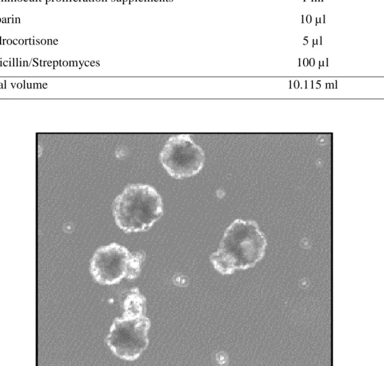
Anti-CSC activity was qualitatively evaluated by mean of the mammosphere formation assay technique. A cellular bioassay was used to culture breast cancer cell lines as tumorspheres assay from STEMCELL Technologies. The MCF-7 human breast cell line was seeded in 6-well plate at a density of 50,000 cells in 2 mL of completed medium per well, but for 24-well plate (500 L of completed medium/well) (see the components of MammoCult in Table. 1), and the purified-sample from each fraction was added into the plate and cultured at 37°C in a humidified atmosphere containing 5% CO2for 7 days before imaging and counting.
13
the mammospheres were counted with an automated counting program called NIST’s Intergrated Colony Enumerator (NICE) for further analyzing of sample activities.
Table 1. Mammosphere formation assay using breast cancer stem cell derived
from MCF-7 cells and completed Mammocult medium for 6-wells or 24-wells plate.
Components Amount
Mammocult basal medium 9 ml Mammocult proliferation supplements 1 ml
Heparin 10 µl
Hydrocortisone 5 µl
Penicillin/Streptomyces 100 µl Total volume 10.115 ml
14
RESULTS
1. Purification of active substances from SCE
1.1. The anti-CSCs activity of solvent-partitioning fractions
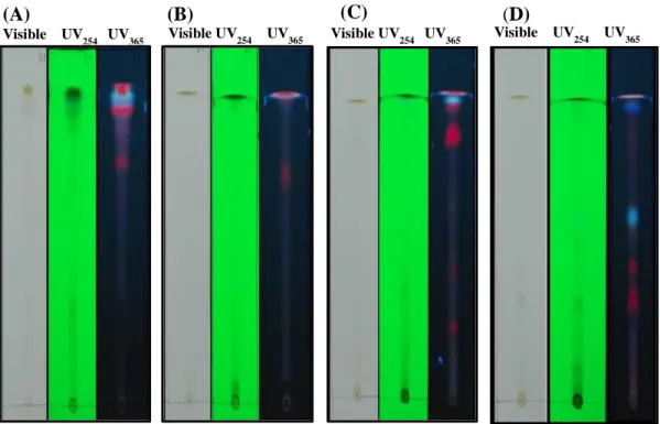
To identify the active substances exerting the anti-CSCs activity, SCE was partitioned with ethyl acetate organic solvents as given the overall and briefly scheme (Fig.2 & Fig.3). The anti-CSCs activity was evaluated by using mammospheres formation assay to examine each fraction (Fig.4). It was shown that the ethyl acetate-soluble fraction presented a very high anti-CSCs activity (not shown). Further isolate anactive substance, SCE was sorted out the mobility of the compounds on the TLC plate with various types of solvent conditions (chloroform: methanol = 4:1, 10:1, 20:1 and 30:1, v/v) by spot and develop in solvent-saturated chamber. TLC plates were dried and detected under UV light (254 and 365nm) (Fig.5). As a result, the TLC plate was shown the best separation in chloroform: methanol (30:1, v/v) for further purified the elements, respectively (Fig. 5D).
15
Figure 5. Thin layer chromatography analysis of ethyl acetate extracts from Saururus chinensis. Ethyl acetate: water equally (1:1,v/v)–extracted samples from S. chinensis were analyzed by spotting on silica gel 60 F254 TLC plates. TLC plates
were developed in solvent-saturated chamber after the spotting of samples. The chromatogram was dried and detected under a UV lamp with wavelength UV254nm
and UV365nm. (A) Chloroform: methanol=4:1; (B) Chloroform: methanol=10:1; (C)
Chloroform: methanol=20:1; (D) Chloroform: methanol = 30:1.
(C) (D) Visible UV 254 UV365 UV Visible UV 254 UV365 UV Visible UV254 UV365 UV (A) (B) Visible UV 254 UV365 UV
16
1. 2. The anti-CSCs activity of fractions obtained from ethyl acetate aqueous
The active parts in the ethyl acetate fraction were separated by silica gel column chromatography and yielded 12 subfractions (F1 to F12) (Fig. 6A). Then, each fraction was further analyzed by TLC with UV light to visible and capture the signal on the TLC plate(Fig. 6B). Last, the anti-CSCs activity of each fraction was evaluated by the mammosphere formation assay. In addition, the cell growth of mammosphere formation cells were suppressed by the fractions from F5 to F8 by 88% to 99% (Fig.6C & 6D). Moreover, because the fraction #6 (F6) was acquired with a proportionately high activity, the fraction was consequently subjected for further purification. On the other hand, F6 was seem to be very cloudy because the sample was performed crystal after treatment, which was made the scanner to be confused; however, this result can be assumed through the inverted microscope. As a result, F6 was convinced to be further investigated in the next step.
18 R e la ti v e g r o w th r a te ( % ) C o n t r o l F 1 F 2 F 3 F 4 F 5 F 6 F 7 F 8 F 9 F 1 0 F 1 1 F 1 2 0 2 0 4 0 6 0 8 0 1 0 0 1 2 0
Figure 6. The purification procedure of CSCs inhibitor derived from S. chinensis using SiO2 gel chromatography. (A) Sample was isolated by using
SiO2 gel chromatography with a solvent mixture CHCl3: MeOH (30:1, v/v) to elute
fraction sample and 12 subfractions were obtained. (B) TLC plate’s analysis of the purified sample. Each of the eluate fractions was spotted on TLC plate and developed in solvent-saturated chamber in CHCl3:MeOH (30:1, v/v). The TLC
plates were dried; then, the spots on TLC plates were analyzed and detected under a UV lamp. (C) The eluates (F1 to F12) were dried up and reconstituted with methanol. Mammospheres formation assay using breast cancer stem cell derived from MCF-7 cells; Cells were treated with 1 L and 2 L of each fraction. After a week incubation (5% CO2, humidified incubator at 37°C, 7 days), the
mammospheres were scanned under the Umax scanner and counting the mammospheres under NICE program. (D) The graphic summary of the activities from SiO2 isolated fractions. Relative growth rate to control (closed bar) was
shown. Control cells were negative control without any treatment.
19
1.3. Open ODS column chromatography of the anti-CSCs activity compound from F6
ODS open column chromatography (C18) was employed to investigate the
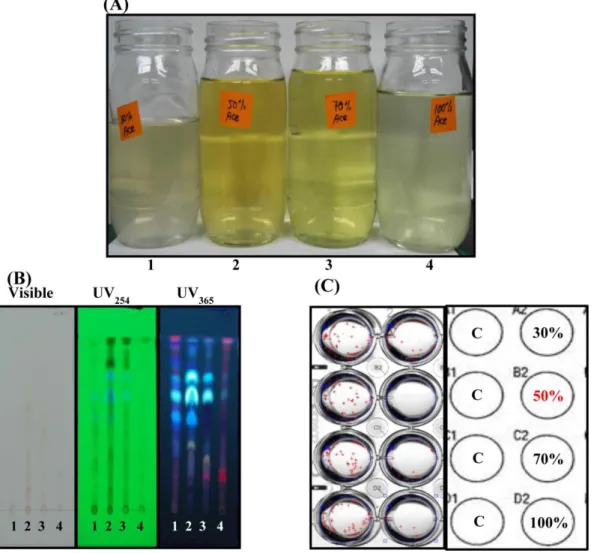
most active compound. F6 was concentrated to dryness in order to remove methanol, and dissolved in the mixture solvents in water and acetonitrile (1=30%, 2=50%, 3=70%, and 4=100%), which gave a yield as depended on the percentage of the solvent system (Fig. 7A). Purified components were classified the target activity with a relatively to purity, the fraction was further figured out by TLC. One among all, Fraction #2 (2 =50% acetonitrile) was consisted of the activity which determined by the dark spot on the upper side near the front line with Rf =
0.76 through UV wavelength 254nm (Fig.7B). Furthermore, in order to confirm the identical spot from TLC, mammosphere formation assay was used to discover the bioactivity materials. As shown in Fig. 7C, three fractions (2 =50% acetonitrile, 3 =70% acetonitrile, and 4 =100% acetonitrile) were shown the activities; however, only fraction #2 (50% acetonitrile) was existed significantly inhibited activity on breast cancer stem cell as bioactive-guided assay, indicating that the identical spot from the TLC was assigned to be the best candidate for further isolation.
20
Figure 7. The purification procedure of CSCs inhibitor in Saururus chinensis using Reversed-Phase C18 (ODS) gel chromatography. (A) ODS open
column chromatography (C18) eluted with CH3CN: H2O (1=30%, 2=50%, 3=70%
and 4=100% of CH3CN) using the partial-purified sample. (B) Thin-layer
chromatography analysis of the partial-purified sample from ODS resin, purified-sample was spotted and developed in chloroform: methanol (30:1, v/v). UV light was used to detect the pigments. (C) Mammosphere formation assay using breast cancer stem cell derived from MCF-7.
21
1.4. Separation of the anti-CSCs activity compound from 50% of acetonitrile elutes using Sephadex LH-20 column
The highest inhibition of F2 (50% MeCN) was further purified by using a gel filtration column chromatography onto a Sephadex LH-20 column; pre-equilibrated with 100% methanol and eluted with the same solvent. There were four subfractions obtained, and each fraction was isolated by its color and amount-dependence (figure 8A and 8B.Table). Individual fraction was analyzed by TLC for further investigation of the active fraction based on the spot as described from previous sessions in C18 ODS open column chromatography. Single fraction was spotted on TLC plate and developed in the solvent gradient system of chloroform: methanol (30:1, v/v). The plate was dried and detected under UV light 254nm and 365nm (Fig.8C). The band was determined under UV light 254nm with consisting of dark spot on the upper side near the front line (Rf value = 0.79) in F2. To be
assured the dark spot as revealed at the beginning by TLC on its anti-CSCs activity, mammosphere formation assay was engaged (Fig.8D). As a result, the mammosphere formation assay defined that the F2 was subsisted of the target band through its highest activities, although it was seen more red cross (counting sign) because of the sample crystal performance. Both the TLC and the bioassay were observed on its highest bioactivity, then F2 was led to further purification. In addition, F1 was also existed of the activity, this may indicate that F1 may certainly comprise of a target band from F2 with a small amount that could not be detected the spot. Thus, it was finally combined with F2. In short, the identical components from gel filtration chromatography (F1 & F2) were further purified for an anti-CSCs activity inhibitor.
22
Figure 8. Purification procedure of CSCs inhibitor in Saururus chinensis using Sephadex LH-20 gel chromatography. (A) Sephadex LH-20 column
chromatography eluted with methanol. (B) Image of sample’s color and table of the sample fractions. (C) Thin-layer chromatography analysis of the eluted fractions of S. chinensis which was developed in chloroform: methanol (30:1, v/v). (D) Mammosphere formation assay using breast CSCs derived from MCF-7 cells using partial-purified sample.
23
1.5. Isolation of the anti-CSCs activity compound from compounds separated using Preparative TLC
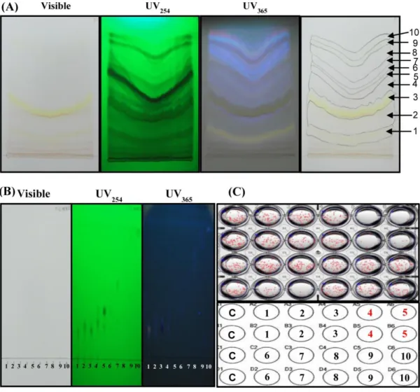
In order to separate successfully these mixtures of active compound using preparative TLC, a biphasic solvent system was selected on the basis of TLC analysis. Therefore, the preparative TLC separation of Sephadex LH-20-purified fraction was first performed in a diverse solvent system of chloroform and methanol by gradually decreased down the polarity of the solvent from 1:1 to 4:1, 10:1, 20:1 and 30:1 (v/v) (Data not shown). Thus, the solvent mixture formed by chloroform added with methanol (CHCl3: MeOH = 30:1, v/v) was shown the best
separation of the target band and led to isolate into ten subfractions (Fig. 9A). Then, F5 with an Rf value of 0.36 seemed to contain the most identical spot respectively,
which displayed the highest inhibition of anti-CSCs activities according to TLC analysis with UV light 254nm follow previous observation (Fig. 9B). On the other hand, F4 was also seemed to attain a small amount of the identical spot from F5. So, through this observation, those two fractions could be illuminated that it might involve a potential element of anti-CSCs activity. Individual fraction was alternatively assayed on cell in two types of concentration (5 L and 10 L from stock 1 mL in methanol); the activities were exposed after a week. Therefore, F5 was found almost 98% exhibitions of the anti-CSCs activities either in low or high concentration compare to negative control without treatment, so it means that the identical compound which was notified in the TLC as describe above was presented, this may indicate that the assume compound was consisted of to be further isolated. In addition, F4 was also subsisted of the activity similar to F5, but based on the analytical TLC chromatogram, this fraction was contained the
24
identical spot from F5, so it might presume to incorporate the F4 with F5 (Fig.9B). Thus, F4 was not further investigated. As a result, only active component from F5 could be a potential for further purification.
25
Figure 9. Purification procedure of CSCs inhibitor in Saururus chinensis using a preparative thin layer chromatography. (A) Preparative Thin-Layer
Chromatography of F1& F2 fractions in Sephadex LH-20 gel chromatography, 10 subfractions were analyzed after the development with chloroform and methanol (30:1, v/v); detected under UV light 254 and 365 nm. (B) TLC analysis of the Prep-TLC bands after scraped, and purified. Individual band was spotted and developed in chloroform: methanol (30:1, v/v). The plate was dried and visualized under UV light 254 and 365 nm. (C) Mammosphere formation assay using breast cancer stem cell derived from MCF-7 cells, individual band from purified samples were done in the assay.
2 2 3 4 4 5 7 7 88 99 1 0 1 0
26
1.6. Purification of active substance in SCE using Preparative HPLC
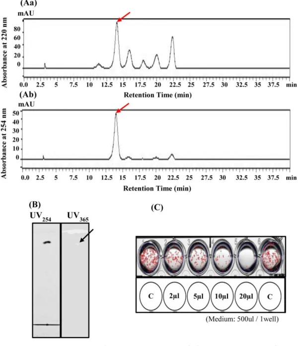
The biological activity components of F5 were found to be the highest quantity of designing component with TLC established; therefore, this fraction was targeted for isolation of anti-CSCs compounds. The chromatogram effluents were pooled together based on TLC analysis and on-line chromatogram; altogether, a total of 5 peaks were recorded in aqueous S. chinensis extracts during the scan at wavelengths ranging from 190 to 254nm. Furtherscanning, two types of the HPLC profile were used to investigate the peaks, detected with 220 and 254 nm. Consequently, based on the 220 nm profile, five main peaks were obtained (Fig. 10Aa), but through the UV detection of 254nm was shown the only dominant peak that is to be identified for further HPLC purification (Fig. 10Ab). Although the assertive peak was displayed as the leading peak, the rest of the peaks were also collected and tested for their activity. Thus, the only preeminent peak was shown the best activity (Data not shown).Furthermore, the ultimate peak as seen in either 220 or 254 nm was addressed to isolate as a major compound of anti-CSCs activity. Moreover, to demonstrate the purity of isolated compound, designed as HP-1, TLC was achieved (Fig. 10B). As a result,the isolate compound (HP-1) might be pure enough to be further studied. However, to be sure for the isolated compound of the HP-1 was the active compound that have been investigated, mammosphere assay was used in order to confirm the target compound (Fig. 10C). Therefore, the HPLC isolated compound know as HP-1 was obtained the activity against CSCs cells compare to negative control, this demonstrated that the purified sample could be further study of structure identification by NMR.
27
Figure 10. Fractionation of the preparative TLC fraction using HPLC. (A)
HPLC chromatogram was detected in UV220nm and UV254nm. Preparative TLC
fraction was fractionated by reversed-phase HPLC: mobile phase, two solvents: A, H2O and B, MeCN with a linear gradient as follows: 45% B (0-10 min), 45-60% B
(10-30 min), 60-100% B (30-40 min), absorbance was monitored at 254 nm with an elution program at 3 mL min-1 and 5 subfractions (F1 to F5) were obtained; however, only the major peak was collected and identified the agent as HP-1. (B) HPLC purified-sample was analyzed by TLC with pre-coated silica gel and developed with (CHCl3: MeOH= 30:1, v/v). The spot was detected with UV light
28
(254 and 365 nm). A single spot was observed in the HPLC fraction. (C) Mammosphere formation assay using CSCs cell derived from MCF-7 cells using the partial purified-sample. The purified-sample was treated on cell with various concentrations. Arrow bar indicates a target compound exhibiting the highest anti-tumor inhibitory activity.
29 2. Identification of active substance in SCE.
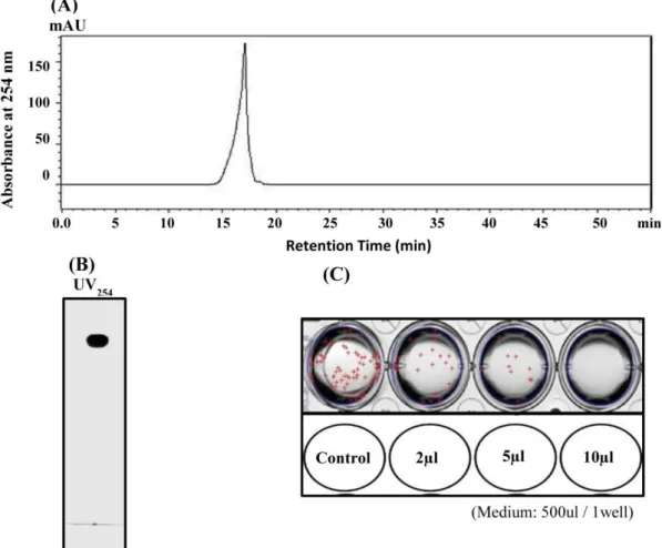
To confirm the purification of the target active compound that was separated through RP-HPLC with MeCN-H2O (45%-60%, and 60%-100%) as eluent to yield
pure compound, HP-1 was manifested the single chromatogram with the absorbance at 254 nm and retention time at 17 min (tR = 17 min) (Fig.11A). In
addition, TLC was also employed to examine the HP-1 compound from RP-HPLC purified (Fig.11B). To elucidate, both the HPLC chromatogram and the TLC chromatogram were clearly shown the outstanding purity of the HP-1 compound. Furthermore, the activity of the HP-1 was also performed in mammosphere formation assay in various concentrations (Fig.10C). As a result, either in low or high in concentration, the activity was presented compare to the negative control. Furthermore, the UV spectrum of the identical compound from HPLC fractionated named as HP-1 was exhibited (Fig. 12), respectively. The UV spectra of the HP-1 compound from the aerial parts of S. chinensis revealed characteristic absorption peaks at 202 nm and 261 nm.
30
Figure 11. The main peak was fractionated and collected by using High Performance Liquid Chromatography. HPLC condition: mobile phase, two
solvents: A, H2O and B, MeCN with a linear gradient as follows: 45% B (0-10
min), 45-60% B (10-30 min), 60-100% B (30-40 min), 100-100% B (40-50 min), 100-45% B (50-55 min); absorbance was monitored at 254 nm with elution program at 3 mL min-1. (A) Chromatogram of the purified-sample from RP-HPLC with UV light 254 nm detected; the sample was eluted with MeCN: H2O =67%
(tR=17 min). (B) TLC analysis of the RP-HPLC sample. The
purified-sample was spotted on TLC plate and developed in the solvent-saturated chamber with a solvent system CHCl3: MeOH (30:1, v/v). TLC plate was dried and detected
under UV lamp 254 nm. (C) Mammosphere formation assay using breast CSCs cells derived from MCF-7 cells. This assay was used to investigate the active compound from RP-HPLC purified-sample, designed as HP-1.
31 Figure 12. UV spectrum of the purified HP-1.
32
DISCUSSION
General stem cells have the ability to differentiate into many different cells. However, abnormal stem cells will develop into abnormal cell populations. These abnormal stem cells are CSCs. These CSCs share many characteristics with normal stem cells, including self-renewal and differentiation39); and CSCs can survive in a nutrient- and oxygen- deficient environment40) and have drug resistance, they are considered as one reason that cancer cannot be completely cured. The existence of CSCs has been implicated in many tumor types including breast cancer. The presence of CSCs, either intrinsic or therapy induced, has been attributed to the progression and poor prognosis in breast cancer patients41-42).
Recently, many biological active compounds from the aerial parts of Saururus
chinensis have been discovered to have anti-inflammatory3-7), antioxidant11-13), and anti-cancer activities15-16). However, only a few researches were demonstrated on breast cancer study with the active components from this plant25,36-37). Conversely, the active phytochemical constituent from this plant have not been elucidated its activity on human breast cancer stem cells (BCSCs). Therefore, in the present study, we have explored, for the first time to isolate anti-CSCs properties using MCF-7 human breast cancer cells as a tool to investigate anti-CSCs material from
S. chinensis plant.
Furthermore, there have been only a few reports regarding the anticancer effects of the lignan24) and the neolignans25,32-33) compounds obtained from the aerial parts of S. chinensis. Our result in this study, we isolated the novel
33
compound HP-1 that exhibits an inhibitory effect against human breast cancer stem cells (BCSCs). The compound is similar to lignan and neolignans compounds from other plants that have anticancer effects43). HP-1 was effective against human breast cancer stem cells. On the basis of RP-HPLC analysis, the total amount of HP-1 in the aerial parts of S. chinensis is approximately 27mg/kg. Here, we have discovered the most abundant compound in Sam-baekcho (Saururus chinensis); new constituents of HP-1 that show a strong inhibitory effect against human breast cancer stem cells.
Taken together, our finding result would be valuable for exploiting the potential of Saururus chinensis extraction (SCE) in the therapy of breast malignancy. The CSCs activity effects of SCE and its component on human breast cancer stem cells will provide an evidence to develop a chemotherapeutic drug against breast cancer cell. However, further works would be required to determine the details chemical formular of this plant by NMR. In addition, the anticancer activity of SCE to determine the details mechanism of action of these compound in vitro and in vivo is needed to be examined in the future.
34
CONCLUSION
CSCs are drug-resistant and radiation-resistant cancer cells to be responsible for tumor progression, maintenance and recurrence of cancer, and metastasis. Thus, the breast CSCs inhibitor that inhibits cancer stem cell derived from MCF-7 cells was isolated from the aerial parts of Saururus chinensis. The ethyl acetate fraction obtained from methanol extracts exhibited the outstanding inhibitory effect on mammospohere formation. From the ethyl acetate fraction, a new compound named HP-1 was purified through Silica gel column chromatography, ODS open column chromatography, Sephadex LH-20 column chromatography, preparative thin-layer chromatography, and reversed-phase high performance liquid chromatography. The UV spectra of the HP-1 revealed characteristic absorption peaks at 202 and 261 nm. The HP-1 isolated compound is highly effective against mammosphere cell tested, and could be used in the control of cancer cell. Therefore, the isolated compound exhibited a strong anti-CSCs activity, which may contribute to the interpretation of the pre-pharmacological and clinical effects of S.chinensis.
35
REFERENCES
1) Lyle E. Craker, James E. Sinom. “Herbs, Spices, and Medicinal Plants”.
Recent Advances in Botany, Horticulture, and Pharmacology, 4, p. 135
(1990).
2) Chung BS, Shin MG. “Dictionary of Korean Folk Medicine”. Young Lim
Sa, Seoul, pp. 813–814 (1990).
3) Kim RG, Shin KM, Kim YK, Jeong HJ, Ha J, Choi JW, Park HJ, Lee KT. “Inhibition of methanol extract from the aerial parts of Saururus chinensis on lipopolysaccharide-induced nitric oxide and prostagladin E2 production from murine macrophage RAW 264.7 cells”. J. Biol. Pharm. Bull., 26(4), 481–486 (2003).
4) Lee S.U, Choi CH, Kim YS, Min YK, Rhee M, Kim SH. “Anit-resorptive saurolactam exhibits in vitro anti-inflammatory activity via ERK-NF-kB signaling pathway”. J. Int. Immunopharmacol., 10, p.298-303 (2010). 5) Meng X, Kim I, Jeong YJ, cho YM, Kang SC. “Anti-inflammatory effects
of Saururus chinensis aerial parts in murine macrophages via induction of heme oxygenase-1”. J. Exp Biol Med (Maywood), 241(4), 396-408 (2016). 6) Kim BW, Koppula S, Park SY, Hwang JW, Park PJ, Lim JH, Choi DK.
“Attenuation of inflammatory-mediated neurotoxicity by Saururus chinensis extract in LPS=induced BV-2 microglia cells via regulation of NF-kB signaling and anti-oxidant properties”. J. BMC Compl Alternative
Med., 14, 502 (2014).
7) Kwon OE, Lee HS, Lee SW, Chung MY, Bae KH, Rho MC, Kim YK. “Manassantin A and B isolated from Saururus chinensis inhibit TNF-alpha-induced cell adhesion molecule expression of human umbilical vein endothelial cells”. J. Arch Pharm Res. 28(1), 55-60 (2005).
36
8) Yoo HJ, Kang HJ, Jung HJ, Kim K, Lim CJ, Park EH. “Anti-inflammatory, anti-angiogenic and anti-nociceptive activities of Saururus chinensis extract”. J Ethnopharmacol, 120(2), 282-286 (2008).
9) Lee E, Haa K, Yook JM, Jin MH, Seo CS, Son KH, Kim HP, Bae KH, Kang SS, Son JK, Chang HW. “Anit-asthmatic Activity of an Ethanol Extract from Saururus chinensis”. J. Biol Pharm Bull., 29(2), 211-215 (2006).
10) Quan Z, Lee YJ, Yang JH, Lu Y, Li Y, Lee YK, Jin M, Kim JY, Choi JH, Son JK, Chang HW. “Ethanol extracts of Saururus chinensis suppress ovalbumin-sensitization airway inflammation”. J. Ethnopharmacol.,
132(1), 143-9 (2010).
11) Cho HY, Cho CW, Song YS. “Antioxidative and Anti-Inflammatory Effects of Saururus chinensis Methanol Extract in RAW 264.7 Macrophages”. J.
Medicinal Food., 8(2), 190-197 (2005).
12) Jung JY, Lee K, Lee MY, Jung D, Cho ES, Son HY. “Antioxidant and antiasthmatic effects of saucerneol D in a mouse model of airway inflammation”. J. Int immunopharmacol., 11(6), 698-705 (2011).
13) Kang CS, Lee MJ, Park CB, Bang IS. “Study on the Antioxidative and Physiological Activities of Saururus chinensis Extract”. J. Life Science.,
22(6), 807-814 (2012).
14) Chio MS, Kim EC, Lee HS, Kim SK, Choi HM, Park JH, Han JB, An HJ, Um JY, Kim HM, Han AR, Hong MC, Bae H, Min BI. “Inhibitory effects of Saururus chinensis (LOUR.) BAILL on the development of atopic dermatitis-like skin lesions in NC/Nga mice”. J. Biol Pharm Bull., 31(8), 51-6 (2008).
15) Son ES, Choi JU, Lee KJ, Park HR. “Selective cytotoxicity of Saururus
chinensis in glucose-deprived HT-29 human colon cancer cells”. J. Food Sci. Biotechnol., 23(2), pp.609-613 (2014).
37
16) Daifuku M, Nishi K, Nishiwaki H, Kim I-H, Kim S, Sugahara T. “Antiproliferative acitivity and its underlying mechanisms of methanol extract from leaves of Saururus chinensis (Lour.) Biall. On mouse colon-26 carcinoma cells”. Int. J. Applied Research in Natural Products (IJARNP).,
7(3), pp.17-27 (2014).
17) Zhuang T, Li F, Huang LR, Liang JY, Qu W. “Secondary Metabolites from the Plants of the Family Saururaceae and Their Biological Properties”. J.
Chem. Biodi., 12(2), p.194-220 (2015).
18) Sung SH, Kwon SH, Cho NJ, Kim YC. “Hepatoprotective Flavonol glycosides of Suarurus chinensis Herbs”. J. Phytotherapy Res., 11, p.500-503 (1997).
19) Wang E.C, Shih M.H, Liu M.C, Chen M.T, Lee G.H. “Studies on constituents of Saururus chinensis”. J. Heterocycles., 43(5), p.969-975 (1996).
20) Chen HJ, Li X, Chen JW, Guo S, Cai BC. “Simultaneous determination of eleven bioactive compounds in Saururus chinensis from different harvesting seasons by HPLC-DAD”. J. Pharm Biomed Anal., 51(5), 1142-6 (2010).
21) Moon TC, Seo CS, Haa KM, Kim JC, Hwang NK, Hong TG, Kim JH, Kim DH, Son JK, Chang HW. “Meso-dihydroguaiaretic acid isolated from Saururus chinensis inhibits cyclooxygenase-2 and 5-lipoxygenase in mouse bone narrow-derived mast cells”. J. Arch Pharm Res., 31(5), pp. 606-610 (2008).
22) Song JW, Seo CS, Kim TI, Moon OS, Won YS, Son HY, Son JK, Kwon HJ. “Protective Effects of Manassantin A against Ethanol-Induced Gastric Injury in Rats”. J. Biol. Pharm. Bull. 39, 221-229 (2016).
23) Han HJ, Li M, Son JK, Seo CS, Song SW, Kwak SH, Bae HB. “Sauchinone, a lignan from Saururus chinensis, attenuates neutrophil
pro-38
inflammatory activity and acute lung injury”. J. Int. Immunopharmacol, 17, 471-477 (2013).
24) Bae HB, Li M, Son JK, Seo CS, Chung SH, Kim SJ, Jeong CW, Lee HG, Kim WM, Park HC, Kwak SH. “Sauchinone, a lignan from Saururus chinensis, reduces tumor necrosis factor-α production through the inhibition of c-raf/ MEK1/2/ERK 1/2 pathway activation”. J. Int.
Immunopharmacol., 10 (9), p. 1022-1028 (2010).
25) Hahm J.C, Lee I.K, Kang W.K, Kim S.U, Ahn Y.J. “Cytotoxicity of Neolignans Identified in Saururus chinensis towards Human Cancer Cell Lines”. J. Planta Med, 71(5), p.464-469 (2005).
26) Kang T.H, Cho H, Oh H, Sohn D.H, Kim Y.C. “Flavonol glycosides with free radical-scavenging activity of Saururus chinensis”. J. Fitoterapia., 76, p.115-117 (2005).
27) Kim YW, Kim YM, Yang YM, Kim TH, Hwang SJ, Lee JR, Kim SC, Kim SG. “Inhibition of SREBP-1c-mediated hepatic steatosis and oxidative stress by sauchinone, an AMPK-activating lignan in Saururus chinensis”. J.
Freeradbiomed, 48 (4), 567-578 (2010).
28) Lee J, Huh MS, Kim YC, Hattori M, Otake T. “Lignan, sesquilignans and dilignans, novel HIV-1 protease and cytopathic effect inhibitors purified from the rhizomes of Saururus chinensis”. J. Antiviral Res., 85(2), 425-428 (2010).
29) Kaur R, Kharb R. “Anti-HIV potential of Medicinally Important plants”. Int. J. Pharma. Bio Sciences., 2(3), p.387-398 (2011).
30) Wang C, Wang P, Chen X, Wang W, Jin Y. “Saururus chinensis (Lour.) Biall blocks enterovirus 71 infection by hijacking MEK1-ERK signaling pathway”. J. Antiviral Res., 119, p. 47-56 (2015).
31) Lee WS, Lee D-W, Baek Y-I, An S-J, Cho K-H, Choi Y-K, Kim H-C, Park H-Y, Bae K-H, Jeong T-S. “Human ACAT-1 and -2 inhibitory activities of
39
saucerneol B, manassantin A and B isolated from Saururus chinensis”. J.
Bioorganic & Medicinal Chemistry Letters., 14(12), p.3109-3112 (2004).
32) Song SY, Lee I, Park C, Lee H, Hahm JC, Kang WK. “Neolignans from Saururus chinensis inhibit PC-3 prostate cancer cell growth via apoptosis and senescence-like mechanisms”. Int. J. Mol. Med., 16(4), p. 517-523 (2005).
33) Lee YJ, Kim J, Yi JM, Oh SM, Kim NS, Kim H, Oh DS, Bang OS, Lee J. “ Anti-proliferative neolignans from Saururus chinensis against human cancer cell lines”. J. Biol Pharm Bull., 35(8), 1361-6 (2012).
34) Jeong HJ, Koo BS, Kang T, Shin HM, Jung S, Jeon S. “Inhibitory effects of Saururus chinensis and its components on stomach cancer cells”. J.
Phytomedicine., 22, 256-261 (2015).
35) Feng Y, Wang N, Zhu M, Feng Y, Li H, Tsao S. “Recent progress on anticancer candidates in patents of herbal medicinal products”. J. Recent
Pat Food Nutr Agric., 3(1), 30-48 (2011).
36) Eun-Yi Ko, Aree Moon. “Natural Products for Chemoprevention of Breast Cancer”. J. Cancer Prev., 20(4), 223-231 (2015).
37) Kim HY, Choi TW, Kim HJ, Kim SM, Park KR, Jang HJ, Lee EH, Kim CY, Jung SH, Shim BS, Ahn KS. “A methylene chloride fraction of Saururus
chinensis induces apoptosis through the activation of caspase-3 in prostate
and breast cancer cells”. J. Phytomedicine., 18, p. 567-574 (2011).
38) Chen K, Huang Y-H, Chen J-I. “Understanding and targeting cancer stem cells: therapeutic implications and challenges”. J. Acta Pharmacologica
Sinica., 34, p. 732-740 (2013).
39) Lobo N.A, Shimono Y, Qian D, Clarke M.F. “The biology of cancer stem cells”. J. Annu Rev Cell Dev Biol., 23, p. 675-699 (2007).
40
between cancer stem cells and their microenvironment”. J. Tumor Biol.,
35(5), p.3945-51 (2014).
41) Bozorgi A, Khazaei M, Rasool Khazaei M. “ New Findings on Breast Cancer Stem Cells: A review”. J. Breast Cancer., 18(4), p.303-312 (2015). 42) Velosco-Velazquez M.A, Homsi N, De La Fuente M, Pestell R.G. “Breast
cancer stem cells”. Int. J. Biochem. Cell Biol., 44, p.573-577 (2012).
43) Cuong T.D, Lim C.J, Thu-Trang T.T, Bae Y.H, Thu N.V, Tung N.T, Hung T.M, Woo M.H, Choi J.S, Min B.S. “Compounds from the seeds of
Myristica fragrans and their cytocoxic activity”. Nat. Prod. Sci., 18, p.
41
ACKNOWLEDGEMENTS
During my academic life at Jeju National University, I have got tremendously warm and comfortable stay till I accomplish my degree. Firstly, I would like to express my sincere gratitude to the College of Applied Life Sciences, JNU for letting me fulfill my dream of being a student here through my professor’s introduction. In addition, I would also like to thank the Biotechnology Faculty officer and assistant for giving me the opportunity to write an honors thesis. Secondly, I have got a full scholarship from my professor Dong-Sun Lee, and those support were included school fee, dormitory fee, living expenses, and study materials, so I would like to express the deepest appreciation to my Professor Dong-Sun Lee, who has the attitude and the substance of a genius: he continually and convincingly conveyed a spirit of adventure in regard to research and scholarship, and as excitement in regard to teaching. Without his guidance and persistent help this thesis would not have been possible. Moreover, to Dr. Hack Sun Choi, I am extremely grateful for your assistance advises and suggestions throughout my experiment process and thesis revising; and also to Dr. Kyung Hwan Boo for his helping. Finally, to my laboratory researchers, Kang Jiyoung, Kim Jihyang, Kim Solim, Mao Romnea and Deung Hong Won who have assisted me in the experiment. As well as, to all my friends and family for helping me survive all the stress from these years and not letting me give up.