저작자표시-비영리-변경금지 2.0 대한민국 이용자는 아래의 조건을 따르는 경우에 한하여 자유롭게 l 이 저작물을 복제, 배포, 전송, 전시, 공연 및 방송할 수 있습니다. 다음과 같은 조건을 따라야 합니다: l 귀하는, 이 저작물의 재이용이나 배포의 경우, 이 저작물에 적용된 이용허락조건 을 명확하게 나타내어야 합니다. l 저작권자로부터 별도의 허가를 받으면 이러한 조건들은 적용되지 않습니다. 저작권법에 따른 이용자의 권리는 위의 내용에 의하여 영향을 받지 않습니다. 이것은 이용허락규약(Legal Code)을 이해하기 쉽게 요약한 것입니다. Disclaimer 저작자표시. 귀하는 원저작자를 표시하여야 합니다. 비영리. 귀하는 이 저작물을 영리 목적으로 이용할 수 없습니다. 변경금지. 귀하는 이 저작물을 개작, 변형 또는 가공할 수 없습니다.
A Thesis
For the Degree of Master of Science in Medicine
Cytoprotective Effect of 3,4-Dihydroxybenzoic Acid
Isolated from Cladophora wrightiana Harvey Against
Ultraviolet B Radiation-Mediated Oxidative Damage
in HaCaT Keratinocytes
Ji Won Cha
Department of Medicine
Graduate School
Jeju National University
자외선 B에 의한 산화적 피부세포 손상에 대한
Cladophora wrightiana
Harvey에서 추출한
3,4-Dihydroxybenzoic Acid의 세포 보호효과
지도교수 현 진 원 강 희 경
차 지 원
이 논문을 의학 석사학위 논문으로 제출함
2015년 2월
차지원의 의학 석사학위 논문을 인준함
심사위원장 ○
印위 원 ○
印위 원 ○
印제주대학교 대학원
2015년 2월
Cytoprotective Effect of 3,4-Dihydroxybenzoic Acid
Isolated from Cladophora wrightiana Harvey Against
Ultraviolet B Radiation-Mediated Oxidative Damage
in HaCaT Keratinocytes
Ji Won Cha
(Supervised by Professor Jin-Won Hyun and Hee-Kyoung Kang)
A thesis submitted in partial fulfillment of the requirement for
the degree of Master of Science in Medicine
2015. 02.
This thesis has been examined and approved.
Department of Medicine
GRADUATE SCHOOL
JEJU NATIONAL UNIVERSITY
ABSTRACT
The purpose of the present study was to elucidate the protective effect of 3,4-dihydroxybenzoic acid (DBA) isolated from Cladophora wrightiana Harvey (a green algae) against ultraviolet B (UVB)-induced damage on HaCaT cells. DBA exhibited scavenging activity against the 1,1-diphenyl-2-picrylhydrazyl radical (DPPH), the superoxide anion and the hydroxyl radical. Furthermore, DBA diminished the levels of intracellular reactive oxygen species (ROS) generated by hydrogen peroxide or UVB treatment of the cells. DBA also decreased the UVB-augmented levels of phospho-histone H2A.X and the extent of comet tail formation, which are both indications of damaged DNA. In addition, the compound prevented keratinocytes from UVB-induced injury by reversing the production of apoptotic bodies, overturning the disruption of mitochondrial membrane potential, increasing the expression of the anti-apoptotic protein, B-cell lymphoma 2 (Bcl-2) and decreasing the expression of the pro-apoptotic proteins, Bcl-2-associated X (Bax) and cleaved caspase-3. Taken together, these results demonstrate that DBA isolated from a green algae protects human keratinocytes against UVB-mediated oxidative stress and apoptosis.
Keywords3,4-Dihydroxybenzoic acid ∙Cladophora wrightiana Harvey ∙ Human keratinocytes
CONTENTS
ABSTRACT………...Ӏ
CONTENTS………...II LIST OF FIGURES………...………...IV 1. Introduction………....1 2. Materials and Methods………...3
2-1. Materials 2-2. Cell Culture
2-3. Cell Viability Assay
2-4. Detection of the DPPH Radical 2-5. Detection of the Superoxide Anion 2-6. Detection of the Hydroxyl Radical 2-7. Detection of Intracellular ROS 2-8. Western Blot Analysis
2-9. Single-Cell Gel Electrophoresis (Comet Assay) 2-10. Nuclear Staining with Hoechst 33342
2-11. Analysis of Mitochondrial Membrane Potential 2-12. Statistical Analysis
3. Results………....8
3-1. Assessment of DBA Cytotoxicity in HaCaT Cells 3-2. Direct Radical Scavenging Activity of DBA 3-3. Scavenging Activity of DBA Against H2O2- or UVB-Generated Intracellular ROS 3-4. Protective Effect of DBA Against UVB-Induced DNA Damage 3-5. Cytoprotective Effect of DBA Against UVB-Induced Apoptosis 4. Discussion………...19
5. Reference………...21
6. Abstract in Korean………...26
LIST OF FIGURES
Figure 1………...……....8 Figure 1A. DBA doesn't show cytotoxicity in keratinocytes.
Figure 1B. DBA scavenges DPPH radical. Figure 1C. DBA scavenges superoxide anion. Figure 1D. DBA scavenges hydroxyl radical.
Figure 2………...……...12 Figure 2A. DBA scavenges intracellular ROS induced by H2O2 in HaCaT keratinocytes.
Figure 2B. DBA scavenges intracellular ROS induced by UVB in HaCaT keratinocytes.
Figure 3………...……...14 Figure 3A. DBA safeguards keratinocytes from elevated phospho-histone H2A.X level. Figure 3B. DBA safeguards keratinocytes from UVB-induced DNA damage.
Figure 4………...……...17 Figure 4A. DBA mitigates the formation of apoptotic bodies in UVB-irradiated cells. Figure 4B. DBA alleviates the disruption of mitochondrial membrane potential in UVB-
irradiated cells.
1. Introduction
Recent documents report that phenolic compounds derived from green seaweeds are promising antioxidant and antibacterial agents [Sabeena Farvin, K. H., & Jacobsen, C., 2013; Senthilkumar, P., & Sudha, S., 2011; Vinayak, R. C. et al., 2011]. Phenolic acids are substances that contain a phenolic ring and exhibit an organic carboxylic acid function. The phenolic hydrogen in the phenolic acids acts as a hydrogen-donating radical scavenger, conferring antioxidant properties to these molecules [Rice-Evans, C. A. et al., 1996]. Their scavenging capacity against free radicals and other reactive oxygen species (ROS) is used in the management of many chronic diseases, including diabetes mellitus, atherosclerosis, cancer and cardiovascular disease [Andreasen, M. F. et al., 2001; Yu, L. et al., 2002; Yu, L. et al., 2003].
Excessive sun exposure, closely related with induction of ROS, leads to harmful influence on skin. Acute or chronic exposure of the skin to Ultraviolet (UV) radiation results in oxidative stress, DNA damage and the development of inflammation leading to several skin disorders including hyperpigmentation, premature aging or photoaging of the skin, melanoma and non-melanoma skin cancers [De Gruijl, F. R., 1999; Katiyar, S. K., & Mukhtar, H., 2001; Kligman, L. H., 1986; Miller, D. L., & Weinstock, M. A., 1994]. In side of molecular mechanisms on UV radiation, it also induces caspase activation and disruption of mitochondrial membrane potential during UV radiation-induced apoptosis which is major cellular protective response for skin from the carcinogenic effects of sunlight [Denning, MF. et al., 2002]. Ultraviolet B (UVB) radiation (280–320 nm), especially, induces pivotal ROS in the skin and cultured skin cells, leading to gene mutations and abnormal cellular proliferation [Ahmed, N. U. et al., 1999; Hattori, Y. et al., 1996]. These ROS are generated by transferring electromagnetic energy from UVB radiation to molecular oxygen. A minimum of 50% of the damage caused by UVB light is due to ROS formation [Dinkova-Kostova, A. T., 2008]. UVB radiation as a potent ROS inducer can permeate through the epidermis to the dermis, contributing to skin wrinkling, freckling and the
development of skin cancers [Amaral, S. et al., 2013; Lee, C. H. et al., 2013].
The phenolic compound employed in this study, 3,4-dihydroxybenzoic acid (DBA, protocatechuic acid), was extracted from the green seaweed, Cladophora wrightiana Harvey, which is distributed in the North Pacific [Harvey, W. H., 1860]. Many researches have proved that DBA has an antioxidant activities [Sroka, Z., & Cisowski, W., 2003; Tung, Y. T. et al., 2009; Tung, Y. T. et al., 2007]. One study demonstrated that DBA shows much more effective antioxidant activities than standard antioxidant ‘Trolox’ in vitro through various antioxidant assay including DPPH, ABTS, reducing power (Fe3+ or Cu2+), superoxide anion radical-scavenging, hydroxyl radical-radical-scavenging, chelating ability (Fe2+ or Cu2+) [Li, X. C. et al., 2011]. Although there are many studies regarding antioxidant activities of DBA, however, little is known about the protective effects against UVB radiation-induced damage in skin cells. Therefore, the present study investigated the abilities of DBA to safeguard human HaCaT keratinocytes from UVB-induced oxidative stress and apoptosis as well as confirmed the scavenging activities of DBA against various ROS.
2. Materials and Methods
2-1. Materials
DBA was provided by Professor Nam Ho Lee of Jeju National University (Republic of Korea). The 1,1-diphenyl-2-picrylhydrazyl radical (DPPH), N-acetylcysteine (NAC), 5,5-dimethyl-1-pyrroline-N-oxide (DMPO), 2′,7′-dichlorodihydrofluorescein diacetate (DCF-DA), [3-(4,5-dimethylthiazol-2-yl)-2,5-diphenyltetrazolium] bromide (MTT), Hoechst 33342 dye and antibody against actin were purchased from Sigma-Aldrich Inc. (St. Louis, MO, USA). The primary antibodies against B-cell lymphoma 2 (Bcl-2), Bcl-2-associated X (Bax) was purchased from Santa Cruz Biotechnology, Inc. (Santa Cruz, CA, USA) and the primary antibodies against caspase-3, phospho-histone H2A.X, were purchased from Cell Signaling Technology, Inc. (Danvers, MA, USA). All other chemicals and reagents were of analytical grade.
2-2. Cell Culture
The human keratinocyte cell line, HaCaT, was obtained from the Amore Pacific Company (Gyeonggi-do, Republic of Korea) and maintained at 37°C in an incubator with a humidified atmosphere of 5% CO2. The cells were cultured in Dulbecco’s modified Eagle’s medium
containing 10% heat-inactivated fetal calf serum, streptomycin (100 μg/mL) and penicillin (100 U/mL).
2-3. Cell Viability Assay
The effect of DBA on the viability of HaCaT cells was assessed as follows. Cells were seeded into a 96-well plate at a density of 0.5×10⁵ cells/mL and treated 16 h later with 5, 10, 20, 40, 80, 160 μM DBA or 1 mM NAC. After 24 h incubation, MTT stock solution (50 μL, 2 mg/mL) was added to each well to yield a total reaction volume of 250 μL. Four hours later, the supernatants
were aspirated. The formazan crystals in each well were dissolved in dimethyl sulfoxide (150 μL) and the absorbance at 540 nm was read on a scanning multi-well spectrophotometer [Carmichael, J. et al., 1987].
2-4. Detection of the DPPH Radical
DBA (5, 10, 20, 40, 80, or 160 μM) or NAC (1 mM) was added to methanol containing 0.1 mM DPPH and the resulting reaction mixture was shaken vigorously. After 3 h, the amount of unreacted DPPH was measured spectrophotometrically at 520 nm.
2-5. Detection of the Superoxide Anion
The superoxide anion was generated via the xanthine/xanthine oxidase system and then reacted with DMPO. The DMPO/·OOH adducts were detected using a JES-electron spin resonance (ESR) spectrometer (JEOL, Tokyo, Japan) [Kohno, M. et al., 1994; Ueno, I. et al., 1984;]. Briefly, ESR signal was recorded 2 min after 20 μL of xanthine oxidase (0.25 U/mL) was mixed with 20 μL each of xanthine (5 mM), DMPO (3 M) and DBA (80 μM). The ESR spectrometer parameters were as follows: a magnetic field of 336.8 mT, power of 5.00 mW, frequency of 9.4380 GHz, modulation width of 0.2 mT, amplitude of 500, sweep time of 0.5 min, sweep width of 10 mT, time constant of 0.03 sec and temperature of 25°C. Superoxide anion generation was defined as the detected signal value.
2-6. Detection of the Hydroxyl Radical
The hydroxyl radical was generated by the Fenton reaction (H2O2+FeSO4) and then reacted
with DMPO. The resultant DMPO/·OH adducts were detected by ESR spectrometry [Li, L. et al., 2004; Li, L. et al., 2003]. The ESR spectrum was recorded immediately after a phosphate buffer solution (pH 7.4) was mixed with 20 μL each of DMPO (0.3 M), FeSO4 (10 mM), H2O2
field of 336.8 mT, power of 1.00 mW, frequency of 9.4380 GHz, modulation width of 0.2 mT, amplitude of 100, sweep time of 0.5 min, sweep width of 10 mT, time constant of 0.03 sec and temperature of 25°C. Hydroxyl radical generation was defined as the detected signal value.
2-7. Detection of Intracellular ROS
The DCF-DA assay was used to detect intracellular ROS generated by H2O2 or UVB in H2O2-
or UVB-treated HaCaT cells [Rosenkranz, A. R. et al., 1992]. The cells were seeded into 96-well plates at a density of 1.0×10⁵ cells/mL and treated with DBA (80 μM). After a 1 h incubation at 37°C, the cells were treated with H2O2 (1 mM) or UVB (30 mJ/cm
2
) and the plates were again incubated for 30 min (H2O2) or 30 h (UVB) at 37°C. A DCF-DA solution (50 μM)
was then added to the cells. Ten minutes later, the fluorescence of the 2′,7′-dichlorofluorescein product was detected and quantified using a PerkinElmer LS-5B spectrofluorometer (PerkinElmer, Waltham, MA, USA).
2-8. Western Blot Analysis
Harvested cells were lysed by incubation on ice for 10 min in 150 μL of a lysis buffer (120 mM NaCl, 40 mM Tris (pH 8) and 0.1% NP-40). The cell lysates were then centrifuged at 13,000×g for 5 min. The supernatants were collected and protein concentrations were determined. Aliquots of the lysates (15 μg of protein) were boiled for 5 min and electrophoresed in a 12% sodium dodecyl sulfate–polyacrylamide gel. The electrophoresed proteins were transferred onto nitrocellulose membranes and the membranes were subsequently incubated with the appropriate primary antibodies specific for each protein. Following the reaction with the primary antibodies, the membranes were further incubated with secondary anti-immunoglobulin-G-horseradish peroxidase conjugates (Pierce, Rockford, IL, USA). Protein bands were detected using an enhanced chemiluminescence Western blotting detection kit (Amersham, Little Chalfont, Buckinghamshire, UK) according to the manufacturer’s
instructions.
2-9. Single-Cell Gel Electrophoresis (Comet Assay)
The degree of oxidative DNA damage was determined in a Comet assay [Rajagopalan, R. et al., 2003; Singh, N. P., 2000]. The cell suspension was mixed with 75 μL of 0.5% low-melting agarose at 39°C and the mixture was spread onto a fully frosted microscopic slide pre-coated with 200 μL of 1% normal-melting agarose. After solidification of the agarose, the slide was covered with another 75 μL of 0.5% low-melting agarose and then immersed in a lysis solution (2.5 M NaCl, 100 mM Na–ethylenediaminetetraacetic acid (Na-EDTA), 10 mM Tris, 1% Triton X-100 and 10% dimethyl sulfoxide, pH 10) for 1 h at 4°C. The slides were subsequently placed in a gel electrophoresis apparatus containing 300 mM NaOH and 10 mM Na-EDTA (pH 13) for 40 min to allow for DNA unwinding and the expression of alkali-labile damage. An electrical field was then applied (300 mA, 25 V) for 20 min at 4°C to draw the negatively charged DNA towards the anode. The slides were washed three times for 5 min at 4°C in a neutralizing buffer (0.4 M Tris, pH 7.5), stained with 40 μL of ethidium bromide (10 μg/mL) and observed under a fluorescence microscope equipped with an image analyzer (Kinetic Imaging, Komet 5.5, UK). The percentage of the total cellular fluorescence in the comet tails and the tail lengths of 50 cells per slide were recorded.
2-10. Nuclear Staining with Hoechst 33342
HaCaT cells were treated with DBA (80 μM) and exposed to UVB radiation (30 mJ/cm2
) 3 h later. After an additional 24 h incubation at 37°C, the DNA-specific fluorescent dye Hoechst 33342 (1 μL of a 20 mM stock) was added to each well of a 96-well plate and the cells were incubated for 10 min at 37°C. The stained cells were visualized under a fluorescence microscope equipped with a CoolSNAP-Pro color digital camera. The degree of nuclear condensation was evaluated and the apoptotic cells were quantified.
2-11. Analysis of Mitochondrial Membrane Potential
Cells were treated with DBA (80 μM), exposed to UVB radiation (30 mJ/cm2
) and incubated at 37°C for 12 h. They were then stained with JC-1 (5 μM) to detect mitochondrial polarity and analyzed by flow cytometry [Troiano, L. et al., 2007] and confocal microscopy with the laser scanning microscope 5 PASCAL program (Carl Zeiss, Jena, Germany).
2-12. Statistical Analysis
All measurements were performed in triplicate and all values are expressed as the mean ± the standard error of the mean. The results were subjected to an analysis of variance and Tukey’s post hoc test to analyze differences between means. In each case, a p value of <0.05 was considered statistically significant.
3. Results
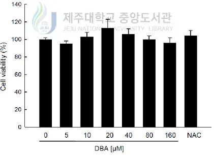
3-1. Assessment of DBA Cytotoxicity in HaCaT Cells
The effects of DBA and NAC, a well-known antioxidant, on HaCaT cell viability were assessed via the MTT assay. DBA was not cytotoxic to the keratinocytes at any of the concentrations employed (5, 10, 20, 40, 80 and 160 μM; Figure 1A). Similar results were obtained with NAC (1 mM; Figure 1A). The lack of cytotoxicity signifies that DBA might be useful as antioxidant reagent.
Figure 1A. DBA doesn’t show cytotoxicity in keratinocytes. HaCaT cells were treated
with DBA (0, 5, 10, 20, 40, 80, or 160 μM) or NAC (1 mM). After 24 h, the cells were incubated with the MTT reagent (2 mg/mL) for another 4 h. Cell viability was measured spectrophotometrically at 540 nm. NAC was used as the positive control.
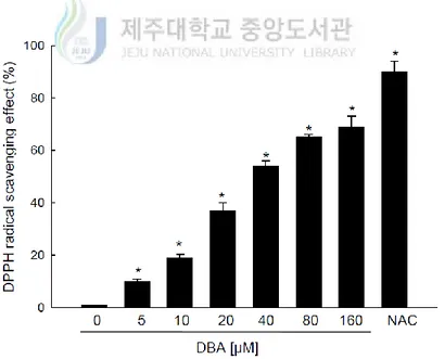
3-2. Direct Radical Scavenging Activity of DBA
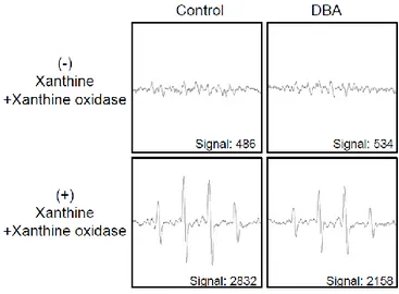
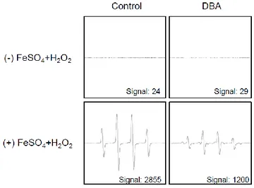
Next, it was conducted to measure the direct radical scavenging activity of DBA using the DPPH assay. DBA significantly scavenged 10, 19, 37, 54, 65 and 69% of the DPPH radical in a dose-dependent manner at 5, 10, 20, 40, 80 and 160 μM, respectively. These results can be compared with 90% for NAC (Figure 1B). From these data, it was determined to 80 μM DBA as the optimal concentration for further study. Treatment with 80 μM DBA decreased the levels of the superoxide anion generated by the xanthine–xanthine oxidase system, with a signal value of 2,832 in xanthine–xanthine oxidase system and 2,158 in the DBA-treated xanthine–xanthine oxidase system (Figure 1C). DBA also scavenged the hydroxyl radical generated by the Fenton reaction, with a signal value of 2,855 in the positive control Fenton reaction and 1,200 in the DBA-treated Fenton reaction (Figure 1D).
Figure 1B. DBA scavenges free radicals. DPPH was added to methanol along with DBA (0,
5, 10, 20, 40, 80, or 160 μM) or NAC (1 mM) for 3 h. The radical scavenging actions of DBA were determined spectrophotometrically at 520 nm. Asterisk significantly different from control (DBA-untreated cells; p<0.05).
Figure 1C. DBA scavenges free radicals. The xanthine/xanthine oxidase system was used
to investigate the scavenging effect of DBA against the superoxide anion. DMPO (3 M) and DBA (80 μM) were mixed with the xanthine/xanthine oxidase system. The resultant DMPO/·OOH adducts were then detected by ESR spectrometry. The peak heights correspond to the amount of superoxide anion generated. Asterisk significantly different from control (p<0.05) and number sign significantly different from the superoxide anion (p<0.05).
Figure 1D. DBA scavenges free radicals. The Fenton reaction system (H2O2+FeSO4) was
used to investigate the scavenging actions of DBA against the hydroxyl radical. DMPO (0.3 M) and DBA (80 μM) were mixed with the Fenton reaction system. The resultant DMPO/·OH adducts were detected by ESR spectrometry. The peak heights correspond to the amount of hydroxyl radical generated. Asterisk significantly different from control (p<0.05) and number sign significantly different from the hydroxyl radical (p<0.05).
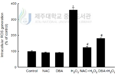
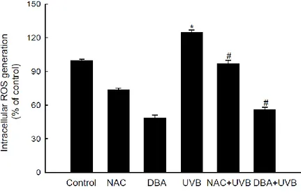
3-3. Scavenging Activity of DBA Against H
2O
2- or UVB-Generated Intracellular ROS
Next, it was carried out DCF-DA assay to assess the actions of DBA and NAC against intracellular ROS generated by H2O2 or UVB radiation in HaCaT keratinocytes. Intercellular
ROS levels were set at 100% in control, untreated cells. This value increased dramatically to 359% in H2O2-treated cells. However, DBA and NAC treatment both significantly decreased the
intracellular ROS content in H2O2-treated-cells (181% for DBA versus 124% for NAC; Figure
2A). Furthermore, the mean intracellular ROS value was 125% in UVB-irradiated keratinocytes, which was significantly decreased by DBA and NAC (56% for DBA versus 97% for NAC; Figure 2B).
Figure 2A. DBA scavenges intracellular ROS in HaCaT keratinocytes. DBA (80 μM) or
NAC (1 mM) was added to HaCaT cells for 1 h. The cells were then incubated with H2O2
for 30 min, followed by DCF-DA for 10 min. Intracellular ROS levels generated by H2O2
were detected spectrofluorometrically. Asterisk significantly different from control cells (p<0.05) and number sign significantly different from H2O2-treated cells (p<0.05).
Figure 2B. DBA scavenges intracellular ROS in HaCaT keratinocytes. DBA (80 μM) or
NAC (1 mM) was added to HaCaT cells for 1 h. Following 30 h incubation after exposure to UVB (30 mJ/cm2), the cells were treated with DCF-DA for 30 min. Intracellular ROS levels generated by UVB radiation were detected spectrofluorometrically. Asterisk significantly different from control cells (p<0.05) and number sign significantly different from UVB-irradiated cells (p<0.05).
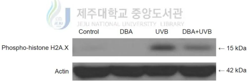
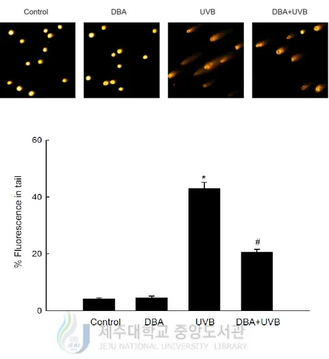
3-4. Protective Effect of DBA Against UVB-Induced DNA Damage
Histone H2A.X is required for checkpoint-mediated cell cycle arrest and DNA repair triggered by double-stranded DNA breaks [Yuan, J. et al., 2010]. Within a few minutes following DNA damage, H2A.X is phosphorylated on Ser139 at the sites of the damage [Burma, S. et al., 2001; Rogakou, E. P. et al., 1999; Rogakou, E. P. et al., 1998]. Figure 3A demonstrates that UVB-irradiated cells expressed significantly higher levels of phospho-histone H2A.X (Ser139) than control cells; however, DBA treatment decreased its expression. Moreover, exposure of the keratinocytes to UVB increased the number of DNA breaks, as assessed by a concomitant increase in the percentage of cellular DNA in the tails of the comet-like structures. The mean percentage of cellular DNA in comet tails was 43% in UVB-treated cells and 21% in DBA/UVB-treated cells (Figure 3B).
Figure 3A. DBA safeguards keratinocytes from elevated phospho-histone H2A.X level.
DBA (80 μM) was added to HaCaT cells for 1 h. Following 5 h incubation after exposure to UVB (30 mJ/cm2), the cells were harvested and proteins were extracted from the cell lysates. Phospho-histone H2A.X (Ser139) was detected on a Western blot via immunoreaction with a specific antibody.
Figure 3B. DBA safeguards keratinocytes from UVB-induced DNA damage. DBA (80 μM)
was added to HaCaT cells for 1 h. Following 1 h incubation after exposure to UVB (30 mJ/cm2), the cells were harvested and mixed with 0.5% low-melting agarose. Gel electrophoresis was performed after the mixture has been harden in a slide. The images were observed with a fluorescence microscope and analyzed by an image analyzer ‘Komet 5.5’. Representative images and the percentage of total cellular DNA fluorescence in the comet tails of ethidium bromide-stained cells are shown. Asterisk significantly different from control cells (p<0.05) and number sign significantly different from UVB-irradiated cells (p<0.05).
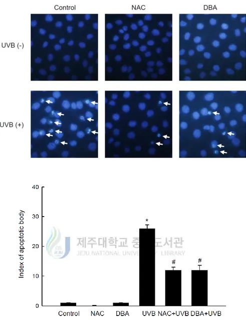
3-5. Cytoprotective Effect of DBA Against UVB-Induced Apoptosis
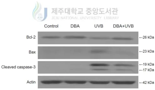
UVB-induced apoptosis can be mediated by DNA damage, mitochondrial membrane depolarization, death receptor activation and the generation of intracellular ROS [Kulms, D. et al., 2002]. Therefore, DNA damage was further assessed by investigating the presence of apoptotic bodies via Hoechst 33342 staining. Figure 4A demonstrates the formation of apoptotic bodies in UVB-treated cells. On the other hand, the staining was diminished by DBA treatment (Figure 4A). Mitochondrial membrane potential was next evaluated by staining with the membrane permanent dye, JC-1. The depolarized membrane regions of UVB-exposed cells were clearly shown by the green fluorescence of the JC-1 monomer, but the fluorescence intensity was decreased by DBA treatment (Figure 4B). Finally, it was investigated to observe the expression levels of the anti-apoptotic protein, B-cell lymphoma 2 (Bcl-2), as well as the pro-apoptotic proteins, Bcl-2-associated X protein (Bax) and cleaved caspase-3. Bcl-2 expression was attenuated in UVB exposed cells, but restored in DBA/UVB-treated cells (Figure 4C). Furthermore, expression of Bax was increased in UVB-irradiated cells and decreased in DBA/UVB-treated cells (Figure 4C). Moreover, the expression of cleaved caspase-3, a key executer of apoptosis, was also decreased by DBA treatment of UVB-exposed cells (Figure 4C).
Figure 4A. DBA mitigates the formation of apoptotic bodies in UVB-irradiated cells. DBA
(80 μM) or NAC (1 mM) was added to HaCaT cells for 1 h. Following 18 h incubation after exposure to UVB (30 mJ/cm2), cells were stained with Hoechst 33342 for 30 min and imaged via fluorescence microscopy. Apoptotic body formation (arrows) was then quantified. Asterisk significantly different from control cells (p<0.05) and number sign significantly different from UVB-irradiated cells (p<0.05).
Figure 4B. DBA alleviates the disruption of mitochondrial membrane potential in
UVB-irradiated cells. DBA (80 μM) was added to HaCaT cells for 1 h. Following 12 h incubation after exposure to UVB (30 mJ/cm2), cells were stained with JC-1 which is the mitochondrial membrane potential dye and fluorescence was observed via confocal laser scanning microscopy. A representative image shows JC-1 monomers (green) near mitochondria in UVB-irradiated cells.
Figure 4C. DBA modulates the expression of apoptotic regulators in UVB-irradiated cells.
DBA (80 μM) was added to HaCaT cells for 1 h. Following 7 h incubation after exposure to UVB (30 mJ/cm2), the cells were harvested and proteins were extracted from the cell lysates. Bcl-2, Bax and cleaved caspase-3 were detected on Western blots by immunoreaction with specific antibodies against each protein.
4. Discussion
UV light commonly consists of radiation of three wavelengths: UVA radiation (∼320–400 nm), UVB radiation (∼280–320 nm) and UVC radiation (∼100–280 nm). This study focused on UVB radiation, given that it is absorbed in greater amounts by the human epidermis and keratinocyte DNA than UVA or UVC radiation [Liebel, F. et al., 2012]. Excessive exposure to UV radiation occurs under various conditions, such as premature skin aging (photoaging), local and systemic immunosuppression and ultimately photo-carcinogenesis [De Gruijl, F. R. et al., 1993; Fisher, G. J. et al., 1997; Ryoo, I. et al., 2010]. Recently, many research groups have searched for herbal compounds and extracts with a protective effect against UVB-induced oxidative stress. One of these compounds is DBA, a well-known green algae-derived antioxidant containing a simple phenolic acid that potently suppresses lipid peroxidation and acts as scavenger of H2O2 and the DPPH radical [Sroka, Z., & Cisowski, W., 2003; Tseng, T. H.
et al., 1996]. DBA not only exerts antioxidant effects, but it is also a strong chemoprevention agent that can suppress carcinogenesis [Kawamori, T. et al., 1994; Nakamura, H. et al, 2000; Tanaka, T. et al., 1995; Tanaka, T. et al., 1993]. Furthermore, DBA selectively induces apoptosis in human carcinoma cells [Babich, H. et al., 2002; Lin, H. H. et al., 2007]. Here, the results show that DBA wields its antioxidant actions via direct quenching of the superoxide anion and the hydroxyl radical and also by scavenging intracellular ROS. UV radiation induces various kinds of intracellular ROS, including the superoxide anion, H2O2, the hydroxyl radical and
singlet oxygen [Fang, Y. et al., 2012; Yasui, H., & Sakurai, H., 2000]. UVB-induced DNA damage is particularly notorious for its association with apoptotic cell death, because cells with lethal or irreparable damage to nuclear DNA are removed by apoptosis to limit the incidence and propagation of defective cells [Vostalova, J. et al., 2010]. The phospho-histone H2A.X is critical for DNA repair following double-stranded DNA breaks. The expression of this protein
was considerably augmented by UVB radiation in human HaCaT keratinocytes. However, DBA reduced phospho-histone H2A.X expression in irradiated cells. This observation signifies that the compound can protect cells from UVB-evoked DNA damage. At the same time, DBA also prevented apoptosis in keratinocytes resulting from UVB-related oxidative stress. The electron transport chain in the inner mitochondrial membrane, which is required for the major functions of these organelles, is also affected by UVB. Inhibition of electron transport not only leads to mitochondrial membrane depolarization, a decrease in mitochondrial oxygen uptake and reduced phosphorylation of ADP to generate ATP, but also to an increase in ROS production following the incomplete reduction of oxygen (O2) [Paz, M. L. et al., 2008]. Outer membrane
localized JC-1 monomers emitted strong fluorescence in UVB-treated keratinocytes, indicative of mitochondrial injury and oxidative stress. However, DBA pretreatment decreased JC-1 fluorescence in irradiated cells. Finally, Western blotting analysis showed that DBA increased expression of anti-apoptotic factors, such as Bcl-2, in UVB-stressed cells, while simultaneously decreasing the expression of pro-apoptotic factors, such as Bax and cleaved caspase-3.
In conclusion, DBA has a preventative capacity to safeguard human HaCaT keratinocytes from excessive ROS generated by UVB exposure, as well as a protective effect against UVB-provoked DNA damage and apoptosis. Thus, DBA may find utility as a therapeutic agent to mitigate the effects of excessive sun exposure and the ensuing oxidative stress.
The main contents and experimental data of the thesis have been published on ‘Applied
Biochemistry and Biotechnology’ with ‘DOI: 10.1007/s12010-013-0711-3 in January 2014’,
entitled ‘Protective Effect of 3,4-Dihydroxybenzoic Acid Isolated from Cladophora
wrightiana Harvey Against Ultraviolet B Radiation-Induced Cell Damage in Human HaCaT Keratinocytes’.
5. References
Ahmed, N. U., Ueda, M., Nikaido, O., Osawa, T., & Ichihashi, M. (1999). The British Journal
of Dermatology, 140, 226–231.
Amaral, S., Redmann, K., Sanchez, V., Mallidis, C., Ramalho-Santos, J., & Schlatt, S. (2013).
Andrology, 1, 707–714.
Andreasen, M. F., Kroon, P. A., Williamson, G., & Garcia-Conesa, M. T. (2001). Free Radical
Biology & Medicine, 31, 304–314.
Babich, H., Sedletcaia, A., & Kenigsberg, B. (2002). Pharmacology & Toxicology, 91, 245–253. Burma, S., Chen, B. P., Murphy, M., Kurimasa, A., & Chen, D. J. (2001). The Journal of
Biological Chemistry, 276, 42462–42467.
Carmichael, J., DeGraff, W. G., Gazdar, A. F., Minna, J. D., & Mitchell, J. B. (1987). Cancer
Research, 47, 943–946.
De Gruijl, F. R. (1999). European Journal of Cancer. 35, 2003–2009.
De Gruijl, F. R., Sterenborg, H. J., Forbes, P. D., Davies, R. E., Cole, C., Kelfkens, G., et al. (1993). Cancer Research, 53, 53–60.
Denning, MF., Wang, Y., Tibudan, S., Alkan, S., Nickoloff, BJ., & Qin, J-Z. (2002). Cell Death
and Differentiation, 9, 40–52.
Dinkova-Kostova, A. T. (2008). Planta Medica, 74, 1548–1559.
Fang, Y., Hu, X. H., Jia, Z. G., Xu, M. H., Guo, Z. Y., & Gao, F. H. (2012). The Australasian
Fisher, G. J., Wang, Z. Q., Datta, S. C., Varani, J., Kang, S., & Voorhees, J. J. (1997). The New
England Journal of Medicine, 337, 1419–1428.
Harvey, W. H. (1860). Proceedings of the American Academy of Arts and Sciences, 4, 327–335. Hattori, Y., Nishigori, C., Tanaka, T., Uchida, K., Nikaido, O., Osawa, T., et al. (1996). The
Journal of Investigative Dermatology, 107, 733–737.
Katiyar, S. K., & Mukhtar, H. (2001). Journal of Leukocyte Biology, 69, 719–726.
Kawamori, T., Tanaka, T., Kojima, T., Suzui, M., Ohnishi, M., & Mori, H. (1994). Japanese
Journal of Cancer Research, 85, 686–691.
Kligman, L. H. (1986). Dermatologic Clinics, 4, 517–528.
Kohno, M.,Mizuta, Y., Kusai,M., Masumizu, T., & Makino, K. (1994). Bulletin of the Chemical
Society of Japan, 67, 1085–1090.
Kulms, D., Zeise, E., Poppelmann, B., & Schwarz, T. (2002). Oncogene, 21, 5844–5851.
Lee, C. H., Wu, S. B., Hong, C. H., Yu, H. S., & Wei, Y. H. (2013). International Journal of
Molecular Sciences, 14, 6414–6435.
Li, L., Abe, Y., Kanagawa, K., Usui, N., Imai, K., Mashino, T., et al. (2004). Analytica Chimica
Acta, 512, 121–124.
Li, L., Abe, Y., Mashino, T., Mochizuki, M., & Miyata, N. (2003). Analytical Sciences: the
International Journal of the Japan Society for Analytical Chemistry, 19, 1083–1084.
Li, X. C., Wang, X. Z., Chen, D. F., & Chen, S. Z. (2011). Functional Foods in Health and
Disease, 7, 232-244.
Liebel, F., Kaur, S., Ruvolo, E., Kollias, N., & Southall, M. D. (2012). The Journal of
Lin, H. H., Chen, J. H., Huang, C. C., & Wang, C. J. (2007). Journal International du Cancer,
120, 2306–2316.
Miller, D. L., & Weinstock, M. A. (1994). Journal of the American Academy of Dermatology, 30, 774–778.
Nakamura, H., Nishikawa, A., Furukawa, F., Kasahara, K., Miyauchi, M., Son, H. Y., et al. (2000). Anticancer Research, 20, 3423–3427.
Paz, M. L., Gonzalez Maglio, D. H., Weill, F. S., Bustamante, J., & Leoni, J. (2008).
Photodermatology Photoimmunology and Photomedicine, 24, 115–122.
Rajagopalan, R., Ranjan, S. K., & Nair, C. K. (2003). Mutation Research, 536, 15–25.
Rice-Evans, C. A., Miller, N. J., & Paganga, G. (1996). Free Radical Biology & Medicine, 20, 933–956.
Rogakou, E. P., Boon, C., Redon, C., & Bonner, W. M. (1999). The Journal of Cell Biology, 146, 905–916.
Rogakou, E. P., Pilch, D. R., Orr, A. H., Ivanova, V. S., & Bonner, W. M. (1998). The Journal of
Biological Chemistry, 273, 5858–5868.
Rosenkranz, A. R., Schmaldienst, S., Stuhlmeier, K. M., Chen, W., Knapp, W., & Zlabinger, G. J. (1992). Journal of Immunological Methods, 156, 39–45.
Ryoo, I., Moon, E. Y., Kim, Y. H., Lee, I., Choo, S. J., Bae, K. H., et al. (2010). Biomolecules &
Therapeutics, 18, 300–307.
Sabeena Farvin, K. H., & Jacobsen, C. (2013). Food Chemistry, 138, 1670–1681. Senthilkumar, P., & Sudha, S. (2011). Jundishapur Journal of Microbiology, 5, 411–415.
Mutagenesis, 455, 111–127.
Sroka, Z., & Cisowski, W. (2003). Food and Chemical Toxicology, 41, 753–758. Tanaka, T., Kojima, T., Kawamori, T., & Mori, H. (1995). Cancer, 75, 1433–1439.
Tanaka, T., Kojima, T., Kawamori, T., Yoshimi, N., & Mori, H. (1993). Cancer Research, 53, 2775–2779.
Troiano, L., Ferraresi, R., Lugli, E., Nemes, E., Roat, E., Nasi, M., et al. (2007). Nature
Protocols, 2, 2719–2727.
Tseng, T. H., Wang, C. J., Kao, E. S., & Chu, H. Y. (1996). Chemico-Biological Interactions,
101, 137–148.
Tung, Y. T., Wu, J. H., & Chang, S. T. (2007). Bioresource Technology, 98, 1120–1123.
Tung, Y. T., Wu, J. H., Huang, C. Y., Kuo, Y. H., & Chang, S. T. (2009). Bioresource Technology,
100, 509–514.
Ueno, I., Kohno, M., Yoshihira, K., & Hirono, I. (1984). Journal of Pharmacobio-Dynamics, 7, 563–569.
Vinayak, R. C., Sudha, S. A., & Chatterji, A. (2011). Journal of the Science of Food and
Agriculture, 91, 2471–2476.
Vostalova, J., Zdarilova, A., & Svobodova, A. (2010). Archives of Dermatological Research,
302, 171–181.
Yasui, H., & Sakurai, H. (2000). Biochemical and Biophysical Research Communications, 269, 131–136.
Yu, L., Haley, S., Perret, J., Harris, M., Wilson, J., & Qian, M. (2002). Journal of Agricultural
Yu, L., Perret, J., Harris, M., Wilson, J., & Haley, S. (2003). Journal of Agricultural and Food
Chemistry, 51, 1566–1570.
6. Abstract in Korean
이 연구는 녹조류인 Cladophora wrightiana Harvey로부터 추출한 3,4dihydroxybenzoic acid (DBA)가 자외선 B로 인해 유발된 HaCaT 세포의 손상에 있어 보호효과를 나타 낸다는 것을 증명하기 위해 진행되었다. DBA는 1,1-diphenyl-2-picrylhydrazyl radical (DPPH), 과산화물 음이온, 히드록실 라디칼에 대한 소거능을 보였다. 이뿐만 아니라, DBA는 세포에 과산화수소 또는 자외선 B에 노출시켰을 때 생성되는 세포 내 활성 산소의 양을 감소시켰다. 또한 DBA는 자외선 B에 의해 증가된 ‘DNA 손상 표지’인 phospho-histone H2A.X의 발현을 약화시키고 comet tail의 형성을 경감시켰다. 게다가 이 화합물은 자외선 B에 의해 유도된 피부손상으로부터 세포자멸사체의 형성과 미 토콘드리아 막전위의 상실을 막고, anti-apoptotic protein인 B-cell lymphoma 2 (Bcl-2) 발 현을 상향 조절하며 pro-apoptotic protein인 Bcl-2-associated X (Bax)와 cleaved caspase-3 의 발현을 하향 조절하여 HaCaT 세포를 보호한다. 결론적으로 이러한 결과들은 녹 조류로부터 추출된 DBA가 자외선 B로 인한 산화적 스트레스와 세포사멸로부터 인 간의 피부세포를 보호한다는 것을 증명한다.