저작자표시-비영리-동일조건변경허락 2.0 대한민국 이용자는 아래의 조건을 따르는 경우에 한하여 자유롭게 l 이 저작물을 복제, 배포, 전송, 전시, 공연 및 방송할 수 있습니다. l 이차적 저작물을 작성할 수 있습니다. 다음과 같은 조건을 따라야 합니다: l 귀하는, 이 저작물의 재이용이나 배포의 경우, 이 저작물에 적용된 이용허락조건 을 명확하게 나타내어야 합니다. l 저작권자로부터 별도의 허가를 받으면 이러한 조건들은 적용되지 않습니다. 저작권법에 따른 이용자의 권리는 위의 내용에 의하여 영향을 받지 않습니다. 이것은 이용허락규약(Legal Code)을 이해하기 쉽게 요약한 것입니다. Disclaimer 저작자표시. 귀하는 원저작자를 표시하여야 합니다. 비영리. 귀하는 이 저작물을 영리 목적으로 이용할 수 없습니다. 동일조건변경허락. 귀하가 이 저작물을 개작, 변형 또는 가공했을 경우 에는, 이 저작물과 동일한 이용허락조건하에서만 배포할 수 있습니다.
A Thesis for the Degree of Master of Science
Control of
Cronobacter sakazakii
ATCC 29544 by
Newly Isolated Bacteriophage CSP1 Recognizing
Lipopolysaccharide as a Receptor
지질다당체를 수용체로 인식하는 신규 박테리오파지
CSP1을 이용한 크로노박터 사카자키균의 병원성 저감
August, 2018
Mooseung, Kim
Graduate School of Agriculture and Life Science
Seoul National University
Department of Agricultural Biotechnology
Control of
Cronobacter sakazakii
ATCC 29544 by
Newly Isolated Bacteriophage CSP1 Recognizing
Lipopolysaccharide as a Receptor
지질다당체를 수용체로 인식하는 신규 박테리오파지
CSP1을 이용한 크로노박터 사카자키균의 병원성 저감
지도교수 유 상 열
이 논문을 석사학위논문으로 제출함
2018년 8월
서울대학교 대학원
농생명공학부
김무성
김무성의 석사학위논문을 인준함
2018년 6월
위 원 장
강 동 현 (인)
부위원장
유 상 열 (인)
위 원
장 판 식 (인)
ABSTRACT
Mooseung Kim
Department of Agricultural BiotechnologyCollege
The Graduate School
Seoul National University
Cronobacter sakazakii is an opportunistic foodborne pathogen causing fatal necrotizing enterocolitis and sepsis particularly in infants. Here, a virulent bacteriophage CSP1 specific for C. sakazakii
was analyzed to use as an antimicrobial agent for controlling this pathogen. Morphologically, CSP1 belongs to the family Siphoviridae
having an icosahedral head and a long, non-contractile tail. Complete genome of the phage was composed of 60,222 base-pairs of dsDNA (44.96% GC content), and 99 open reading frames with no tRNAs were predicted from the genome. Screening of Tn5 random mutant library of C. sakazakii ATCC 29544 revealed that disruption of genes involved in lipopolysaccharide (LPS) biosynthesis is associated with the CSP1 resistance. Deletion of waaG gene, which plays a role in LPS outer core biosynthesis, protected C. sakazakii
ATCC 29544 from CSP1 infection, and the complementation fully recovered the phage sensitivity. These results implied that LPS, particularly beyond the inner core of LPS, might be served as a receptor for CSP1. Spontaneous CSP1-resistant C. sakazakii
mutants possessed truncated LPS similar to the waaG deletion mutant and exhibited an impaired invasion ability against Caco-2 cells. Taken together, phage CSP1 could be applied as alternative biocontrol agents for C. sakazakii.
Keywords: Cronobacter sakazakii, bacteriophage, receptor, LPS,
waaG, virulence
CONTENTS
ABSTACT∙∙∙∙∙∙∙∙∙∙∙∙∙∙∙∙∙∙∙∙∙∙∙∙∙∙∙∙∙∙∙∙∙∙∙∙∙∙∙∙∙∙∙∙∙∙∙∙∙∙∙∙∙∙∙∙∙∙∙∙∙∙∙∙∙∙∙∙∙∙∙∙∙∙∙∙∙∙∙∙∙∙∙∙∙∙∙∙∙∙∙∙∙ⅰ
CONTENTS∙∙∙∙∙∙∙∙∙∙∙∙∙∙∙∙∙∙∙∙∙∙∙∙∙∙∙∙∙∙∙∙∙∙∙∙∙∙∙∙∙∙∙∙∙∙∙∙∙∙∙∙∙∙∙∙∙∙∙∙∙∙∙∙∙∙∙∙∙∙∙∙∙∙∙∙∙∙∙∙∙∙∙∙∙∙∙∙∙∙ⅲ
List of Figure∙∙∙∙∙∙∙∙∙∙∙∙∙∙∙∙∙∙∙∙∙∙∙∙∙∙∙∙∙∙∙∙∙∙∙∙∙∙∙∙∙∙∙∙∙∙∙∙∙∙∙∙∙∙∙∙∙∙∙∙∙∙∙∙∙∙∙∙∙∙∙∙∙∙∙∙∙∙∙∙∙∙∙∙∙∙∙∙ⅵ
List of Table∙∙∙∙∙∙∙∙∙∙∙∙∙∙∙∙∙∙∙∙∙∙∙∙∙∙∙∙∙∙∙∙∙∙∙∙∙∙∙∙∙∙∙∙∙∙∙∙∙∙∙∙∙∙∙∙∙∙∙∙∙∙∙∙∙∙∙∙∙∙∙∙∙∙∙∙∙∙∙∙∙∙∙∙∙∙∙∙∙ⅶ
Ⅰ. INTRODUCTION∙∙∙∙∙∙∙∙∙∙∙∙∙∙∙∙∙∙∙∙∙∙∙∙∙∙∙∙∙∙∙∙∙∙∙∙∙∙∙∙∙∙∙∙∙∙∙∙∙∙∙∙∙∙∙∙∙∙∙∙∙∙∙∙∙∙∙∙∙∙∙∙∙∙∙∙1
Ⅱ. MATERIALS AND METHODS∙∙∙∙∙∙∙∙∙∙∙∙∙∙∙∙∙∙∙∙∙∙∙∙∙∙∙∙∙∙∙∙∙∙∙∙∙∙∙∙∙∙∙∙∙∙∙∙∙∙∙∙∙5
1. Bacterial strains and growth condition∙∙∙∙∙∙∙∙∙∙∙∙∙∙∙∙∙∙∙∙∙∙∙∙∙∙∙5
2. Bacteriophage isolation and propagation∙∙∙∙∙∙∙∙∙∙∙∙∙∙∙∙∙∙∙∙∙∙∙∙5
3 Determination of phage host range∙∙∙∙∙∙∙∙∙∙∙∙∙∙∙∙∙∙∙∙∙∙∙∙∙∙∙∙∙∙∙∙∙∙∙7
4. Morphological analysis by Transmission electron
microscopy∙∙∙∙∙∙∙∙∙∙∙∙∙∙∙∙∙∙∙∙∙∙∙∙∙∙∙∙∙∙∙∙∙∙∙∙∙∙∙∙∙∙∙∙∙∙∙∙∙∙∙∙∙∙∙∙∙∙∙∙∙∙∙∙∙∙∙∙∙∙∙∙∙∙∙∙∙∙∙∙∙∙∙∙∙∙∙∙∙∙∙∙∙8
5. Bacteriophage DNA extraction∙∙∙∙∙∙∙∙∙∙∙∙∙∙∙∙∙∙∙∙∙∙∙∙∙∙∙∙∙∙∙∙∙∙∙∙∙∙∙∙∙9
6. Bacteriophage genome sequencing and bioinformatics
analysis∙∙∙∙∙∙∙∙∙∙∙∙∙∙∙∙∙∙∙∙∙∙∙∙∙∙∙∙∙∙∙∙∙∙∙∙∙∙∙∙∙∙∙∙∙∙∙∙∙∙∙∙∙∙∙∙∙∙∙∙∙∙∙∙∙∙∙∙∙∙∙∙∙∙∙∙∙∙∙∙10
7. Bacteriophage adsorption assay∙∙∙∙∙∙∙∙∙∙∙∙∙∙∙∙∙∙∙∙∙∙∙∙∙∙∙∙∙∙∙∙∙∙∙∙∙10
8. Bacterial challenge assay∙∙∙∙∙∙∙∙∙∙∙∙∙∙∙∙∙∙∙∙∙∙∙∙∙∙∙∙∙∙∙∙∙∙∙∙∙∙∙∙∙∙∙∙∙∙∙∙∙11
9. Bacteriophage heat stability test∙∙∙∙∙∙∙∙∙∙∙∙∙∙∙∙∙∙∙∙∙∙∙∙∙∙∙∙∙∙∙∙∙∙∙∙12
10. Constructing transposon random mutant library and
selection of phage CPS1-resistant mutant∙∙∙∙∙∙∙∙∙∙∙∙∙∙∙∙∙∙∙∙∙∙∙∙∙∙∙∙∙∙∙∙∙∙∙12
11. Construction of waaG deletion mutant and obtaining
spontaneous CSP1 resistance mutant∙∙∙∙∙∙∙∙∙∙∙∙∙∙∙∙∙∙∙∙∙∙∙∙∙∙∙∙∙∙∙∙∙∙∙∙∙∙∙∙∙∙∙∙14
12. Constructing complementation strain of waaG mutant
and complementation test∙∙∙∙∙∙∙∙∙∙∙∙∙∙∙∙∙∙∙∙∙∙∙∙∙∙∙∙∙∙∙∙∙∙∙∙∙∙∙∙∙∙∙∙∙∙∙∙∙∙∙∙∙∙∙∙∙∙∙∙∙∙∙∙∙16
13. Virulence test of phage CSP1-resistant mutant∙∙∙∙∙∙23
Ⅲ. RESULTS∙∙∙∙∙∙∙∙∙∙∙∙∙∙∙∙∙∙∙∙∙∙∙∙∙∙∙∙∙∙∙∙∙∙∙∙∙∙∙∙∙∙∙∙∙∙∙∙∙∙∙∙∙∙∙∙∙∙∙∙∙∙∙∙∙∙∙∙∙∙∙∙∙∙∙∙∙∙∙∙∙∙∙∙∙∙∙25
1. Bacteriophage isolation, morphological analysis and
host range determination∙∙∙∙∙∙∙∙∙∙∙∙∙∙∙∙∙∙∙∙∙∙∙∙∙∙∙∙∙∙∙∙∙∙∙∙∙∙∙∙∙∙∙∙∙∙∙∙25
2. Whole genome sequencing and bioinformatic analysis
of phage CSP1∙∙∙∙∙∙∙∙∙∙∙∙∙∙∙∙∙∙∙∙∙∙∙∙∙∙∙∙∙∙∙∙∙∙∙∙∙∙∙∙∙∙∙∙∙∙∙∙∙∙∙∙∙∙∙∙∙∙∙∙∙∙∙∙∙∙∙30
3. Bacterial challenge assay and phage adsorption
assay∙∙∙∙∙∙∙∙∙∙∙∙∙∙∙∙∙∙∙∙∙∙∙∙∙∙∙∙∙∙∙∙∙∙∙∙∙∙∙∙∙∙∙∙∙∙∙∙∙∙∙∙∙∙∙∙∙∙∙∙∙∙∙∙∙∙∙∙∙∙∙∙∙∙∙∙∙∙∙∙∙∙∙33
4. Heat stability of phage CSP1∙∙∙∙∙∙∙∙∙∙∙∙∙∙∙∙∙∙∙∙∙∙∙∙∙∙∙∙∙∙∙∙∙∙∙∙∙∙∙∙∙∙37
5. Constructing transposon random mutant library and
6. Determination
of
the
phage
receptor
by
complementation test∙∙∙∙∙∙∙∙∙∙∙∙∙∙∙∙∙∙∙∙∙∙∙∙∙∙∙∙∙∙∙∙∙∙∙∙∙∙∙∙∙∙∙∙∙∙∙∙∙∙∙∙∙∙42
7. Virulence test of phage CSP1-resistant mutant∙∙∙∙∙∙∙∙49
Ⅳ. DISCUSSION∙∙∙∙∙∙∙∙∙∙∙∙∙∙∙∙∙∙∙∙∙∙∙∙∙∙∙∙∙∙∙∙∙∙∙∙∙∙∙∙∙∙∙∙∙∙∙∙∙∙∙∙∙∙∙∙∙∙∙∙∙∙∙∙∙∙∙∙∙∙∙∙∙∙∙∙∙∙∙∙∙51
Ⅴ. REFRENCES∙∙∙∙∙∙∙∙∙∙∙∙∙∙∙∙∙∙∙∙∙∙∙∙∙∙∙∙∙∙∙∙∙∙∙∙∙∙∙∙∙∙∙∙∙∙∙∙∙∙∙∙∙∙∙∙∙∙∙∙∙∙∙∙∙∙∙∙∙∙∙∙∙∙∙∙∙∙∙∙∙∙55
Ⅵ. 국문초록∙∙∙∙∙∙∙∙∙∙∙∙∙∙∙∙∙∙∙∙∙∙∙∙∙∙∙∙∙∙∙∙∙∙∙∙∙∙∙∙∙∙∙∙∙∙∙∙∙∙∙∙∙∙∙∙∙∙∙∙∙∙∙∙∙∙∙∙∙∙∙∙∙∙∙∙∙∙∙∙∙∙∙∙∙∙∙∙∙∙63
List of Figure
Figure 1. Electron microscopic image of negatively stained phage CSP1
∙∙∙∙∙∙∙∙∙∙∙∙∙∙∙∙∙∙∙∙∙∙∙∙∙∙∙∙∙∙∙∙∙∙∙∙∙∙∙∙∙∙∙∙∙∙∙∙∙∙∙∙∙∙∙∙∙∙∙∙∙∙∙∙∙∙∙∙∙∙∙∙∙∙∙∙∙∙∙∙∙∙∙∙∙∙
29 Figure 2. Genomic map of phage CSP1∙∙∙∙∙∙∙∙∙∙∙∙∙∙∙∙∙∙∙∙∙∙∙∙∙∙∙∙∙∙∙∙∙∙∙∙∙∙∙∙∙∙∙∙∙∙∙
32 Figure 2. The growth of C. sakazakii∙∙∙∙∙∙∙∙∙∙∙∙∙∙∙∙∙∙∙∙∙∙∙∙∙∙∙∙∙∙∙∙∙∙∙∙∙∙∙∙∙∙∙∙∙∙∙∙∙∙
35 Figure 3. Adsorption of phage CSP1 to the host C. sakazakii ATCC29544 cells
∙∙∙∙∙∙∙∙∙∙∙∙∙∙∙∙∙∙∙∙∙∙∙∙∙∙∙∙∙∙∙∙∙∙∙∙∙∙∙∙∙∙∙∙∙∙∙∙∙∙∙∙∙∙∙∙∙∙∙∙∙∙∙∙∙∙∙∙∙∙∙∙∙∙∙
36 Figure 4. Thermal stability of CSP1∙∙∙∙∙∙∙∙∙∙∙∙∙∙∙∙∙∙∙∙∙∙∙∙∙∙∙∙∙∙∙∙∙∙∙∙∙∙∙∙∙∙∙∙∙∙∙∙∙∙∙∙
38 Figure 5. Scheme of transposon inserted sites of CSP1 resistantATCC 29544 mutant
∙∙∙∙∙∙∙∙∙∙∙∙∙∙∙∙∙∙∙∙∙∙∙∙∙∙∙∙∙∙∙∙∙∙∙∙∙∙∙∙∙∙∙∙∙∙∙∙∙∙∙∙∙∙∙∙∙∙∙∙
41 Figure 6. General scheme of core oligosaccharide of lipopolysacchride∙∙∙∙∙∙∙∙∙∙∙∙∙∙∙∙∙∙∙∙∙∙∙∙∙∙∙∙∙∙∙∙∙∙∙∙∙∙∙∙∙∙∙∙∙∙∙∙∙∙∙∙∙∙∙∙∙∙∙∙∙∙∙∙∙∙∙∙∙∙∙∙∙∙∙∙∙∙∙∙∙∙
42 Figure 7. CSP1 spotting assay∙∙∙∙∙∙∙∙∙∙∙∙∙∙∙∙∙∙∙∙∙∙∙∙∙∙∙∙∙∙∙∙∙∙∙∙∙∙∙∙∙∙∙∙∙∙∙∙∙∙∙∙∙∙∙∙∙∙∙∙∙∙
46 Figure 8. Motility test∙∙∙∙∙∙∙∙∙∙∙∙∙∙∙∙∙∙∙∙∙∙∙∙∙∙∙∙∙∙∙∙∙∙∙∙∙∙∙∙∙∙∙∙∙∙∙∙∙∙∙∙∙∙∙∙∙∙∙∙∙∙∙∙∙∙∙∙∙∙∙∙∙∙∙∙
47 Figure 9. DOC-PAGE∙∙∙∙∙∙∙∙∙∙∙∙∙∙∙∙∙∙∙∙∙∙∙∙∙∙∙∙∙∙∙∙∙∙∙∙∙∙∙∙∙∙∙∙∙∙∙∙∙∙∙∙∙∙∙∙∙∙∙∙∙∙∙∙∙∙∙∙∙∙∙∙∙∙∙
48 Figure 10. Invasion assay∙∙∙∙∙∙∙∙∙∙∙∙∙∙∙∙∙∙∙∙∙∙∙∙∙∙∙∙∙∙∙∙∙∙∙∙∙∙∙∙∙∙∙∙∙∙∙∙∙∙∙∙∙∙∙∙∙∙∙∙∙∙∙∙∙∙∙∙∙
50List of Table
Table 1. Primers used in this study∙∙∙∙∙∙∙∙∙∙∙∙∙∙∙∙∙∙∙∙∙∙∙∙∙∙∙∙∙∙∙∙∙∙∙∙∙∙∙∙∙∙∙∙∙∙∙∙21
Table 2. Host range of the phage CSP1∙∙∙∙∙∙∙∙∙∙∙∙∙∙∙∙∙∙∙∙∙∙∙∙∙∙∙∙∙∙∙∙∙∙∙∙∙∙∙∙∙∙27
Ⅰ. INTRODUCTION
Cronobacter sakazakii causes serious fatal necrotizing enterocolitis and sepsis through invasive infection in human epithelial cell, particularly of neonatal, commonly associated with powdered milk. C. sakazakii has highly desiccation-tolerance and can survive in the powdered milk. Due to the threat of C. sakazakii
infection, WHO (World Health Organization) recommend that the infant formula need to be dissolved in 70℃ of hot water. However, as the fortified infant formula products are increasing, the formular dissolving temperature are recommendedat 50~60℃ of warm water by the manufacturer for preventing some nutrients and probiotic loss, despite of the infection. The different in position between manufacturer and government causes confusion to consumer and threat public health.
Recently, the antibiotic resistant bacteria have been increasing and threatening public health. Twenty-three thousands of people are died in USA for a year, and will increasing to 1,000 people in world in the future (Ministry of Health & Welfare, 2016). It also applies to Korea, one of the highest antibiotics using country.
The antibiotics prescription has been decreasing in Korea, but still it is much higher than OECD (Organization for Economic Cooperation and Development) average. That causes increasing of antibiotic resistant bacteria in medical industry and livestock industry. Especially, antibiotics resistant bacteria in livestock industry is directly connected to food industry.
Bacteriophages are noticed as an alternative agent of antibiotics with the emergence of problems with antibiotic resistant bacteria. The bacteriophages are ubiquitous and diverse as many as hosts. The phages could be used directly to food without extra confirmation, because the phages are known as GRAS (Generally Recognized As Safe). The sources for the bacteriophage isolation are obtained from dirty place where lots of the host bacteria are present. The animal pathogens are commonly colonized in intestine, and the bacteria would come out in the stool. Naturally, bacteriophages are settled in such places with plenty of prey.
Bacteriophages utilize specific sites, called as receptor, of host bacteria like O-antigen and flagella, for injecting their genome into the host cell. The receptors are diverse according to host bacteria and are the factor of determining host range. Peptidoglycan
of phage infected bacteria lysed when the phages are bursting out after propagation, and eventually the bacteria will die. The bacteria gain phage resistant after surviving from phage infection. The receptor modification is one of the major defense mechanisms of the host bacteria against the phages. Although the modification is a good method to evading the phage infection, it cannot defense other phage which use different receptor. Phage cocktail treatment is treating mixed phages with having different receptor for solving the problem. Moreover, the modification also often reduces virulence of the bacteria for modifying virulence factor like LPS and K-antigen (Marcela León et al, 2015). Therefore, screening variety of phages, which have different receptor, would contribute to controlling food-borne pathogens more effectively.
Genome of bacteriophage is much smaller than the bacterial DNA, very closed to bacterial genome. The phage contains not only genes of replicating its genome in bacteria but also genes of lysine for bursting out from inside of host bacteria. The phage genome also contains bacterial genes in their DNA, and often the phage contributes to horizontal gene transfer (HGT) by transferring the genes to other bacteria. Sometimes, bacteriophage could contain
virulence gene of host bacteria while replicate their DNA, and expressing the virulence factor in the host bacteria. That possibly causes changing non-pathogenic bacteria to pathogenic bacteria, and rather increasing threats by bacteria. Particularly, temperate phage, which forms lysogen with host bacterial chromosome, have relatively high possibility of HGT of virulence factor to host bacteria, and turns the host bacteria to pathogenic bacteria. The genomic analysis of the phage would be necessary for confirming types of life cycle and existence of virulence gene.
At this study, we isolated C. sakazakii infection bacteriophage from chicken feces, and investigating biocontrol of C. sakazakiiby analyzing its genome and characteristics.
Ⅱ. METERIALS AND METHODS
2.1 Bacterial strains and growth condition.
Thirty-three bacterial strains (Table 2) were used to determine the host range of bacteriophage. Laboratory strain of
Escherichia coli MG 1655 was firstly used for isolation bacteriophages from chicken fecal samples, Cronobacter sakazakii
ATCC 29544 was used for propagation of C. sakazakii-infecting phage. Bacteria were aerobically grown in Luria-Bertani (LB) (1% tryptone peptone; 1% NaCl; 0.5% yeast extract) broth medium (BD Difco) and in Luria-Bertani (LB) agar (BD Difco). The final concentration of 50 ug/ml of kanamycin and 100 ug/ml of carbenicillin were used as selectable marker for mutant and complementation strains.
2.2 Bacteriophage isolation and propagation
Chicken fecal samples were collected from Buchen-si, Gyeonggi-do, South Korea. To isolate bacteriophages, 25 g of sample was mixed with 225 ml of sodium chloride-magnesium
sulfate (SM) buffer (100 mM NaCl, 10 mM MgSO4•7H2O and 50 mM
Tris•HCl, pH 7.5) in a sterile bag. After homogenization using stomacher (Bagmixer, interscience), the aqueous part of homogenized sample was collected. After centrifugation at 12,000 g for 5 min in 4℃, supernatant was filtered by syringe filter (0.22-um pore size, Minisart, Sartorius) to remove bacteria and its debris. Twenty-five milliliters of the sample were mixed with equal volumes of 2X LB broth, and 500 ul of E. coli MG 1655 overnight culture was inoculated E. coli for the bacteriophage enrichment. After aerobic incubation at 37℃ for overnight, the culture was centrifuged and filter-sterilized as above. The filtrated bacterial lysate was serially diluted in 10-fold with SM buffer, and spotted on lawns of E. coli MG 1655 strain, which was prepared by solidification of E. coli MG 1655-inoculated top LB agar (0.4% agar) on a plain bottom LB agar (1.5% agar) plate E. coli. The plates were incubated at 37℃, and individual plaques on the lawn were examined on the next day. A single phage plaque was picked from the lawn using sterilized pipette tips, and was suspended in 1 ml of SM buffer. After vigorous vortex mixing, filter-sterilized (0.22-um pore size, Minisart, Sartorius) suspension containing phage particles were used
to form the phage plaques on the lawns. This‘plaqueing and picking’ procedure was repeated at least three times to purify the single phage.
For a propagation of the phage, E. coli MG 1655 culture was infected with the suspension of single phage plaque at an MOI of 10 and incubated aerobically (220 rpm) at 37℃ for 4 hours. Bacterial debris were removed by centrifugation and filtration as above, and the bacterial lysate was stored at 4℃. Sequential 10-fold scale-up was applied to obtain more than 400 ml of bacterial lysates. After propagation, the phage particles were precipitated by polyethylene glycol (PEG) 6,000 (Sigma) treatment in the presence of NaCl. Cesium chloride (Biotech)-density gradient (step density = 1.3, 1.45, 1.50 and 1.70 g/ml) ultracentrifugation (himac CP 100β, Hitachi, Japan) was conducted at 25,000 g for 2 h at 4℃. Bluish phage bands were collected by pipetting, and were dialyzed against standard dialysis buffer (5 M NaCl, 1 M MgCl2 and 1 M Tris•HCl at
pH 8.0) for 2 h. This highly concentrated phage stock was stored at 4℃ in glass vials before further uses.
Lawns of different bacterial strains were prepared on LB agar plates as above. After complete solidification of top bacterial lawns, 10 ul of serially diluted phage lysates were spotted, and the plates were incubated overnight at 37℃. Sensitivity of the bacterial strain to the phage was determined by degrees of plaque clarity and presence of halo around the plaque.
2.4 Morphological analysis by Transmission electron
microscopy
Morphology of phage CSP1 was analyzed by Energy-Filtered Transmission Electron Microscope (EF-TEM) in Rural development administration, Jeonju-si, South Korea. Pure phage samples were diluted with distilled water, and 5 ul of diluted sample was dropped on the surface of carbon-coated copper grid. Aqueous uranyl acetate (2%) was applied to negatively stain the specimens, and washed with distilled water. After careful removal of excessive distilled water by filter paper, the grid was observed by EF-TEM (LEO 192 AB, Karl Zeiss, Germany) at 100-kV voltage. The morphology of phage CPS1 was identified and classified by
guidelines of the International Committee on Taxonomy of Viruses (Fauquet, International Committee on Taxonomy of Viruses).
2.5 Bacteriophage DNA extraction
The phage DNA was manually extracted from 109 PFU/ml of
phage lysates as previously described. Briefly, DNaseⅠ (1 mg/ml) and RNase A (10 mg/ml) were firstly treated to the phage lysates and incubated at 37℃ for 30 min to eliminate other contaminated DNAs and/or RNAs. After addition of EDTA (final concentration of 20 mM), proteinase K (50 ug/ml; final concentration), and sodium dodecyl sulfate (SDS) (0.5%; final concentration), samples were incubated at 65℃ for 15 min. Then, equal volumes of phenol were added, mixed by inverting, and centrifuged at 5,000 g for 5 min. To the collected aqueous phase equal volumes of phenol:chloroform:isoamylalchol solution (25:24:1, Sigma) were treated as above, and then, chloroform was lastly applied to obtain a protein-free, DNA-containing aqueous phase. Sodium acetate (final conc. 0.3 M, pH 5.2, Sigma) and 100% ethyl alcohol (EMSURE®) were added and incubated at -80℃ for 20 min to precipitate the DNA. The precipitated DNA was washed with 70% ethyl alcohol, and
then re-suspended with TE buffer (pH 8.0, GeneDEPOT). DNA was stored at -20℃ before further uses.
2.6
Bacteriophage
genome
sequencing
and
bioinformatic analysis
The extracted phage DNA was sequenced by Illumina Miseq (v2.4) NGS technique (LabGenomics, South Korea) and the raw sequence data were assembled through an A5-miseq pipeline. ORFs (Open reading frames) from the assembled whole genome were predicted using GenemarkS (Georgia Institute of Technology), FGENESB (softberry), and GLIMMER-3 (NCBI, National Center for Biotechnology Information). Predicted ORFs were annotated using CLC Genomics Workbench 9.0 based on the results of NCBI BLAST (Basic Local Alignment Search Tool) and interproscan-5.13-52.0. The whole genome sequence with the annotated information of the phage was deposited to NCBI genbank.
2.7 Bacteriophage adsorption assay
was 10-fold diluted with fresh LB broth, and 10 ul of chloramphenicol was treated to prevent phage propagation by blocking protein synthesis in the host cell. The prepared bacterial culture was infected with the pre-wormed (37℃) phages at an MOI of 1.0, and then, dispensed in 1 ml into 6 microtubes. During incubation for 15 min at 37℃, one microtube was centrifuged every 3 min to remove the bacterial cells. Unbound free phages in the filtered supernatant were enumerated by standard plaqueing assay using C. sakazakiiATCC 29544 as indicator strain.
2.8 Bacterial challenge assay
C. sakazakii ATCC 29544 cells at the early exponential phase (OD600 = 0.5) were infected with the CSP1 at an MOI of 0.1
or 1, and was cultured aerobically at 37℃ for 24 hours. The growth of bacterial cells was periodically monitored by measuring OD600 nm
using spectrophotometer (Orion Aquamate-8000, Thermofisher) and counting CFU (colony forming unit) per milliliter culture.
CSP1 phages in SM buffer (106 PFU/ml) was aliquoted in 50
ul into flat cap PCR tubes, and heat-shocked (37, 45, 50, 55, 60, 65, 70 and 75℃)using thermal cycler (C1000 TouchTM, BIORAD). After
heating, the phage were cooled in 4℃ for 1 h and the infectivity of phage particles was determined through a standard plaqueing assay using C. sakazakiiATCC 29544 as indicator strain.
2.10 Selection of phage CPS1-resistant mutants from
a transposon random mutant library of
C. sakazakii
Transposon random mutant library of C. sakazakii ATCC 29544 was constructed using a conjugative plasmid pRL27 that carrying a Tn5 transposon. Donor E. coli MFDpir harboring pRL27
and recipient C. sakazakii ATCC 29544 were mixed in ratio of 3:1 (v/v) after three times of washing with 3 ml of 10 mM MgSO4.
Mixed cells were harvested in 50 ul of 10 mM MgSO4, and then spotted on a fresh LB agar plate containing diaminopimelic acid (DAP; 30 mM, final concentration). After incubation at 37℃ for 19 h, the grown bacterial spot was scraped using a sterilized loop and suspended in 1 ml of LB broth. Serially diluted cell suspension was
mixed with an equal volume of phage CSP1 (MOI ≥ 1000), and then incubated at 37℃ for 30 min prior the plating on LB-kanamycin agar plate. After overnight incubation, 60 colonies were randomly selected and purified through three times of subculture in fresh LB-kanamycin agar plates. The phage sensitivity of collected mutants was tested by phage spotting assay. The genomic DNA of phage resistance bacteria were extracted by G-spin™ Genomic DNA Extraction Kit (for Bacteria) (Intronbio) and restriction enzyme digestion with BamHⅠ(Takara), which enzyme site of Tn5 transposon does not containing. The digested DNA were self-ligased by Rapid DNA ligation kit (Roche), and the self-ligased DNA, products from self-ligation, were inserted in E. coliDH5αpir by heat
shock treatment. Transposon containing plasmid-like DNA inserted DH5αpir clones were selected by LB-kanamycin agar, and plasmids
of the clones were extracted with DNA Spin plasmid DNA Purification kit (Intronbio). The sequence of extracted plasmid-like DNA was analyzed by Macrogen, and the results were assembled with sequence of wild type C. sakazakii ATCC 29544 by CLC Main Workbench 7.7.2.
2.11 Construction of
waaG
deletion mutant and
obtaining spontaneous CSP1 resistance mutant
(1) Constructing waaG deletion mutant
Gene deletion was performed by following method of lambda red recombination and used primers were in the Table 1. pKD46 was inserted in C. sakazakii ATCC 29544 by electroporation (MicroPulser™ Electroporator, BIORAD) and induced with 1 M L-arabinose. The kanamycin resistance (Kmr) cassette of pKD13 was
amplified using primers waaGLambda_F which composed of 40 base of waaG downstream start from 15 base before start codon following the priming site 1 of pKD13, and waaGLambda_R which composed of 40 base of waaG upstream start from 40 base before stop codon following the priming site 4 of pKD13. Whole seqeunce of the waaG
cannot be deleted, because the start codon and stop codon of the
waaG were overlapped with front (CSK29544_RS05120) and rear (waaC) gene, therefore, the primers were designed covering slightly small part of whole waaG. The amplified DNA was inserted into induced C. sakazakii ATCC 29544 that harboring pKD46 by electroporation. Size of the waaG gene was confirmed with PCR with
confirm primer, waaG_confirmF and waaG_confirmR, which designed for covering whole waaG. Kmr was removed as inserting pCP20 by electroporation. waaG deleted C. sakazakii ATCC 29544 was confirmed with size by PCR and CSP1 sensitivity by phage spotting assay. pCP20 was removed by incubating the mutant bacteria in 37℃.
(2) Obtaining spontaneous CSP1 resistance mutant
One hundred microliter of overnight culture was inoculated into 5 ml of LB soft agar (0.4%) and CSP1 was infected in it (MOI = 1000). The soft agar was poured and solidified on LB agar for making the bacterial lawn. The plate was incubated in 37℃ for 48 h. The colonies from the transient phage and bacteria lawn were collected, and CSP1 resistance was confirmed by phage spotting assay. The sequence information of the used primers is listed in table 1.
2.12 Constructing complementation strain of
waaG
mutant and complementation test
Wild type waaG gene was amplified by PCR using primers, CSK29544_F_RS05130 (EcoRⅠ) and CSK29544_R_RS05130 (Pst Ⅰ), with EcoRⅠ and PstⅠ enzyme sites were flanked at each side. The insert DNA and pUHE21-2lacIq were enzyme double digested
in 37℃ heat block for 1.5 h. Ligation of two cut products were performed with Rapid DNA ligation kit and cloned in DH5α. The clones were confirmed by PCR with confirm primer, and the plasmid was extracted by DNA Spin plasmid DNA Purification kit. The extracted plasmid was inserted into C. sakazakii ATCC 29544 waaG
deleted mutant strain by electroporation. The sequence information of the used primers is listed in table 1.
(2) CSP1 sensitivity test
The complementation strain was incubated in 3 ml of LB broth with IPTG (Isopropyl β-D-1-thiogalactopyranoside) (Sigma) as waaG inducer (0 mM, 0.1 mM, 0.2 mM, 0.5 mM and 1.0 mM). Ten microliter of CSP1 was spotted on bacterial lawns of each differently induced bacterium and incubated 37℃ for overnight.
(3) Motility assay
Wild type, waaG mutant, and 0 mM and 1.0 mM of IPTG induced complementation strains of C. sakazakii ATCC 29544 were
incubated in 37℃ with shaking until OD600 reached 1.0. Two
microliter of the bacteria were inoculated in LB soft agarose plate (0.3%) by pricking inside of the agarose. The plates were incubated in 37℃ for 10 h. The results were pictured with Gel DOCTM EZ
Imager (BIORAD).
(4) Lipopolysaccharide (LPS) extraction and DOC-PAGE (deoxycholate-polyacrylamide gel electrophoresis)
LPS extraction
Because role of waaG is involved in synthesizing outer core of LPS, bacterial LPS was examined by DOC-PAGE. LPS was extracted from wild type, waaG transposon mutant, knock out mutant, complementation strain (IPTG = 0 mM) and two spontaneous CSP1 resistant mutants of C. sakazakii ATCC 29544. The bacteria were incubated at 37℃ with shaking (220 rpm) until OD600 became 1.5 (2.5 h, wild type; 3.5 h, knock out mutant; 3 h,
transposon mutant; 3 h, complementation strain; 3.5 h, spontaneous mutants). Two milliliter of each cultures was centrifuged at 13,000 g for 1 min in 4℃ and suspended with 1ml of DPBS buffer (Dulbecco’ s Phosphate-Buffered Saline without calcium and Magnesium, genDEPOT) including 0.5 mM CaCl2 and 0.5 mM MgCl2.
Centrifugation again and re-suspended the pellet in 100 ul DPBS buffer. The bacteria cells were broken by the sonication (BIORUPTOR, COSMO BIO) at medium power for 20 min with 30 second interval. Proteinase K was treated to digest bacterial protein and incubated 1 h at 37℃. Then 300 ul of pre-heated phenol solution (pH 8.0, Sigma) and 200 ul of sterile water were treated with vortexing. The mixtures were incubated at 68℃ of water for 15 min with vortexing in every 5 min. The mixtures were cooled in ice for 5 min after incubation and centrifuged at 10,000 g for 5 min in 4℃. Two hundred fifty microliter of supernatant were collected and add 250 ul of sterile water in mixtures with vortexing. Another 250 ul of supernatant were collected after repeating prior steps. Five hundred microliter of 1 M NaOAC and 10 ml of 95% ethyl alcohol were added and incubated in -80℃ for overnight. The mixtures were centrifuged at 10,000 g for 5 min in 4℃ and the pellets were re-suspended with 100 ul of sterile water and 1 ml of 95% ethyl alcohol. The mixtures were incubated in -80℃ for overnight again. The pellets were collected by prior steps and re-suspended with 50 ul of sterile water. The extracted LPS were stored at -80℃.
DOC-PAGE
DOC-PAGE of LPS was processed with Pro-Q® Emerald 300 Lipopolysaccharide Gel Stain Kit (Thermo Fisher). One liter of DOC-PAGE running buffer was made with dissolving 2.5 g of sodium deoxycholate (Sigma), 4.5 g of tris ultrapure (Duchefa Biochemical), and 21.7 g of glycine (Sigma) in distilled water. Separation gel for PAGE was made by mixing 5 m of solution A (250 ml; dissolving 75 g of acrylamide and 2 g of N,N’-methylenebisacrylamide in distilled water), 2 ml of solution B (1.875 M Tris•HCl, pH 8.8), 3 ml of distilled water, 17.5 ul of APS (ammonium persulfate) and 8.75 ul of TEMED (tetramethylethylenediamine, Sigma). Stacking gel was made by mixing 330 ul of solution A, 500 ul of solution C (0.635 M Tris•HCl, pH 6.8), 12.5 ul of APS and 6.25 ul of TEMED. Gel casting cassette was assembled with 0.75 mm. The PAGE was pre-run at 15 mA for an hour, and LPS from various bacteria were loaded. The LPS were mixed with same volume of DOC-PAGE sample buffer (solution C; bromophenol blue, Wako; glycerol, Sigma; sterile water) before being loaded. The electrophoresis was run for 50 min when the blue band went down enough. All of next processes were carried on with
rocking by rocker (VS-95RK, VISiON). The gel was fixed by 100 ml of fixing buffer (100 ml of methyl alcohol, 10 ml of acetic acid, and 90 ml of distilled water) twice for 45 min, and washed with 100 ml of washing buffer (3% acetic acid) two times for 20 min. Twenty milliliter of oxidation solution was added after washing buffer was removed, and incubated for 30 min. After washing for 20 min three times, 25 ml of staining buffer with 500 ul of Pro-Q Emerald 300 stock solution (Pro-Q Emerald 300 stock dissolved in 6 ml of N,N-dimethylformamide) was treated for 2 h in dark. Then the fluorescence was analyzed with Gel DOCTM EZ Imager after washing
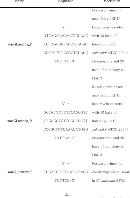
Table 1. Primers used in this study
Name Sequence Description
waaGLambda_F 5’-GTCAGACAGACCTGGAAC CCTAACGGCGGCGCGGAG CGCTGTGTAGGCTGGAGC TGCTTC-3’
Forward primer for amplifying pKD13 kanamycin cassette with 40 base of homology to C. sakazakii ATCC 29544 chromosome and 20 base of homology to Pkd13 waaGLambda_R 5’-GCCATTCTTTCCAGCGTC CAGGGCTCTGCGGTGCGT CGCGCTGTCAAACATGAG AATTAA-3’
Reverse primer for amplifying pKD13 kanamycin cassette with 40 base of homology to C. sakazakiiATCC 29544 chromosome and 20 base of homology to Pkd13 waaG_confirmF 5’-TGGATGCGATGGAGCAGG TGTTCC-3’
Forward primer for confirming size of waaG in C. sakazakiiATCC
29544 chromosome
waaG_confirmR
5‘-GAGCGCTATCGATACTCT GCTGCTG-3’
Reverse primer for confirming size of waaG inC. sakazakiiATCC 29544 chromosome CSK29544_F_RS05130 5’-ATAGAATTCGATACTCT CTGCTGGAAAC-3’
Forward primer for amplifying wild type waaGto make complementation plasmid CSK29544_R_RS05130 5’-ATACTGCAGTGTATGCT GGATGCGATGGA-3’
Reverse primer for amplifying wild type waaGto make complementation plasmid
2.13 Virulence test of phage CSP1-resistant mutant
Virulence of C. sakazakii ATCC 29544 wild type and the CSP1 resistant mutant strains were tested with invasiveness of the bacteria to epithelial cell, Caco-2. Caco-2 epithelia cell was cultured in EMEM (Eagle's Minimum Essential Medium) (ATCC) with 20% FBS (Fetal bovine serum) (Gibco®) in 5.0% CO2 at 37℃
incubator. The -196℃ (liquid nitrogen)-stored cell line was melting in water bath at 37℃ and cultured in EasYFlask 25cm2 Cell
Culture Flask (Themo fisher) with EMEM with 20% FBS adding penicillin-streptomycin (Sigma). Completely grown Caco-2 cells were separated from flask wall by treating 0.25% Trypsin-EDTA (Gibco®) at 37℃ for 4 min, and the separated cells were cell down with 130 g at room temperature. The pellet was re-suspended as EMEM with 20% FBS adding penicillin-streptomycin, and the lysate was moved to EasYFlask 75cm2 Cell Culture Flask with EMEM with
20% FBS The culture was sub-cultured to bigger flask EasYFlask 75cm2 Cell Culture Flask, (Thermo fisher) with EMEM with 20%
FBS adding penicillin-streptomycin. After separating Caco-2 from wall of the flask, the cells were cell down with 130 g at room temperature and re-suspended as EMEM with 20% FBS. The cells
were stained with trypan blue, and counted by microscope. Eight milliliter of Caco-2 cells (2*105) were dispensed as 500 ul in the each well of 24 well plate. The plate was incubated for a 24 h. Confirming the cells were properly grown, the broth was sucked with suction and refilled with fresh broth. After 1 h incubation, CFU of 5 types of C. sakazakii ATCC 29544 (OD600 = 1.5) (wild type, waaG knock out mutant, complementation strain, spontaneous mutant 1, spontaneous mutant 2) were checked by spotting 50 ul of the bacteria culture before 100 ul of the bacteria were inoculated to three wells each. The plate was incubated for 1.5 h, and the broth were replaced with 500 ul of 100 ug/ml gentamycin (Sigma). After 1.5 h incubation, gentamycin was completely washed out with PBS, and Triton X-100 (Sigma) + PBS solution was treated for 10 min in the incubator. Separated cells were collected and 50 ul of the cells were spotting on LB agar plate after dilution. Then the bacterial CFU of before and after invasion were compared. The virulence was statistically validated by student’s t-test between wild type and other strains.
Ⅲ. RESULTS
3.1 Bacteriophage isolation, morphological analysis and
host range determination
Seven bacteriophages infecting E. coli MG 1655 were isolated from chicken fecal samples from Buchen-si, Gyeonggi-do, South Korea. Among them, phage CSP1 can also infect C. sakazakii
ATCC 29544 as well. Interestingly, purified CSP1 formed two distinguished types of plaque on host bacterial lawn: clear big plaques without halo and relatively turbid plaques with halo was exhibited with E. coli MG 1655. Although similar types of plaques were also formed on the lawns of C. sakazakii ATCC 29544, no enclosed halo was found around the turbid plaques. TEM analysis of the phage CSP1 particles indicates that CSP1 belonged to the family
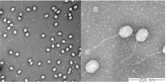
Siphoviridaehaving A slightly elongated icosahedral head of 78.6 nm in diameter and a 187.3 nm-long, non-contractile tail (n = 5) (Fig. 1). Only single type of phage particle was observed through the TEM analysis, supporting that both of the clear and turbid plaques were originated from phage CSP1.
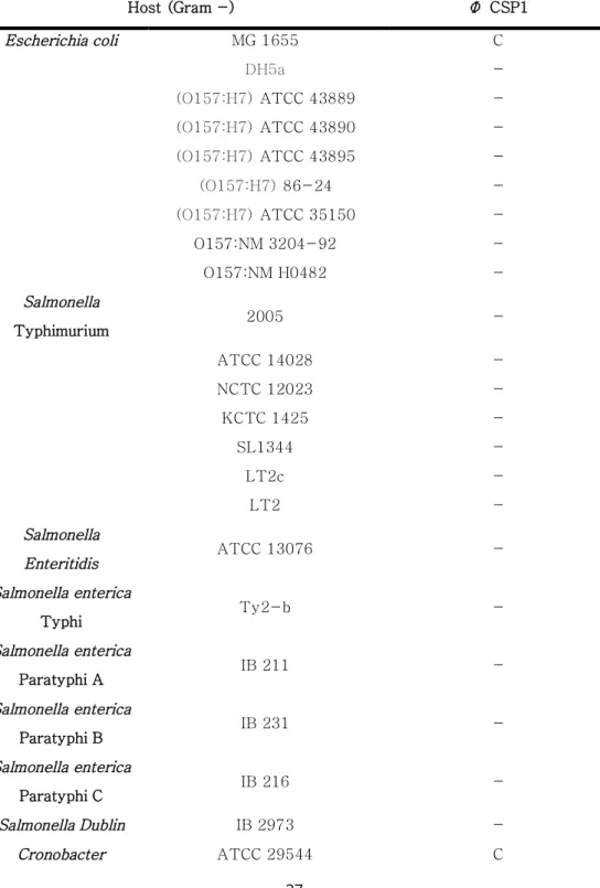
The host range of CSP1 was shown in Table 2. Several gram positive bacteria including Staphylococcus aureus, Listeria monocytogenes, Bacillus cereus, and Enterococcus faecalis were not susceptible to the CSP1 infection. Gram negative bacteria in the genus Salmonella and Shigella tested were also resistant to the CSP1. Four out of six strains of C. sakazakiiand two non-virulent E. coli strain tested could support the formation of CSP1 plaques. Based on these results, C. sakazakii, particularly strain ATCC 29544 whose genome was completely sequenced, was selected as the host bacteria for phage CSP1 throughout the study.
Table 2. Host range of the phage CSP1 Host (Gram -) Φ CSP1 Escherichia coli MG 1655 C DH5a -(O157:H7) ATCC 43889 -(O157:H7)ATCC 43890 -(O157:H7)ATCC 43895 -(O157:H7)86-24 -(O157:H7)ATCC 35150 -O157:NM 3204-92 -O157:NM H0482 -Salmonella Typhimurium 2005 -ATCC 14028 -NCTC 12023 -KCTC 1425 -SL1344 -LT2c -LT2 -Salmonella Enteritidis ATCC 13076 -Salmonella enterica Typhi Ty2-b -Salmonella enterica Paratyphi A IB 211 -Salmonella enterica Paratyphi B IB 231 -Salmonella enterica Paratyphi C IB 216 -Salmonella Dublin IB 2973 -Cronobacter ATCC 29544 C
C, clear zone with single plaque in low dilution factor; I, inhibition zone without single plaque; -, no susceptibility to the CSP1
NCTC, National Collection of Type Cultures; ATCC, American Type Cultures Collection; KTCT, Korea Type Culture Collection
sakazakii ATCC 51329 -BAA-894 -ATCC 33426 C NCTC 11467 C KCTC 2949 C
Shigella flexneri 2a strain 2457T
-Shigella boydii IB 2474 -Shigella dysenteriae IB 2476 -Shigella sonnei IB 707 -Pseudomonas aeruginosa ATCC 27853 -Host (Gram +) Φ CSP1 Staphylococcus aureus ATCC 29213 -ATCC 35983 -Listeria monocytogenes ATCC 19114
-Bacillus cereus ATCC 13061
-ATCC 23857
-Enterococcus
faecalis ATCC 29212
-Figure 1. Electron microscopic image of negatively stained phage CSP1. The phage has a slightly elongated icosahedral head and a long, non-contractile tail.
3.2
Whole genome sequencing and bioinformatic
analysis of phage CSP1
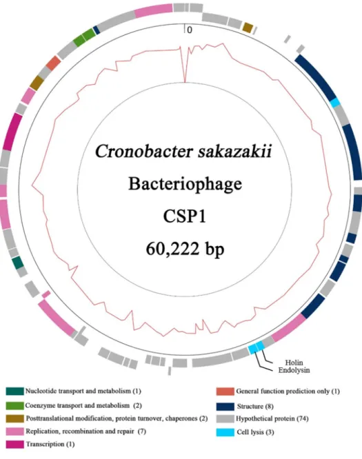
Sequenced reads of CSP1 DNA were assembled into a single contig. Complete genome of CSP1 is composed of 60,222 base-pairs DNA with 44.96% of G+C contents. Total 99 ORFs were predicted from the genome without any noticeable tRNA and rRNA genes. The ORFs were consisted of 74 hypothetical proteins, 8 structural proteins, 7 DNA replication, recombination, and repair proteins, 3 host lysis-related proteins, 2 coenzyme transport and metabolism proteins and 1 transcription, general function predicted, and nucleotide transport and metabolism protein. Genes related with lysogenic cycle were not found from the BLAST result, suggesting that phage CSP1 could be considered as a virulent phage. Interestingly, the phage genome was resembled a portion of
Salmonella enterica subsp. diarizonae genome but no virulence factors were found. Moreover, E. coli phages Seurat and CAjan isolated in Texas A&M University, College Station, Texas, USA were also highly homologous to the phage CSP1 in genomic level (equal 94% of identity to each), and showed equal 85% of identity to
each in protein level. In particular, endolysins from three phages were consisted with exactly same amino acid sequences (100% identity). The major differences between each two phages and CSP1 was in structural proteins, and some unknown proteins. The genomic information of CSP1 was deposited at the NCBI Genbank under the accession number KY982929.
Figure 2. Genomic map of phage CSP1. Genes with their predicted functions are represented as color boxes. Cell lysis-related genes, endolysin and holin, were indicated in the genome map.
3.3 Bacterial challenge assay and phage adsorption
assay
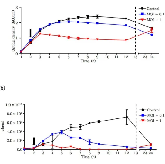
To test bacterial growth inhibition activity of phage CSP1, growth of C. sakazakii ATCC 29544 was periodically observed after treatment of CSP1 at an MOI of 0.1 or 1.0. When the exponentially growing C. sakazakii was infected by CSP1 at MOI of 1.0, bacterial growth inhibition was shown at 1 hour post-infection and was continued for 10 hours (Fig. 2a). When a lower number of CSP1 was treated (MOI=0.1), inhibitory activity was exhibited at about 4 hours post-infection and also continued for more than 10 hours. However, bacterial growth was resumed in prolonged incubation, presumably due to the emergence of spontaneous CSP1-resistant mutants. Similar results were also obtained with the bacterial cell counting: the number of living bacteria was significantly reduced after the addition of CSP1, but the number was start to recover at 24-hours of incubation (Fig. 2b).
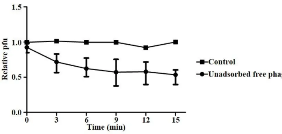
Initial binding kinetics of phage particles to the host C. sakazakii cell was assessed through an adsorption assay. Although phages were not efficiently bind to the cells when a low MOI (MOI =
0.001) was applied (data not shown), approximately half of virions were rapidly adsorbed to the C. sakazakii cells at a higher MOI (MOI = 1) within 15 min. These results were in accordance with the bacterial challenge assay where treatment of CSP1 at a higher MOI led earlier decreasing of C. sakazakiiviability.
(a)
(b)
Figure 2. The growth of C. sakazakii was periodically observed by measuring optical density at 600 nm (a) or viable counting (b). Vertical arrow indicates treatment of phage CSP1 at an indicated MOI. SM buffer instead of CSP1 was added as negative control.
Figure 3. Adsorption of phage CSP1 to the host C. sakazakiiATCC 29544 cells. CSP1 was mix with C. sakazakiicells at MOI of 1 at 37℃, and unadsorbed free phage particles were periodically
enumerated (Pt) by plaqueing assay. Same experiment without cells
was conducted in parallel to use as control (Pc). Adsorption rate was
3.4 Heat stability of phage CSP1
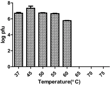
To test a heat stability of phage CSP1, phage was exposed thermal stress for 30 min, and the number of survived phage was enumerated by spotting assay using C. sakazakii ATCC 29544 cells as indicator strain. CSP1 phage stably maintained its infectivity at 37 to 55℃. At 60℃, a slight but non-significant loss of infectivity was observed. The infectivity of CSP1 was completely vanished 65℃ or more. Powdered milk, the main source of C. sakazakii infection outbreaks, is generally prepared with 60℃ of warm water. Therefore, CSP1 that can maintain the specific infectivity at this temperature has a potential to be developed as a novel biological agent for controlling of C. sakazakii contamination in food.
37 45 50 55 60 65 70 75 0 2 4 6 8 Temperature(°C) lo g p fu
Figure 4. Thermal stability of CSP1 from 37℃ to 75℃. CSP1 completely lost its infectivity more than 65℃.
3.5 Construction of transposon random mutant library
and selection of phage CSP1-resistant mutants
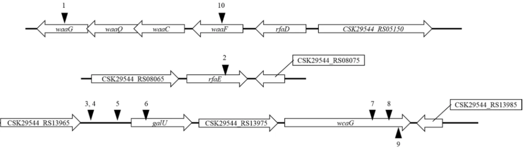
Transposon random mutagenesis by using conjugation between donor E. coli MFDpir harboring pRL27 and recipient C. sakazakii ATCC 29544 was performed to screen the phage resistant bacteria. Interestingly, some library colonies were surrounded by extracellular polysaccharide (EPS)-like matrix, and it might be the strategy to survive from phage attacks (ref). Therefore, colonies without the EPS-like matrix were randomly picked from the three hundred thousand of total transformants, and the transposon insertion site was determined after the another verification step of phage resistance through the spotting assay. Coding sequence of
waaG, waaF, rfaE, putative galU, and putative wcaG gene and an `upstream region of operon consisting of galU to wcaG were disrupted by Tn5 insertion. Glucosyltransferase I encoded by waaG
gene plays a role in the construction of LPS outer core oligosaccharide by transferring the first glucose residue to the outer core. waaF gene product transfers the last glucose residue of inner core oligosaccharide (Zhou Wang et al, 2016). rfaE gene product is
involved in the biosynthesis of heptose that consisting the inner core oligosaccharide (Valvan Ma et al, 2000). The operon from galU to
wcaG is also involved in the carbohydrate biosynthesis which eventually used for construction of LPS core oligosaccharide. Altogether, the influenced genes by Tn5 insertions in the screened mutants are involved in the LPS core oligosaccharide construction, suggesting that the CSP1 might utilize the LPS, particularly beyond the inner core oligosaccharide, as a receptor.
Figure 5. Schematic representation of transposon insertion sites in the CSP1-resistant C. sakazakii
ATCC 29544 mutants. The location of trasposon insertions is marked with black arrowheads. Unnamed genes are reprsented with a locus tag. waaG, LPS glucosyltransferase I; waaF, ADP-heptose-LPS heptosyltransferase; rfaE, bifunctional heptose 7-phosphate kinase/heptose 1-phosphate adenyltransferase; galU, UTP--glucose-1-phosphate uridylyltransferase; wcaG, NAD-dependent epimerase.
Figure 6. General structure of lipopolysaccharides in Cronobacter sakazakii. Carbohydrate residues are expressed as hexagons, and the relevant biosynthetic genes are indicated with arrows. Dashed line divides inner core part and outer core part of core oligosaccharide.
3.6 Determination of the phage CSP1 receptor
(1) CSP1 susceptibility test
To confirm that the CSP1 resistance in Tn5 mutants was derived from the defection of host receptor, one of Tn5-disrupted gene waaG was complemented with the IPTG-inducible vector pUHE21-2 lacIq in waaG::Tn5 mutant. The phage susceptibility of
the complementation strain was restored and increased in an IPTG concentration-dependent manner up to 0.5 mM, and an efficiency of infection (EOP) was similar to WT level even without the IPTG induction (Fig. 7). Interestingly, induction with 1.0 mM of IPTG decreased the phage susceptibility of complementation strain suggesting that the adequate amount of waaG gene product is required for the normal biosynthesis and efficient physiological functions of LPS as phage receptor. Taken together, proper waaG
gene expression involved in the LPS core oligosaccharide biosynthesis is critical for the phage CSP1 infection probably providing the host receptor.
(2) Motility assay
recent study has shown that waaG gene contributes to the flagella formation in E. coli (Wang Z et al, 2016). Therefore, the motility of
C. sakazakii ATCC 29544 waaG mutant and its complementation strain was tested to confirm the waaG gene complementation. As shown in Fig. 8, the mutant strain showed dramatically decreased motility compare to the WT. The complementation strain without IPTG induction, which has the most similar CSP1 susceptibility to the WT level, exhibited the recovered motility as much as wild type, confirming the complementation of waaGgene as expected
(3) DOC-PAGE of LPS
Transposon random mutagenesis suggested that the CSP1 resistance was accrued from mutation in the genes related to the LPS core oligosaccharide biosynthesis. To confirm the LPS defection in Tn mutants, LPS of various bacterial strains was phenol-extracted and electrophoresed in an polyacrylamide gel. Disruption of waaG gene by Tn5 insertion or targeted mutagenesis caused loss of bands responsible for the LPS core region, indicating the truncation of LPS including the core oligosaccharide. In contrast, the ladder pattern of LPS was fully restored to that of WT in the
waaG gene complementation strain. Two of spontaneous CSP1-resistant mutants showed the similar LPS truncation to the waaG
gene defective mutants, suggesting that LPS core oligosaccharide is naturally targeted to be modifying to avoid the CSP1 infection.
(4) Determination of the phage receptor
Above results indicate that the mutation in LPS, particularly beyond the inner core of LPS, could protect the C. sakazakii ATCC 29544 from the CSP1 infection. When we tested the
waaL gene deletion mutant, which has no O-antigens after the outer core, CSP1 can normally formed the plaques (data not shown). Therefore, CSP1 might specifically recognize the outer core part of LPS as host receptors to infect C. sakazakiiATCC 29544.
Figure 7. Phage susceptibility test through spotting assay with C. sakazakii ATCC 29544 WT (a), waaG gene deletion mutant (b),
waaG gene complementation strains with indicated concentrations of IPTG. Overexpression of waaG gene (1.0 mM of IPTG) dramatically decreased the phage sensitivity to the WT level.
Figure 8. Motility assay of C. sakazakii ATCC 29544 WT (a), waaG
gene deletion mutant (b), and waaG complementation strain (c). Boundary of the circle caused by swimming bacteria were indicated with red dotted line. Diameter of the circle is 38.14 mm, 0.51 mm , and 42.41 mm for WT, waaG deletion mutant, and waaG
complementation strain, respectively (n = 3). One representative result from the three independent experiments was shown.
Figure 9. Deoxycholate-polyacrylamide gel electrophoresis of LPS extracted from C. sakazakiiATCC 29544 WT(lane 1), Tn5::waaG
mutant (lane 2), waaG gene deletion mutant (lane 3), waaG gene complementation strain (lane 4), and two spontaneous CSP1-resistant mutants (lanes 5 and 6). Extracted LPS fromE. coli
serotype O55:B5 was used as standard ladder (lane 7). The location of lipid A, core oligosaccharides, and O-antigens is indicated. The CSP1 resistant mutants showed truncated LPS, and the
complementation strain fully recovered the LPS ladder pattern as WT.
3.7 Virulence test of phage CSP1-resistant mutant
C. sakazakii cause invasive infection in human epithelial cells, and LPS is known as one of the virulence factors of C. sakazakii(Ziad W. Jaradat et al, 2014). When the waaG gene deletion mutant was subjected to the invasion assay using human intestinal epithelial Caco-2 cells, significantly reduced number of invaded C. sakazakii
cells compared to WT was observed as expected. The impaired virulence was completely recovered when the waaG gene was complemented (Fig. 10). Although the reduction level was not much as waaG deletion mutant, two spontaneous CSP1-resistant mutants also exhibited the reduced invasion ability, . These results suggested that the truncation of LPS to evade the CSP1 infection could causes the impaired virulence of C. sakazakii..
Figure 10. Invasion assay of various C. sakzakii strains into the human intestinal epithelial Caco-2 cells. The CFU of invaded C. sakazakii strains was represented in relative to that of WT cells.
waaG gene deletion mutant and spontaneous CSP1-resistant mutants showed impaired invasion ability compared to WT. ***, P<0.001; ns, p>0.05..
Ⅳ. DISCUSSION
In this study, bacteriophage CSP1, which utilizes LPS outer core as host receptor, was isolated from chicken feces of Buchen-si, Gyeonggi-do, South Korea using E. coli MG 1655 as a host bacterium. Host spectrum was tested in twenty-seven of gram negative bacteria and six gram positive bacteria. The CSP1 infect gram negative bacteria only but very specific to the various strains of Cronobacter sakazakiiand two E. colistrains. Until the present, C. sakazakii infecting phages have not been studied generally, and the host receptors for these phages were under characterized yet. Biocontrolling of pathogenic bacteria using phage cocktails would be more effective when the cocktail is consisted with various phage that utilize different kinds of receptor.
The CSP1 showed two types of plaque morphology; one is turbid plaque and the other is clear plaque. The two distinct plaque morphology were still appeared after the purification process and were also exhibited with not only E. coli but also C. sakazakii strains. Sometimes different phages were mixed while isolation, but the two types of plaques could be originated from the phage CSP1, because
only one type of virion was found during the TEM analysis. It is also supported that the extracted phage DNA was sequenced and assembled as one contig without foreign DNA.
Analyzing the phage receptor is important to characterize the phage and understand the phage-host interaction. Diverse phage receptors have been reported: O-antigen, flagella, variety of outer membrane pore proteins that worked as nutrition transporting channel and etc. Bacteria evade the phage infection through various mechanisms such as CRISPR/Cas9 system, restriction and modification, abortive infection, blocking adsorption by altering the host receptor and etc. The mutation in host receptor, one of the common way to protect bacterial cells from the phage infection, often also affect the bacterial virulences, that consequently make the host cells attenuated or even avirulent. Phage CSP1 utilizes LPS outer core of the C. sakazakii as host receptor. Because the LPS is commonly known as one of important virulent factors for C. sakazakii pathogenesis, truncation of the LPS in the waaG deletion mutant and the Tn5 insertion mutant exhibited the decreased virulence against mammalian intestinal epithelial cells. Similarly, the spontaneous CSP1 resistant mutants that emerged after the
treatment of C. sakazakii with CSP1 showed a significantly reduced virulence with the truncated LPS, minimizing the problems of phage-resistant C. sakazakii infections Taken all together, novel phage CSP1, a non-virulent and thermostable bacterial virus, could be developed as a useful biocontrol agent to control C. sakazakii
Ⅴ. REFERENCES
Abedon, S. T. (2012). Bacterial 'immunity' against bacteriophages.
Bacteriophage, 2(1), 50-54. doi:10.4161/bact.18609
Castro-Mejia, J. L., Muhammed, M. K., Kot, W., Neve, H., Franz, C. M., Hansen, L. H., . . . Nielsen, D. S. (2015). Optimizing protocols for extraction of bacteriophages prior to
metagenomic analyses of phage communities in the human gut.
Microbiome, 3, 64. doi:10.1186/s40168-015-0131-4 Datsenko, K. A., & Wanner, B. L. (2000). One-step inactivation of
chromosomal genes in Escherichia coli K-12 using PCR products. Proc Natl Acad Sci U S A, 97(12), 6640-6645. doi:10.1073/pnas.120163297
Eriksen, R. S., Svenningsen, S. L., Sneppen, K., & Mitarai, N. (2018). A growing microcolony can survive and support persistent propagation of virulent phages. Proc Natl Acad Sci U S A, 115(2), 337-342. doi:10.1073/pnas.1708954115
Ferrieres, L., Hemery, G., Nham, T., Guerout, A. M., Mazel, D., Beloin, C., & Ghigo, J. M. (2010). Silent mischief:
bacteriophage Mu insertions contaminate products of
Escherichia coli random mutagenesis performed using suicidal transposon delivery plasmids mobilized by broad-host-range RP4 c Abedon, S. T. (2012). Bacterial 'immunity' against bacteriophages. Bacteriophage, 2(1), 50-54.
doi:10.4161/bact.18609
M., Hansen, L. H., . . . Nielsen, D. S. (2015). Optimizing protocols for extraction of bacteriophages prior to
metagenomic analyses of phage communities in the human gut.
Microbiome, 3, 64. doi:10.1186/s40168-015-0131-4 Datsenko, K. A., & Wanner, B. L. (2000). One-step inactivation of
chromosomal genes in Escherichia coli K-12 using PCR products. Proc Natl Acad Sci U S A, 97(12), 6640-6645. doi:10.1073/pnas.120163297
Eriksen, R. S., Svenningsen, S. L., Sneppen, K., & Mitarai, N. (2018). A growing microcolony can survive and support persistent propagation of virulent phages. Proc Natl Acad Sci U S A, 115(2), 337-342. doi:10.1073/pnas.1708954115
Ferrieres, L., Hemery, G., Nham, T., Guerout, A. M., Mazel, D., Beloin, C., & Ghigo, J. M. (2010). Silent mischief:
bacteriophage Mu insertions contaminate products of
Escherichia coli random mutagenesis performed using suicidal transposon delivery plasmids mobilized by broad-host-range RP4 conjugative machinery. J Bacteriol, 192(24), 6418-6427. doi:10.1128/JB.00621-10
Friedemann, M. (2007). Enterobacter sakazakii in food and
beverages (other than infant formula and milk powder). Int J Food Microbiol, 116(1), 1-10.
doi:10.1016/j.ijfoodmicro.2006.12.018
Gao, R., Hu, Y., Li, Z., Sun, J., Wang, Q., Lin, J., . . . Feng, Y. (2016). Dissemination and Mechanism for the MCR-1 Colistin
doi:10.1371/journal.ppat.1005957
Grimm, I., Dumke, J., Dreier, J., Knabbe, C., & Vollmer, T. (2018). Biofilm formation and transcriptome analysis of Streptococcus gallolyticus subsp. gallolyticus in response to lysozyme. PLoS One, 13(1), e0191705. doi:10.1371/journal.pone.0191705 Hsieh, P. F., Lin, H. H., Lin, T. L., Chen, Y. Y., & Wang, J. T. (2017).
Two T7-like Bacteriophages, K5-2 and K5-4, Each Encodes Two Capsule Depolymerases: Isolation and Functional Characterization. Sci Rep, 7(1), 4624. doi:10.1038/s41598-017-04644-2
Jaradat, Z. W., Al Mousa, W., Elbetieha, A., Al Nabulsi, A., & Tall, B. D. (2014). Cronobacter spp.--opportunistic food-borne pathogens. A review of their virulence and environmental-adaptive traits. J Med Microbiol, 63(Pt 8), 1023-1037. doi:10.1099/jmm.0.073742-0
Kim, M., Kim, S., Park, B., & Ryu, S. (2014). Core
lipopolysaccharide-specific phage SSU5 as an Auxiliary Component of a Phage Cocktail for Salmonella biocontrol.
Appl Environ Microbiol, 80(3), 1026-1034. doi:10.1128/AEM.03494-13
Larsen, R. A., Wilson, M. M., Guss, A. M., & Metcalf, W. W. (2002). Genetic analysis of pigment biosynthesis in Xanthobacter autotrophicus Py2 using a new, highly efficient transposon mutagenesis system that is functional in a wide variety of bacteria. Arch Microbiol, 178(3), 193-201.
Lee, H. J., Kim, W. I., Kwon, Y. C., Cha, K. E., Kim, M., & Myung, H. (2016). A Newly Isolated Bacteriophage, PBES 02, Infecting Cronobacter sakazakii. J Microbiol Biotechnol, 26(9), 1629-1635. doi:10.4014/jmb.1605.05020
Lee, J. H., Bai, J., Shin, H., Kim, Y., Park, B., Heu, S., & Ryu, S. (2016). A Novel Bacteriophage Targeting Cronobacter sakazakii Is a Potential Biocontrol Agent in Foods. Appl Environ Microbiol, 82(1), 192-201.
doi:10.1128/AEM.01827-15
Leon, M., & Bastias, R. (2015). Virulence reduction in bacteriophage resistant bacteria. Front Microbiol, 6, 343.
doi:10.3389/fmicb.2015.00343
MacLean, L. L., Pagotto, F., Farber, J. M., & Perry, M. B. (2009). Structure of the antigenic repeating pentasaccharide unit of the LPS O-polysaccharide of Cronobacter sakazakii
implicated in the Tennessee outbreak. Biochem Cell Biol, 87(2), 459-465. doi:10.1139/O09-004
Mitarai, N., Brown, S., & Sneppen, K. (2016). Population Dynamics of Phage and Bacteria in Spatially Structured Habitats Using Phage lambda and Escherichia coli. J Bacteriol, 198(12), 1783-1793. doi:10.1128/JB.00965-15
Nielsen, T. K., Carstens, A. B., Browne, P., Lametsch, R., Neve, H., Kot, W., & Hansen, L. H. (2017). The first characterized phage against a member of the ecologically important sphingomonads reveals high dissimilarity against all other known phages. Sci Rep, 7(1), 13566.
doi:10.1038/s41598-017-13911-1
Ogrodzki, P., & Forsythe, S. (2015). Capsular profiling of the Cronobacter genus and the association of specific
Cronobacter sakazakii and C. malonaticus capsule types with neonatal meningitis and necrotizing enterocolitis. BMC
Genomics, 16, 758. doi:10.1186/s12864-015-1960-z Pires, D. P., Cleto, S., Sillankorva, S., Azeredo, J., & Lu, T. K.
(2016). Genetically Engineered Phages: a Review of Advances over the Last Decade. Microbiol Mol Biol Rev, 80(3), 523-543. doi:10.1128/MMBR.00069-15
Ross, A., Ward, S., & Hyman, P. (2016). More Is Better: Selecting for Broad Host Range Bacteriophages. Front Microbiol, 7, 1352. doi:10.3389/fmicb.2016.01352
Samson, J. E., Magadan, A. H., Sabri, M., & Moineau, S. (2013). Revenge of the phages: defeating bacterial defences. Nat Rev Microbiol, 11(10), 675-687. doi:10.1038/nrmicro3096
Scholl, D., Adhya, S., & Merril, C. (2005). Escherichia coli K1's capsule is a barrier to bacteriophage T7. Appl Environ Microbiol, 71(8), 4872-4874. doi:10.1128/AEM.71.8.4872-4874.2005
Simpson, D. J., Sacher, J. C., & Szymanski, C. M. (2016).
Development of an Assay for the Identification of Receptor Binding Proteins from Bacteriophages. Viruses, 8(1). doi:10.3390/v8010017
Singh, N., Goel, G., & Raghav, M. (2015). Insights into virulence factors determining the pathogenicity of Cronobacter
sakazakii. Virulence, 6(5), 433-440. doi:10.1080/21505594.2015.1036217
Stern, A., & Sorek, R. (2011). The phage-host arms race: shaping the evolution of microbes. Bioessays, 33(1), 43-51.
doi:10.1002/bies.201000071
Sutherland, I. W., Hughes, K. A., Skillman, L. C., & Tait, K. (2004). The interaction of phage and biofilms. FEMS Microbiology Letters, 232(1), 1-6. doi:10.1016/s0378-1097(04)00041-2 Thomsen, L. E., Chadfield, M. S., Bispham, J., Wallis, T. S., Olsen, J.
E., & Ingmer, H. (2003). Reduced amounts of LPS affect both stress tolerance and virulence ofSalmonella entericaserovar Dublin. FEMS Microbiology Letters, 228(2), 225-231. doi:10.1016/s0378-1097(03)00762-6
Wang, L., Hu, X., Tao, G., & Wang, X. (2012). Outer membrane defect and stronger biofilm formation caused by inactivation of a gene encoding for heptosyltransferase I in Cronobacter sakazakii ATCC BAA-894. J Appl Microbiol, 112(5), 985-997. doi:10.1111/j.1365-2672.2012.05263.x
Yang, Y., Cai, L., Ma, R., Xu, Y., Tong, Y., Huang, Y., . . . Zhang, R. (2017). A Novel Roseosiphophage Isolated from the
Oligotrophic South China Sea. Viruses, 9(5). doi:10.3390/v9050109
Yosef, I., Goren, M. G., Globus, R., Molshanski-Mor, S., & Qimron, U. (2017). Extending the Host Range of Bacteriophage Particles for DNA Transduction. Mol Cell, 66(5), 721-728 e723. doi:10.1016/j.molcel.2017.04.025