저작자표시-비영리-변경금지 2.0 대한민국 이용자는 아래의 조건을 따르는 경우에 한하여 자유롭게 l 이 저작물을 복제, 배포, 전송, 전시, 공연 및 방송할 수 있습니다. 다음과 같은 조건을 따라야 합니다: l 귀하는, 이 저작물의 재이용이나 배포의 경우, 이 저작물에 적용된 이용허락조건 을 명확하게 나타내어야 합니다. l 저작권자로부터 별도의 허가를 받으면 이러한 조건들은 적용되지 않습니다. 저작권법에 따른 이용자의 권리는 위의 내용에 의하여 영향을 받지 않습니다. 이것은 이용허락규약(Legal Code)을 이해하기 쉽게 요약한 것입니다. Disclaimer 저작자표시. 귀하는 원저작자를 표시하여야 합니다. 비영리. 귀하는 이 저작물을 영리 목적으로 이용할 수 없습니다. 변경금지. 귀하는 이 저작물을 개작, 변형 또는 가공할 수 없습니다.
농학석사 학위논문
Biodegradation of BTEX Compounds by Massilia
aromaticivorans ML15P13
TIsolated from Arctic
Soil
북극토양에서 분리한 Massilia aromaticivorans
ML15P13
T의
BTEX 물질의 생물학적 분해
연구
2020 년 8 월
서울대학교
대학원
농생명공학부
식물미생물학전공
손
지 관
A THESIS FOR THE DEGREE OF MASTER OF SCIENCE
Biodegradation of BTEX Compounds by Massilia
aromaticivorans ML15P13
TIsolated from Arctic
Soil
BY
JIGWAN SON
Department of Agricultural Biotechnology
The Graduate School of Seoul National University
ABSTRACT
Biodegradation of BTEX Compounds by Massilia
aromaticivorans ML15P13
TIsolated from Arctic
Soil
Jigwan Son Major in Plant Microbiology Department of Agricultural Biotechnology The Graduate School of Seoul National University
BTEX (benzene, toluene, ethylbenzene, and xylene) compounds are the most frequently encountered subsurface contaminants among the various petroleum hydrocarbons. For contamination removal at cold climate sites, bioremediation technology is appealing due to their potential to be more economical and efficient than alternative. The aim of this study is to isolate, identify, and characterize novel bacterial strains from the Arctic and Antarctica soil that could degrade the BTEX compounds at low temperature. A novel aromatic hydrocarbon-degrading bacterial strain, designated ML15P13T, was isolated from Arctic soil at the Svalbard Islands, Norway,
using enrichment culture technique. Cells were Gram-stain-negative, aerobic, motile with multiple flagella at one polar end, and rod-shaped. Growth was observed at 4-35 ℃, pH 6.0-8.0, and 0-0.5% (w/v) NaCl. According to 16S
genus Massilia and closely related to Massilia atriviolacea SODT (98.4%),
Massilia violaceinigra B2T (98.3%), Massilia eurypsychrophila B528-3T
(97.7%), Massilia glaciei B448-2T (97.7%), and Massilia psychrophlia
B115-1T (96.6%). Average nucleotide identity, digital DNA-DNA hybridization,
and average amino acid identity between genome sequences of strain ML15P13T and the closely related species ranged from 75.8 to 84.3%, from
19.6±1.0 to 21.6±0.3%, and from 68.8 to 71.0%. The major fatty acids were C16:0, summed feature 3 (C16:1 ω6c and/or C16:1 ω7c), and summed feature 8
(C18:1 ω7c and/or C18:1 ω6c). Q-8 was the major ubiquinone. The polar lipid
profile showed the presence of phosphatidylethanolamine, phosphatidylglycerol, diphosphatidylglycerol, one unidentified phospholipid, and five unidentified polar lipids. The G+C content of the genomic DNA is 64.2 mol%. The BTEX biodegradation rate was low in MM broth, but adding a small amount of yeast extract, peptone, and tryptone greatly enhanced the biodegradation at low temperature. The ML15P13T utilized 77.3% of BTEX
mixture (initial concentration of each BTEX compounds, 30 mg/L) in MM broth with 0.2% yeast extract for 20 days at 15 ℃. Moreover, the BTEX biodegradation rates were assessed at different temperatures (10, 15, 20, and 28 ℃) in MM broth with 0.05% yeast extract. The ML15P13T degraded 63.2,
64.4, and 67.4% of BTEX mixture at 10, 15, and 20 ℃ for 10 days, respectively. In addition, the MP15P13T was characterized by maximal
biodegradation ability at 28 ℃ and utilized 72.1% of BTEX mixture for 10 days. Based on the results for genotypic and phenotypic study, it is concluded
that strain ML15P13T represents a novel species of the genus Massilia, for
which the name Massilia aromaticivorans sp. nov. is proposed.
Keywords: Massilia aromaticivorans, New Taxa, BTEX compounds, Biodegradation
CONTENTS
ABSTRACT ... page ABSTRACT ... ⅰ
CONTENTS ... iv
LIST OF TABLES ... ⅴi LIST OF FIGURES ... ⅵi Ⅰ. INTRODUCTION ... 1
Ⅱ. MATERIALS AND METHODS ... 4
1. Media and culture condition ... 4
2. Chemicals ... 4
3. Isolation of BTEX-degrading bacteria ... 7
4. Colony Repetitive Extragenic Palindromic-PCR ... 10
5. 16S rRNA gene sequencing and phylogenetic analysis ... 12
6. Whole genome sequencing, assembly, annotation and analysis ... 14
7. Phenotypic and biochemical characteristic ... 16
8. Chemotaxonomic analysis... 18
10. Effect of biodegradation ability on different media and temperature... 20
Ⅲ. RESULTS ... 21
1. Isolation of BTEX-degrading bacteria ... 21
2. Strain identification by 16S rRNA gene sequence analysis and colony REP-PCR analysis ... 22
3. Phylogenetic and whole genome sequencing analysis of ML15P13T ... 25
4. Morphological and phenotypic characteristics of ML15P13T ... 30
5. Chemotaxonomic characteristics of ML15P13T... 37
6. Biodegradation of BTEX by ML15P13T in mineral medium with different conditions. ... 41
Ⅳ. DISCUSSION ... 44
Ⅴ. LITERATURE CITED ... 49
LIST OF TABLES
... ABSTRACTpage Table 1. The composition of bacterial culture media. ... 5 Table 2. PCR conditions for colony REP-PCR. ... 11 Table 3. PCR conditions for 16S rRNA gene amplification. ... 13 Table 4. Possible strains of the novel BTEX-degrading species based on 16S rRNA gene sequence analysis. ... 23 Table 5. Results of average nucleotide identity (ANI, %), genome-to-genome calculations (GGDC, %), and average amino acid identity (AAI, %) from genome comparisons.. ... 28
Table 6. Differential characteristics of strains ML15P13T and other
related species of the genus Massilia. ... 34
Table 7. Cellular fatty acid contents of strains ML15P13T and related
LIST OF FIGURES
ABSTRACT ... page
Figure 1. Structures of BTEX compounds. ... 6
Figure 2. Soil sampling sites in Arctic and Antarctica. ... 8
Figure 3. Isolation of BTEX-degrading bacteria. ... 9
Figure 4. Colony REP-PCR band patterns of isolates. ... 24
Figure 5. ML tree showing the phylogenetic position of the novel species based on 16S rRNA gene sequences. ... 27
Figure 6. Colony morphology and stereo microscope (10X) results after 3 days incubation at 20 ℃... ... 32
Figure 7. Transmission electron micrograph of a 3-day-old culture of strain ML15P13T after negative staining with 0.5% uranyl acetate. ... 33
Figure 8. Two-dimensional TLC showing the total polar lipids of strain ML15P13T detected with molybdophosphoric acid reagent. ... 40
Figure 9. Biodegradation of BTEX (benzene, toluene, ethylbenzene,
and o-, m-, p-xylene 1:1:1:1:1:1) compounds by strain ML15P13T in
Figure 10. Biodegradation of BTEX (benzene, toluene, ethylbenzene,
and o-, m-, p-xylene 1:1:1:1:1:1) compounds by strain ML15P13T in
MM with 0.05% yeast extract at 10 ℃ (A), 15 ℃ (B), 20 ℃ (C), and 28 ℃ (D)... 43
I. INTRODUCTION
The recent economic growth has caused aggressive increase in global petroleum oil consumption (Varjani and Upasani, 2016) and the contamination by petroleum hydrocarbons caused by widespread use of petroleum is a problem in cold climate regions (Ferguson et al., 2020). Petroleum hydrocarbon contamination site in cold sites has significant impacts on surrounding environments and human. Under the same level of contamination, treating contamination in cold regions has been more difficult than other regions because of their harsh condition (Snape et al., 2003).
According to research data, polluted site identified in cold regions that require efficient management and active remediation bordered the ocean, lakes or rivers and threatened by internal pollutants. For example, relatively 407 oil spills were reported, and more than 1000 hydrocarbon contaminated sites have been recorded in Alaska (Camenzuli and Freidman, 2015). Moreover, petroleum hydrocarbon contamination is located to approximately 377 sites in the Canadian Arctic, more than 100 sites in Russia, Iceland, Greenland, Sweden, Norway, Sweden, and Finland, and 200 sites in Antarctica (Filler et al., 2008; Miri et al., 2019).
Petroleum compounds contains four fractions: cycling and linear alkanes, aromatic hydrocarbons, resins and asphaltenes, and poorly characterized compounds. The aromatic hydrocarbons are more difficult to biodegrade because of their stability of the molecules (Mirdamadian et al., 2010). The
symptoms (e.g., headache, dizziness, ataxia, drowsiness, euphoria, hallucinations, tremors, seizures, and coma). In addition, the BTEX compounds are listed as priority pollutants by the United States Environmental Protection Agency (Dean, 1985). Therefore, the BTEX removal techniques have gained increasing attention due to human health and environmental risk.
Under the same level of contamination, treating contamination in cold regions may be more difficult than treating other regions because of their harsh condition. Remediation in the cold climate regions has been applied various technology such as physical treatments, chemical treatments, and bioremediation. Compared to physical and chemical treatments, bioremediation has been more effective and economical option that causes less damage to cold climate environment in removing the BTEX compounds (Pazos et al., 2010).
Low temperatures cause an additional problem for remediation. In cold climate site, the average annual temperature is below 8 ℃ (typically in the range 4-8) and the groundwater temperatures are typically below 10 ℃ or lower (Van Stempvoort and Grande, 2006). Successful bioremediation in this region relies heavily on applicable microorganisms that are biodegradable at low temperatures (Gratia et al., 2009). Furthermore, Biotechnological methods, such as use of biosurfactant, cold adapted enzymes, immobilization methods, biostimulation, and bioaugmentation have been considered to enhance bioremediation ability of microorganisms.
The genus Massilia was first proposed in 1998 by La Scola (La Scola et al., 1998), with Massilia timonae as the type species isolated from blood culture of an immunocompromised patient. The genus was classified into the family Oxalobacteraceae, the order Burkholderiales, the class Betaproteobacteria (https://www.bacterio.net/-classifphyla.html). All species of the genus Naxibacter was reclassified into the genus of Massilia because the two genera shared identical chemotaxonomic properties (Kampfer et al., 2011). At the time of writing, the genus of Massilia comprises 45 species with validly published name (https://www.bacterio.net/genus/massilia). Species of the genus Massilia have been isolated from various environments, such as blood (La Scola et al., 1998), soil (Altankhuu and Kim, 2017; Yang et al., 2019), air (Orthová et al., 2015; Weon et al., 2010), glacier permafrost (Wang et al., 2018), and ice core (Gu et al., 2017; Guo et al., 2016; Shen et al., 2015). Cells of the genus Massilia are Gram-stain-negative, aerobic, rod-shaped, and non-spore-forming cells. Chemotaxonomically, it contained summed feature 3 (C16:1 ω7c and/or iso-C15:0 2-OH) and C16:0 as the major fatty acids, Q-8 as the
predominant isoprenoid quinone and phosphatidylethanolamine (PE), phosphatidylglycerol (PG), and diphosphatidylglycerol (DPG) as the major polar lipid. The DNA G+C contents range from 62.4 to 68.9 mol% (Ren et al., 2018).
In this study, a novel species, designated strain ML15P13T, was isolated
from Arctic soil. The BTEX degradative properties and taxonomic position of the strain were characterized using biochemical and polyphasic approaches.
Ⅱ. MATERIALS AND METHODS
1. Media and culture condition
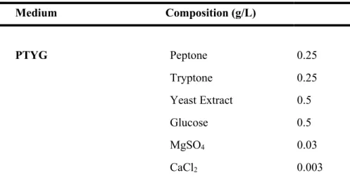
All isolated bacteria were cultured on mineral medium (Park and Ka, 2003) with BTEX mixture (initial concentration of each BTEX compounds, 30 mg/L). Peptone-tryptone-yeast extract-glucose (PTYG) medium containing (per liter) 0.25 g of peptone (Difco), 0.25 g of tryptone (Difco), 0.5 g of yeast extract (Difco), 0.5 g of glucose, 0.03 g of magnesium sulfate, and 0.003 g of calcium chloride (Table 1), Luria-Bertani (LB) medium (Difco), and R2A medium (Difco) were used to isolate strains and routinely culture colonies. All isolates were incubated at 15 ℃ and liquid cultures were aerated by shaking at 150 rpm on a rotary shaker (Vision Co., Korea).
2. Chemicals
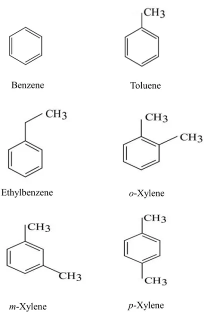
Benzene (Matsunoen), toluene (Methyl benzene, Matsunoen), ethylbenzene (JUNSEI), o-xylene (1,2-Dimethylbenzene, JUNSEI), m-xylene (1,3-Dimethylbenzene, JUNSEI), and p-xylene (1,4-(1,3-Dimethylbenzene, JUNSEI) were used for analytical grade. (Fig. 1).
Table 1. The composition of bacterial culture media. Medium Composition (g/L) PTYG Peptone 0.25 Tryptone 0.25 Yeast Extract 0.5 Glucose 0.5 MgSO4 0.03 CaCl2 0.003 MM Sol. A Na2HPO4 0.71 KH2PO4 0.68 Sol. B (NH4)2SO4 0.3 Sol. C MgSO4·7H2O 0.05 Sol. D CaCl2·H2O 0.001 Sol. E FeSO4·7H2O 0.006 ZnSO4·7H2O 0.0028 MnSO4·7H2O 0.0012 Co(NO3)2·6H2O 0.0017 CuSO4·5H2O 0.0004 (NH4)6Mo7O24·4H2O 0.0002
3. Isolation of BTEX-degrading bacteria
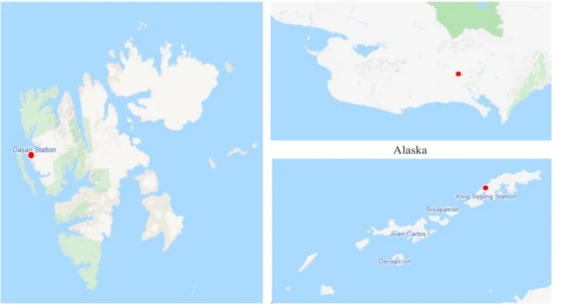
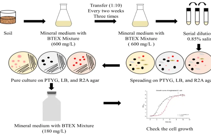
BTEX-degrading bacteria were isolated from the Arctic and Antarctica soil. The soil were collected from the Svalbard Islands (78°54'01.7'' N, 12°04'23.0'' E), Alaska (64°50'47.2'' N, 163°42'41.7'' W), and King Sejong Station (62°13.22'' S, 58°04.384'' W) during July 2016, September 2019, and January 2018, respectively (Fig. 2). To enrich BTEX-degrading bacteria, 10 g Arctic soil sample was transferred to a cotton-plugged 500 mL Erlenmeyer flask containing 100 mL MM broth. A BTEX mixture (benzene, toluene, ethylbenzene, and o-, m-, p-xylene [1:1:1:1:1:1]) corresponding to 600mg/L was added to the flask and the enrichment culture was incubated with shaking (180 rpm) at 15 ˚C. The enrichment culture was transferred (1:10) into 100 mL fresh MM broth containing BTEX mixture three times every two weeks. The final enrichment culture was serial diluted in 0.85% saline and spread onto PTYG, LB, and R2A agar. Finally, the plates were aerobically incubated for 7 days at 15 ˚C. The colonies were picked and subcultured several times to confirm purity onto colony’s optimal medium. Purified colony was cultured in MM broth with BTEX mixture (benzene, toluene, ethylbenzene, and o-, m-, p-xylene [1:1:1:1:1:1]) corresponding to 180mg/L and cell growth was analyzed by NanoDrop 2000c spectrophotometer (Thermo Fisher Scientific). The culture of the final positive tube showing considerable cell growth was selected for strain identification analysis (Fig. 3).
4. Colony Repetitive Extragenic Palindromic-PCR
Colony Repetitive Extragenic Palindromic-PCR (REP-PCR) was performed using BOXA1R primer (5’-CTACGGCAAGGCGACGCTGACG-3’), as described previously (De Bruijn, 1992). Each isolate was grown on their optimal medium agar plate for 48 to 72 hours, and a small amount of cells was resuspended in 25 μl of PCR solution consisting of the following: 5 μl Gitschier buffer [1 M (NH4)2SO4, 1 M Tris-HCl (pH 8.8), 1 M MgCl2, 0.5 M
EDTA (pH 8.8), 14.4 M β-mercaptoethanol], 0.4 μl 0.1% bovine serum albumin, 2.5 μl 100% dimethyl sulfoxide, 8 μl each deoxynucleotide triphosphate (dNTP) at a concentration of 2.5 mM, 5 μl BOXA1R primer (50 pmol/μl), 0.5 μl 5 U Taq DNA polymerase, 3.1 μl distilled water, and 5 μl template DNA (Amoupour et al., 2019). Amplification was carried out by MJ Mini PCR device (BIO RAD) as follows: initial denaturation at 93 ℃ for 7 min, followed by 35 cycles of 1 min for denaturation at 92 ℃, 1 min for annealing at 52 ℃, and 8 min for primer extension at 65 ℃, followed by terminal extension at 65 ℃ for 16 min, and a final soak at 4 ℃ (Table 2). After the reactions, Electrophoresis of PCR products was performed on 1% agarose gel. After electrophoresis, the image was photographed with UV trans-illumination (306 nm).
Table 2. PCR conditions for colony REP-PCR. PCR reaction mixture Gitschier buffer 5.0 μl BSA (0.1%) 0.4 μl DMSO (100%) 2.5 μl dNTP (2.5 mM) 8.0 μl
BOXA1R primer (50 pmol/μl) 0.5 μl
Taq polymerase (5 U/μl) 0.5 μl Distilled water 3.1 μl Template DNA 5.0 μl PCR reaction condition Step 1 93 ℃ 7.0 min Step 2 92 ℃ 1.0 min Step 3 52 ℃ 1.0 min Step 4 65 ℃ 8.0 min Step 2, 3, 4: 35 cycles Step 5 65 ℃ 16.0 min Step 6 4 ℃
5. 16S rRNA gene sequencing and phylogenetic analysis
Total genomic DNA was extracted from the isolate and PCR amplification of 16S rRNA was performed with 27F and 1492R as previously described (Baker et al., 2003) (Table 3). Sequencing was performed using the primer 519R, 926F (Lane, 1991), and 1055R (Lee et al., 1993). The 16S rRNA gene sequences were compiled by using SeqMan software (DNASTAR) and nearly the full length 16S rRNA gene was compared with EzBioCloud database (Yoon et al., 2017). The multiple alignment of the sequences was conducted by SINA (version 1.2.11) according to the SILVA seed alignment (Pruesse et al., 2012). Phylogenetic trees were reconstructed by the neighbour-joining (Saitou and Nei, 1987), parsimony (Fitch, 1971), and maximum-likelihood (Felsenstein, 1981) methods using MEGA X software program (Kumar et al., 2018), and their tree topologies were evaluated through bootstrap value analysis based on 1000 replications.
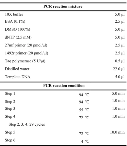
Table 3. PCR conditions for 16S rRNA gene amplification. PCR reaction mixture 10X buffer 5.0 μl BSA (0.1%) 2.5 μl DMSO (100%) 5.0 μl dNTP (2.5 mM) 5.0 μl 27mf primer (20 pmol/μl) 2.5 μl 1492r primer (20 pmol/μl) 2.5 μl Taq polymerase (5 U/μl) 0.5 μl Distilled water 22.0 μl Template DNA 5.0 μl PCR reaction condition Step 1 94 ℃ 5.0 min Step 2 94 ℃ 1.0 min Step 3 55 ℃ 1.0 min Step 4 72 ℃ 1.0 min Step 2, 3, 4: 29 cycles Step 5 72 ℃ 10.0 min Step 6 4 ℃
6. Whole genome sequencing, assembly, annotation and
analysis
The whole genome sequence of strain ML15P13T was obtained by TruSeq
Nano DNA kit (Illumina, Inc.) and sequenced on an Illumina Miseq sequencing platform at Macrogen (Republic of Korea). Genome assembly into contigs was performed by using A5-pipeline. Genomes were annotated using NCBI prokaryotic genome annotation pipeline (PGAP) (Tatusova et al., 2016) and Rapid Annotation using Subsystems Technology (RAST) (Aziz et al., 2008).
For analysis of genomic relatedness, the genome sequences of were obtained from Massilia atriviolacea SODT (RXLQ00000000), Massilia
violaceinigra B2T (CP024608), Massilia eurypsychrophila B528-3T
(PDOC00000000), Massilia glaciei B448-2T (PXWF00000000), and Massilia
psychrophlia B115-1T (PDOB00000000) were obtained from NCBI database.
The average nucleotide identity (ANI) using the BLAST (ANIb), MUMer (ANIm), and the OrthoANIu algorithm was calculated by the JSpecies Web Server (http://jspecies.ribohost.com/jspeciesws) (Richter and Rosselló-Móra, 2009) and the EzGenome web service (https://www.ezbiocloud.net/tools/ani) (Yoon et al., 2017). In addition, digital DNA–DNA hybridization (dDDH) was performed by using GGDC version 2.1 at the genome-to-genome distance calculator (GGDC) website (http://ggdc.dsmz.de/distcalc2.php) (Stackebrandt and GOEBEL, 1994). Furthermore, Average amino acid identity (AAI) was calculated using the AAI calculator by Kostas lab website
(http://enve-omics.ce.gatech.edu/aai/), which used the amino acid FASTA file to calculate both best hits and reciprocal best hits (Medlar et al., 2018).
7. Phenotypic and biochemical characteristic
Cell morphology was observed under stereo microscope (SMZ 445; Nikon), light microscope (AXIO; Zeiss), and transmission electron microscopy (LIBRA 120; Carl Zeiss Co.) after cells grown at 20 ℃ on R2A agar for 3 days. The motility of cells was performed by using hanging-drop technique (Perry, 1973). The type strain of Massilia atriviolacea KCTC 62720T,
Massilia violaceinigra CCM 8877T, Massilia eurypsychrophila JCM 30074T,
Massilia glaciei JCM 30271T, and Massilia psychrophlia JCM 30813T were
obtained for use as reference strains. Gram reaction of strain ML15P13T was
conducted using a Color Gram 2 kit (bioMérieux) according to the manufacturer’s instructions. Catalase and oxidase activity were determined by adding ID Color Catalase Reagent and Oxidase Reagent (bioMérieux), respectively. Anaerobic growth was assessed by cultivation of strain ML15P13T in an anaerobic jar in the presence of an anaerobe atmosphere
generation bag (Sigma). Growth of strain ML15P13T was tested at 20 ℃ for 3
days on R2A agar (Difco), nutrient agar (Difco), Luria-Bertani agar (Difco), trypticase soy agar (Difco), and McConkey agar (Difco). The temperature range for growth was assessed in R2A broth by incubating cultures after 3 days at 4, 10, 15, 20, 25, 28, 30, 35, 40, and 45 ℃. The pH range for growth was determined after 3 days of incubation at 20 ℃ in R2A broth at intervals of 0.5 pH units by using citrate-NaH2PO4 buffer (pH 5.0-6.0), NaH2PO4
-Na2HPO4 buffer (pH 6.5-8.0), Tris-HCl buffer (pH 8.5-9.0), and Na2CO3
2.0% NaCl (w/v). Hydrolysis of casein, starch, DNA, and CM-cellulose was determined using methods of Smibert and Krieg (Smibert et al., 1994) and Tween 80 was tested using method of Lelliott (Lelliott and Stead, 1987). API 20NE and API ZYM strips test were read after incubation at 20 ℃ for 96 hours and 24 hours, respectively.
8. Chemotaxonomic analysis
For fatty acid analysis, cells of strain ML15P13T and all reference strains were
collected after growth on R2A agar for 2 days at 25 ℃. Extraction and analysis of cellular fatty acids were performed according to the instructions of MIDI (Sherlock Microbial Identification System, version 6.3) using gas chromatography.
For analysis of isoprenoid quinone, cells of strain ML15P13T were grown
in R2A medium for 4 days at 20 ℃, and 300 mg of freeze-dried cells was treated with chloroform/methanol (2:1, v/v) for 4 hours. Preparative TLC was filtered by filter paper No. 2 (Whatman) and concentrated with chloroform/methanol (8.5:1.5, v/v). The concentrate was centrifuged at 14000 rpm for 5 min and the supernatant was extracted. The menaquinone was investigated by HPLC (YL9100; YOUNG LIN) equipped with ODS2 column (150 × 4.6 mm; Waters Spherisorb) and a UV detector at 254 nm.
For analysis of polar lipids, cells of strain ML15P13T were grown in R2A
medium for 4 days at 20 ℃. Polar lipids were extracted according to the procedures described by Minnikin (Minnikin et al., 1984). The polar lipids were separated by two-dimensional TLC using the first dimension with chloroform/methanol/water (65:25:3.8, v/v) and the second dimension with chloroform/methanol/acetic acid/water (40:7.5:6:1.8, v/v). All TLC plates were sprayed with ethanolic molybdatophosphoric acid followed by heating at 100 ℃ for 4 min.
9. Biodegradation of BTEX compounds by ML15P13
TThe BTEX biodegradation abilities of strain ML15P13T were tested in sterile
125 mL serum bottles containing 10 mL of MM broth supplemented with 180 mg/L of BTEX mixture (benzene, toluene, ethylbenzene, and o-, m-, p-xylene [1:1:1:1:1:1]). Strain ML15P13T was cultured in R2A broth medium for 48
hours, harvested by centrifugation (15,000 × g, 10 min), washed twice with MM broth and repelleted. Aliquots of resuspended cells were inoculated into 125 mL serum bottles containing 10 mL of MM broth supplemented with BTEX mixture (initial concentration of each BTEX compounds, 30 mg/L) as the sole carbon source. The serum bottles were sealed with Teflon-coated gray butyl rubber septa and aluminum crimp caps and incubated at 15 ℃ on a rotary shaker (180 rpm). Three bottles per sample were sacrificed at specific intervals and the compound concentrations were analyzed using Thermo (Trace 1310/ISQ) gas chromatography (GC) with a DB-VRX column (20 m × 0.18 mm × 1.0 µm) coupled to MSD detector. The residual BTEX compounds was extracted with dichloromethane (1:1, v/v). GC oven temperature was held at 40 ℃ for 2 min and increased at a rate of 8 ℃ per min to 150 ℃ and then at a rate of 30 ℃ per min to a final temperature of 245 ℃, which was held for 5 min.
10.
Effect of biodegradation ability on different media
and temperatures
The effect of additional nutrients biodegradation of BTEX compounds was evaluated with different of nutrients (0.2% yeast extract, 0.2% tryptone, and 0.2% peptone w/v). Biodegradation ability analysis was carried out in 125 mL serum bottles with 10 mL of MM broth supplemented with BTEX mixture (initial concentration of each BTEX compounds, 30 mg/L) with 0.2% yeast extract, 0.2% tryptone, and 0.2% peptone, respectively. The culture serum bottles were incubated with shaking at 180 rpm, 15 ℃ for 10 days intervals.
The effect of temperature on biodegradation of BTEX compounds was tested with different temperatures (10, 15, 20, and 28 ℃). Biodegradation ability analysis was carried out in 125 mL serum bottles with 10 mL of MM broth supplemented with BTEX mixture (initial concentration of each BTEX compounds, 30 mg/L) with 0.05% yeast extract on different temperature for 5 days intervals.
Ⅲ. RESULTS
1. Isolation of BTEX-degrading bacteria
179 bacteria were isolated from the Svalbard Islands soil, 22 were isolated from Alaska soil, and 33 were isolated from King Sejong Station soil. Thus, in total, 234 bacteria were isolated. Among 234 bacterial strains, 27 bacterial strains with identical REP-PCR band were regarded as the same bacterial strains. Finally, only 79 bacteria strains were selected as possible candidates of BTEX-degrading bacteria by cell growth in mineral medium with BTEX mixture (initial concentration of each BTEX compounds, 30 mg/L). Most of the BTEX-degrading bacteria were found from the Svalbard Islands soil.
2. Strain identification by 16S rRNA gene sequence
analysis and colony REP-PCR analysis
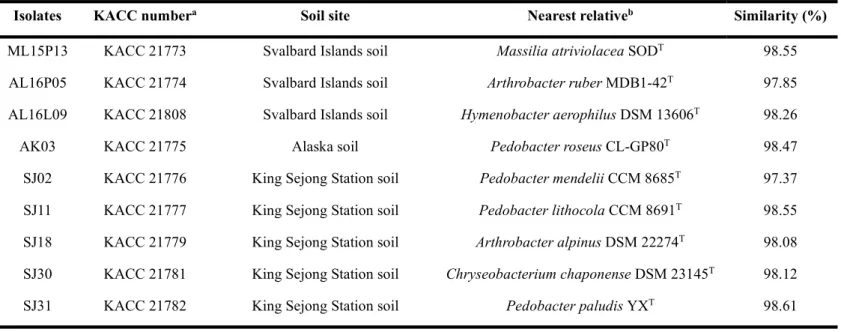
Analysis of 16S rRNA gene sequences revealed that the isolates were related to member of the genera, Arthrobacter, Pseudarthrobacter, Streptomyces, Pseudomonas, Flavobacterium, Massilia, Hymenobacter, Cryobacterium, Chryseobacterium, and Pedobacter, having > 98% sequence similarity to previously reported species. Among the BTEX-degrading bacteria, only 9 strains were less than 98.65% in 16S rRNA gene sequence similarity. On the basis of the threshold value of 98.65% similarity in 16S rRNA gene sequence for bacterial species delineation suggested by Beye (Beye et al., 2018), nine bacterial strain were selected as possible strains of a novel species (Table 4).
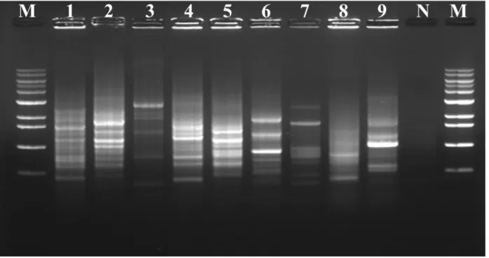
To investigate the genomic relation among the possible strains of a novel species, REP-PCR experiment was performed by PCR amplification with BOX1R primer. It was shown that the nine isolates exhibited different DNA fingerprint patterns (Fig. 4).
Table 4. Possible strains of the novel BTEX-degrading species based on 16S rRNA gene sequence analysis.
Isolates KACC numbera Soil site Nearest relativeb Similarity (%)
ML15P13 KACC 21773 Svalbard Islands soil Massilia atriviolacea SODT 98.55 AL16P05 KACC 21774 Svalbard Islands soil Arthrobacter ruber MDB1-42T 97.85 AL16L09 KACC 21808 Svalbard Islands soil Hymenobacter aerophilus DSM 13606T 98.26
AK03 KACC 21775 Alaska soil Pedobacter roseus CL-GP80T 98.47
SJ02 KACC 21776 King Sejong Station soil Pedobacter mendelii CCM 8685T 97.37 SJ11 KACC 21777 King Sejong Station soil Pedobacter lithocola CCM 8691T 98.55 SJ18 KACC 21779 King Sejong Station soil Arthrobacter alpinus DSM 22274T 98.08 SJ30 KACC 21781 King Sejong Station soil Chryseobacterium chaponense DSM 23145T 98.12
SJ31 KACC 21782 King Sejong Station soil Pedobacter paludis YXT 98.61
a Certificate number of KOREAN Agricultural Culture Collection (KACC). b Based on nearly fully sequences of the 16S rRNA gene.
Figure 4. Colony REP-PCR band patterns of isolates. Lanes: 1, ML15P13; 2, AL16L09; 3, SJ03; 4, AL16P05; 5, SJ18; 6, AK03; 7, SJ02; 8, SJ11; 9, SJ31; M, Maker; N, Negative control.
3. Phylogenetic and whole genome sequencing analysis of
ML15P13
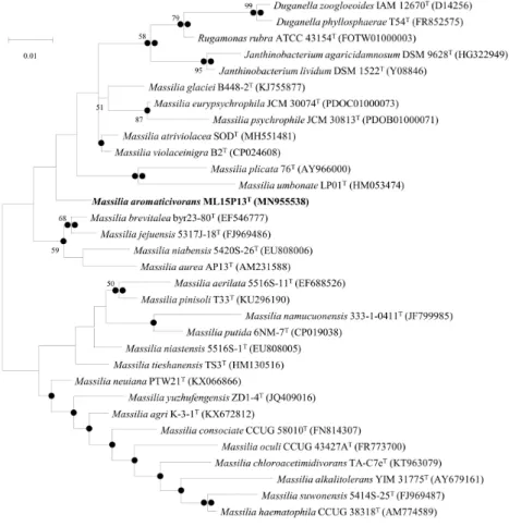
TComparative 16S rRNA gene sequence analysis showed that strain ML15P13T
displayed highest similarity with strains of the genus Massilia (96.5-98.4% similarity). Strain ML15P13T was closely related to Massilia atriviolacea
SODT (98.4%), Massilia violaceinigra B2T (98.3%), Massilia
eurypsychrophila B528-3T (97.7%), Massilia glaciei B448-2T (97.7%), and
Massilia psychrophlia B115-1T (96.6%). The phylogenetic tree with the
maximum-likelihood method supported that strain ML15P13T was grouped
with member of genus Massilia (Fig. 5).
The draft genome of strain ML15P13T consisted of 5,232,923 bp and 24
contigs with an N50 contig length of 748,145 bp. The genome of strain ML15P13T had a genomic G+C content of 64.2 mol%, which was within the
range reported for the genus Massilia (62.4-68.9 mol%) (Kampfer et al., 2011; Yang et al., 2019).
The ANI values of strain ML15P13T when compared with the closely
related strains ranged from based 75.8 to 84.3% on ANIb and ANIm. Moreover, Ortho ANI values were in the range 77.1 to 77.8%. All these values were clearly lower than the criterion (95-96%) for species demarcation (Chun et al., 2018; Kim et al., 2014; Richter and Rosselló-Móra, 2009). The DNA relatedness values between strain ML15P13T and reference strains ranged
the thresholds (95%) generally accepted for species delineation but complied with the 60-80% genus-level similarity (Luo et al., 2014) (Table 5).
Figure 5. ML tree showing the phylogenetic position of the novel species based on 16S rRNA gene sequences. Filled circles indicate that the corresponding branches were recovered when the trees were reconstructed using the NJ and ME algorithms. Bootstrap values of > 50% based on 1000 replication are shown at branch points. Bar, 0.01 substitutions per nucleotide position.
Table 5. Results of average nucleotide identity (ANI, %), genome-to-genome calculations (GGDC, %), and average amino acid identity (AAI, %) from genome comparisons. The error values are standard deviations.
Strain ML15P13T M. atriviolacea SODT M. violaceinigra B2T M. glaciei B448-2T
ANIb Strain ML15P13T 76.3 75.8 76.1 M. eurypsychrophila B528-3T 76.6 78.3 78.1 78.5 M. psychrophila B1555-1T 76.2 78.1 77.7 78.0 ANIm Strain ML15P13T 84.3 84.2 84.1 M. eurypsychrophila B528-3T 84.2 85.2 85.0 85.3 M. psychrophila B1555-1T 81.0 84.9 84.8 84.8 OrthoANI
Strain ML15P13T 77.8 77.1 77.4 M. eurypsychrophila B528-3T 77.8 80.2 79.8 79.9 M. psychrophila B1555-1T 77.4 79.3 78.9 79.0 GGDC Strain ML15P13T 20.8 ± 0.3 19.8 ± 0.7 19.6 ± 1.0 M. eurypsychrophila B528-3T 21.6 ± 0.3 23.0 ± 0.4 22.3 ± 0.5 22.4 ± 0.7 M. psychrophila B1555-1T 20.8 ± 0.3 22.0 ± 0.4 21.3 ± 0.6 21.7 ± 0.6 AAI Strain ML15P13T 70.0 69.6 68.8 M. eurypsychrophila B528-3T 70.8 73.1 72.5 71.9 M. psychrophila B1555-1T 71.0 73.0 72.7 72.1
4. Morphological and phenotypic characteristics of
ML15P13
TColonies of ML15P13T were pale yellow in contrast with other reference
strains (Fig. 6). colonies of ML15P13T were circular, smooth, and convex,
with a diameter of 2-3 mm after 3 days incubation on R2A agar medium at 20 ℃. Cells of ML15P13T were Gram-stain-negative, aerobic, motility, and
rod-shaped with lophotrichous flagellation. The cells were approximately 1.0-1.1 µm wide and 1.7-1.8 µm long (Fig. 7). The strain grew on R2A agar and nutrient agar (NA) but not grew on Luria-Bertani agar (LB), trypticase soy agar (TSA), and McConkey agar. The strain was catalase and oxidase positive. The strain grew at 4-35 ℃ (optimum growth at 25-28 ℃), pH range between 6.0 and 8.0 (optimum growth at pH 7.0), and strain was tolerated under 0.5% (w/v) NaCl (optimum growth at 0% (w/v) NaCl). Casein, starch, Tween 80 were hydrolyzed, and DNA was weakly hydrolyzed in contrast to CM-cellulose. The differential morphological, physiological, and biochemical characteristics of strain ML15P13T with phylogenetically related species are
shown in Table 6. The strain ML15P13T positive for reduction of nitrates to
nitrites but negative for reduction of nitrates to nitrogen. The strain ML15P13T produces alkaline phosphatase, esterase (C4), esterase lipase (C8),
leucine arylamidase, valine arylamidase, trypsin, acid phosphatase, naphthol-AS-BI-phosphohydrolase, glucosidase, cystine arylamidase, and α-chymotrypsin were weakly produced. The strain does not produce lipase (C14), α-galactosidase, ß-galactosidase, ß-glucuronidase, ß-glucosidase,
N-maltose, and potassium gluconate are assimilated. Malic acid was weakly assimilated. D-glucose, D-mannose, D-mannitol, N-acetyl-glucosamine, capric acid, adipic acid, trisodium citrate, and phenylacetic acid are not assimilated. The strain ML15P13T is positive for in test for esculin hydrolysis
but negative for indole production, glucose fermentation, arginine dihydrolase, produce urease, and gelatin hydrolysis.
Figure 6. Colony morphology and stereo microscope (10X) results after 3 days incubation at 20 ℃. a, Massilia aromaticivorans ML15P13T; b, Massilia
violaceinigra CCM 8877T; c, Massilia atriviolacea KCTC 62720T; d,
Massilia eurypsychrophila JCM 30074T; e, Massilia glaciei JCM 30271T; f,
Figure 7. Transmission electron micrograph of a 3-day-old culture of strain ML15P13T after negative staining with 0.5% uranyl acetate. Lophotrichous
Table 6. Differential characteristics of strains ML15P13T and other related species of the genus Massilia.
Strains: 1, ML15P13T (Massilia aromaticivorans sp. nov.); 2, Massilia violaceinigra CCM 8877T (B2T); 3, Massilia atriviolacea KCTC 62720T (SODT); 4, Massilia eurypsychrophila JCM 30074T (B528-3T); 5, Massilia glaciei JCM 30271T (B448-2T); 6, Massilia psychrophila JCM 30813T (B1555-1T). All data were obtained from this study. All strains are positive for catalase, oxidase, alkaline phosphatase, esterase (C4), esterase lipase (C8), leucine arylamidase, valine arylamidase, acid phosphatase, naphthol-AS-BI-phosphohydrolase, glucosidase, and hydrolysis of esculin, but negative for lipase (C14), α-galactosidase, ß-glucuronidase, ß-glucosidase, N-Acetyl-ß-glucosaminidase, α-mannosidase, α-fucosidase, indole production, glucose fermentation, arginine dihydrolase, urease, hydrolysis of CM-cellulose, assimilation of N-Acetyl-glucosamine, capric acid, trisodium citrate, and phenylacetic acid. +, Positive; w, weakly positive; –, negative.
Characteristics 1 2 3 4 5 6
Isolation source* Arctic Soil Glacier permafrosta Soilb Ice corec Ice cored Ice coree
Growth temperature(˚C)* 4-35 4-28a 4-33b 0-25c 4-30d 10-25e
Colour of colonies Pale yellow Purple Purple White White White
Motility + + + + – +
Hydrolysis of: Casein + + + – – – DNA w – + – – – Starch + + + – + – Tween 80 + + + + + – Gelatin – + + – – – Assimilation of: D-Glucose – – + + – – L-Arabinose + – + – – – D-Mannose – – + – – – D-Mannitol – – + – – – D-Maltose + – + + – –
Potassium gluconate + – – – – – Adipic acid – – – w – – Malic acid w – – + – – Enzyme activities Cystine arylamidase w + + + w + Trypsin + + + + – + α-chymotrypsin w w w w – + ß-galactosidase – – – – + –
DNA G+C content (mol%)* 64.4 63.5a 65.4b 66.2c 66.1d 66.4e
5. Chemotaxonomic characteristics of ML15P13
T The cellular fatty acids of strain ML15P13T mainly comprised C16:0 (22%),
summed feature 3 (C16:1 ω6c and/or C16:1 ω7c, 52.2%), and summed feature 8
(C18:1 ω7c and/or C18:1 ω6c, 10.8%). No remarkable differences in fatty acid
profile were found between the reference strains of species of the genus Massilia despite small quantitative differences. For example, strain ML15P13T had a relatively higher proportion of summed feature 3 and a
moderate amount of C12:0 2OH was found in strain ML15P13T but, no C12:0
2OH was found in member of all reference strains (Table 7).
The predominant isoprenoid quinone of ML15P13T was Q-8, which is a
common characteristic of members of the genus Massilia (La Scola et al., 1998; Wang et al., 2018; Yang et al., 2019).
The polar lipids of strain ML15P13T included phosphatidylethanolamine
(PE), phosphatidylglycerol (PG), diphosphatidylglycerol (DPG), unidentified phospholipid (PL), and five unidentified polar lipids (L1-L5) (Fig. 8). The predominant polar lipids of strain ML15P13T were PE, PG, and DPG which
were in accordance with the characteristic for other Massilia species (Shen et al., 2015; Wang et al., 2018; Yang et al., 2019). However, the presence of unidentified phospholipid and five unidentified polar lipids differentiates strain ML15P13T from other Massilia species.
Table 7. Cellular fatty acid contents of strains ML15P13T and related species.
Strains: 1, ML15P13T (Massilia aromaticivorans sp. nov.); 2, Massilia violaceinigra CCM 8877T; 3, Massilia atriviolacea KCTC 62720T; 4, Massilia eurypsychrophila JCM 30074T; 5, Massilia glaciei JCM 30271T; 6, Massilia psychrophila JCM 30813T. All data were obtained from this study. Values are percentages of total fatty acids, and only fatty acids representing more than 1% for at least one of the strains are shown. Major fatty acid components (> 5.0%) are highlighted in bold. –, Not detected; TR, trace amount (< 1.0%).
1 2 3 4 5 6
Saturated Fatty acid:
C12:0 4.2 4.7 4.2 3.0 3.7 3.3
C16:0 22.0 26.0 29.6 25.6 29.1 15.9
Hydroxy fatty acid:
C10:0 3OH 4.3 2.6 2.2 3.4 6.4 4.2
C12:0 2OH 2.8 – – – – –
C12:0 3OH – 3.8 3.3 – – –
Summed feature*:
7 – – – 1.6 2.0 TR
8 10.8 4.6 5.2 7.6 2.0 5.2
* Summed features are groups of two or three fatty acids that cannot be separated by GLC with the MIDI system. Summed feature 3 included C16:1 ω6c and/or C16:1 ω7c; summed feature 7 included C19:1 ω6c, C19:0 cyclo ω10c and/or unknown fatty acid (ECL 18.846); summed feature 8 included C18:1 ω7c and/or C18:1 ω6c.
Figure 8. Two-dimensional TLC showing the total polar lipids of strain ML15P13T detected with molybdophosphoric acid reagent. PE,
phosphatidylethanolamine; PG, phosphatidylglycerol; DPG, diphosphatidylglycerol; PL, unidentified phospholipid; L, unidentified lipid.
6. Biodegradation of BTEX by ML15P13
Tin mineral
medium with different conditions.
The biodegradation ability of the strain ML15P13T was evaluated using
BTEX mixture containing 30 mg/L of each component for 5 days interval in MM broth. In uninoculated-control experiments, BTEX loss during culture was negligible (data not shown). The degradation test showed that strain ML15P13T had the ability to degrade all six BTEX compounds. When the
MM broth did not contain any nutrients, 60.7% of total BTEX mixture (benzene, toluene, ethylbenzene, and o-, m-, p-xylene [1:1:1:1:1:1], 180mg/L) was degraded by strain ML15P13T. As the small amount of nutrients was
added, BTEX degradation improved clearly; in fact, The ML15P13T degraded
77.3, 76.8, and 76.3% of BTEX mixture in MM broth with yeast extract, tryptone, and peptone for 20 days at 15 ℃, respectively (Fig. 9).
The biodegradation ability of the strain ML15P13T for the degradation of
different temperature as 10, 15, 20, and 28 ℃ was verified. The degradation rate of the BTEX mixture increased with increasing temperature. The ML15P13T degraded 63.2, 64.4, and 67.4% of total BTEX mixture (benzene,
toluene, ethylbenzene, and o-, m-, p-xylene [1:1:1:1:1:1], 180mg/L) at 10, 15, and 20 ℃ for 10 days, respectively. In addition, the MP15P13T was
characterized by maximal biodegradation ability at 28 ℃ and utilized 72.1% of BTEX mixture for 10 days (Fig. 10).
Figure 9. Biodegradation of BTEX (benzene, toluene, ethylbenzene, and o-, m-, p-xylene 1:1:1:1:1:1) compounds by strain ML15P13T in MM
Ⅳ. DISCUSSION
Bioremediation technologies using bacterial mechanisms is one of the most efficient, economical, and eco-friendly methods to clean-up the contaminated sites (Fuentes et al., 2014). In addition, the success in removal contamination of Exxon Valdez oil spill in Prince William Sound, Alaska intrigued a significant interest in the possibility of bioremediation technology, especially biodegradation (Atlas and Bartha, 1998). Until today, artificial or synthetic basal media containing a sole carbon compound have usually been used to isolate or enrich microorganisms that have ability of biodegradation (M’rassi et al., 2015; You et al., 2018).
Nine novel bacterial strains that could utilize BTEX compounds as the sole carbon and energy source in artificial synthetic basal media were isolated from Arctic and Antarctica soils. 16S rRNA gene sequencing analysis indicated that the isolates were phylogenetically related to Massilia, Arthrobacter, Hymenobacter, Chryseobacterium, and Pedobacter (Table 4). In several studies, it is shown that bioremediation in cold climate sites could only be accomplished by psychrophilic or psychrotolerant microorganisms. These kinds of bacteria such as Chryseobacterium sp., Pseudomonas sp., Bacillus sp., Rhodococcus sp., Penicillium sp., Arthrobacter sp., Hyphomonas sp., Oleispira sp., Pimelobacter sp., and Mycobacterium sp. could degrade petroleum hydrocarbons at temperature below 20 ℃ (Miri et al., 2019).
Among the nine novel bacterial strains, Massilia sp. ML15P13T isolated
broth containing BTEX mixture (benzene, toluene, ethylbenzene, and o-, m-, p-xylene [1:1:1:1:1:1], 180mg/L) at low temperature.
Massilia aromaticivorans ML15P13T are Gram-stain-negative, aerobic,
motile with multiple flagella at one polar end, and rods shaped (1.0-1.1 µm wide and 1.7-1.8 µm length) (Fig. 7). Colonies on R2A agar after incubation at 20 ℃ for 3 days are circular, smooth, convex, and pale yellow (Fig. 6). Growth occurs on R2A and NA agar, but not on LB, TSA, and McConkey agar. Growth occurs at 4-30 ℃ (optimum, 25-28 ℃), at pH 6.0-8.0 (optimum, pH 7.0), and in the presence of 0-0.5% (w/v) NaCl (optimum, 0%). The strain is positive for catalase and oxidase. Casein, starch, DNA, Tween 80, and esculin are hydrolyzed but, CM-cellulose and gelatin are not hydrolyzed. Positive for catalase and oxidase activities, and reduction of nitrates to nitrites. Negative for indole production, reduction of nitrates to nitrogen, glucose fermentation, arginine dihydrolase, and urease production. Assimilates L-arabinose, D-maltose, and potassium gluconate. Weakly assimilates malic acid but does not assimilates D-glucose, D-mannose, D-mannitol, N-acetyl-glucosamine, capric acid, adipic acid, trisodium citrate, and phenylacetic acid (according to API 20NE test strip). Positive for alkaline phosphatase, esterase (C4), esterase lipase (C8), leucine arylamidase, valine arylamidase, trypsin, acid phosphatase, naphthol-AS-BI-phosphohydrolase, α-glucosidase, crystine arylamidase, and chymotrypsin, but negative for lipase (C14), α-galactosidase, ß-α-galactosidase, ß-glucuronidase, ß-glucosidase, N-Acetyl-ß-glucosaminidase, α-mannosidase, and α-fucosidase (according to API ZYM
(C16:1 ω6c and/or C16:1 ω7c), and summed feature 8 (C18:1 ω7c and/or C18:1 ω6c)
(Table 7). Q-8 is the predominant quinone. The polar lipids comprise phosphatidylethanolamine, phosphatidylglycerol, phosphatidylethanolamine, unidentified phospholipid, and five unidentified polar lipids (Fig. 8).
The degradation tests of strain clearly showed that ML15P13T had
biodegradation ability for all BTEX compounds. The strain ML15P13T
exhibited a higher performance toward ethylbenzene, and o-, m-, p-xylene degradation than other compounds. The strain ML15P13T degraded 69, 69,
and 72% of the ethylbenzene, o-xylene, and m-, p-xylene within 20 days, respectively. However, the strain ML15P13T degraded only 28 and 55% of
benzene and toluene within 20 days, respectively. Therefore, the strain ML15P13T degraded 60.7% of total BTEX mixture (benzene, toluene,
ethylbenzene, and o-, m-, p-xylene [1:1:1:1:1:1], 180mg/L) within 20 days (Fig. 9A).
However, the use of artificial and synthetic basal media as an enrichment medium has often resulted in the enrichment of microorganisms that only grow successfully in laboratory conditions, not in contaminated site (Rodrigo et al., 2009). In addition, although the isolated microorganisms have the capability to degrade organic contaminants under laboratory conditions, they often exhibit a low degradation capability in field conditions because of various biotic and abiotic factors (Johnsen et al., 2005; Van Veen et al., 1997). Therefore, most of the previous studies focused on the biostimulation that was optimize C-N-P relationships with nutrient addition in enrichment medium (Kim et al., 2008; Peltola et al., 2006). When the MM broth did not contain
any nutrients, BTEX degradation rate was slow, especially for benzene and toluene. As the small amount of nutrients was added, BTEX degradation improved clearly. In fact, The ML15P13T degraded 77.3, 76.8, and 76.3% of
BTEX mixture in MM broth with yeast extract, tryptone, and peptone for 20 days at 15 ℃, respectively (Fig. 9B; Fig. 9C; Fig. 9D). Moreover, the strain ML15P13T indicated the most degradation rate in MM broth with 0.02% yeast
extract toward the BTEX compounds. The strain ML15P13T degraded 53, 74,
84, 84, and 84% of the benzene, toluene, ethylbenzene, o-xylene, and m-, p-xylene within 20 days at 15 ℃, respectively (Fig. 9B). The result showed that nutrient additives can be added to stimulate of bioremediation.
Most cold climate sites have large seasonal temperature variation that reduce the efficient of bioremediation of microorganisms (Verma and Jaiswal, 2016). In cold climate site, the average annual temperature is below 8 ℃ (typically in the range 4-8) and the groundwater temperatures are typically below 10 ℃ or lower. However, seasonal temperature variations have been increased due to the recent effects of global warming. The ML15P13T
degraded 63.2, 64.4, and 67.4% of total BTEX mixture (benzene, toluene, ethylbenzene, and o-, m-, p-xylene [1:1:1:1:1:1], 180mg/L) at 10, 15, and 20 ℃ for 10 days, respectively. In addition, the MP15P13T was characterized
by maximal biodegradation ability at 28 ℃ and utilized 72.1% of BTEX mixture for 10 days (Fig. 10). The result indicated that the strain ML15P13T
could adapt to the variable temperatures and could degrade the BTEX compounds at low and moderate temperatures.
Many bacteria can remove petroleum hydrocarbons from the contaminated environment by degradation under aerobic conditions, such as Acinetobacter sp., Bacillus sp., Burkholderia sp., Comamonas sp., Coccobacillus sp., Chryseobacterium sp., Fulvimonas sp., Pseudomonas sp., Serratia sp., Sphingomonas sp., and Terrimonas sp. In this study, the new type strain isolated from Arctic soil at the Svalbard Islands, Norway, and identified as Massilia sp. via taxonomic and 16S rRNA analysis was used for the efficient degradation of BTEX compounds at low and moderate temperatures. Based on the results for genotypic and phenotypic study, it is concluded that strain ML15P13T represents a novel species of the genus Massilia, for which the
name Massilia aromaticivorans sp. nov. is proposed.
Massilia aromaticivorans (a.ro.ma.ti.ci.vo′rans. L. adj. aromaticus aromatic, fragrant; L. pres. part. vorans devouring; N.L. part. adj. aromaticivorans devouring aromatic compounds).
LITERATURE CITED
Altankhuu, K. and Kim, J. 2017. Massilia solisilvae sp. nov., Massilia terrae sp. nov. and Massilia agilis sp. nov., isolated from forest soil in South Korea by using a newly developed culture method. Int. J. Syst. Evol. Microbiol. 67: 3026-3032.
Amoupour, M., Nezamzadeh, F., Bialvaei, A. Z., Sedighi, M., Jazi, F.M. and Alikhani, M.Y. et al. 2019. Differentiation of Brucella abortus and B. melitensis biovars using PCR-RFLP and REP-PCR. New Microbes New Infect. 32: 100589.
Atlas, R. and Bartha, R. 1998. Microbial ecology: fundamentals and applications. 4th ed. Benjamin/Cummings, Redwood City, CA, USA. Aziz, R. K., Bartels, D., Best, A. A., Dejongh, M., Disz, T. and Edwards, R. A.
et al. 2008. The RAST Server: rapid annotations using subsystems technology. BMC genom. 9: 1-15.
Baker, G. C., Smith, J. J. and Cowan, D. A. 2003. Review and re-analysis of domain-specific 16S primers. J. Microbiol. Methods. 55: 541-555.
Beye, M., Fahsi, N., Raoult, D. and Fournier, P. E. 2018. Careful use of 16S rRNA gene sequence similarity values for the identification of Mycobacterium species. New Microbes New Infect. 22: 24-29.
Breznak, J., Costilow, R., Gerhardt, P., Murray, R. and Wood, W. 1994. Methods for general and molecular bacteriology. Physicochemical factors
Camenzuli, D. and Freidman, B. L. 2015. On-site and in situ remediation technologies applicable to petroleum hydrocarbon contaminated sites in the Antarctic and Arctic. Polar Res. 34: 24492.
Chun, J., Oren, A., Ventosa, A., Christensen, H., Arahal, D. R. and Costa, M. et al. 2018. Proposed minimal standards for the use of genome data for the taxonomy of prokaryotes. Int. J. Syst. Evol. Microbiol. 68: 461-466. De Bruijn, F. J. 1992. Use of repetitive (repetitive extragenic palindromic and
enterobacterial repetitive intergeneric consensus) sequences and the polymerase chain reaction to fingerprint the genomes of Rhizobium meliloti isolates and other soil bacteria. Appl. Environ. Microbiol. 58: 2180-2187.
Dean, B. J. 1985. Recent findings on the genetic toxicology of benzene, toluene, xylenes and phenols. Mutat. Res. Rev. Mutat. Res. 154: 153-181. Felsenstein, J. 1981. Evolutionary trees from DNA sequences: a maximum
likelihood approach. J. Mol. Evol. 17: 368-376.
Ferguson, D. K., Li, C., Jiang, C., Chakraborty, A., Grasby, S. E. and Hubert, C. R. J. 2020. Natural attenuation of spilled crude oil by cold-adapted soil bacterial communities at a decommissioned high Arctic oil well site. Sci. Total Environ. 722: 137258.
Filler, D. M., Snape, I. and Barnes, D. L. 2008. Bioremediation of petroleum hydrocarbons in cold regions. Cambridge University Press.
Fitch, W. M. 1971. Toward defining the course of evolution: minimum change for a specific tree topology. Syst. Biol. 20: 406-416.
Fuentes, S., Méndez, V., Aguila, P. and Seeger, M. 2014. Bioremediation of petroleum hydrocarbons: catabolic genes, microbial communities, and applications. Appl. Microbiol. Biotechnol. 98: 4781-4794.
Gratia, E., Weekers, F., Margesin, R., D’amico, S., Thonart, P. and Feller, G. 2009. Selection of a cold-adapted bacterium for bioremediation of wastewater at low temperatures. Extremophiles 13: 763-768.
Gu, Z., Liu, Y., Xu, B., Wang, N., Jiao, N. and Shen, L. et al. 2017. Massilia glaciei sp. nov., isolated from the Muztagh Glacier. Int. J. Syst. Evol. Microbiol. 67: 4075-4079.
Guo, B., Liu, Y., Gu, Z., Shen, L., Liu, K. and Wang, N. et al. 2016. Massilia psychrophila sp. nov., isolated from an ice core. Int. J. Syst. Evol. Microbiol. 66: 4088-4093.
Johnsen, A. R., Wick, L. Y. and Harms, H. 2005. Principles of microbial PAH-degradation in soil. Environ. Pollut. 133: 71-84.
Kampfer, P., Lodders, N., Martin, K. and Falsen, E. 2011. Revision of the genus Massilia La Scola et al. 2000, with an emended description of the genus and inclusion of all species of the genus Naxibacter as new combinations, and proposal of Massilia consociata sp. nov. Int. J. Syst. Evol. Microbiol. 61: 1528-1533.
Kim, J. M., Le, N. T., Chung, B. S., Park, J. H., Bae, J. -W. and Madsen, E. L. et al. 2008. Influence of soil components on the biodegradation of benzene, toluene, ethylbenzene, and o-, m-, and p-xylenes by the newly isolated bacterium Pseudoxanthomonas spadix BD-a59. Appl. Microbiol.
Kim, M., Oh, H. S., Park, S. C. and Chun, J. 2014. Towards a taxonomic coherence between average nucleotide identity and 16S rRNA gene sequence similarity for species demarcation of prokaryotes. Int. J. Syst. Evol. Microbiol. 64: 346-351.
Kumar, S., Stecher, G., Li, M., Knyaz, C. and Tamura, K. 2018. MEGA X: Molecular Evolutionary Genetics Analysis across Computing Platforms. Mol. Biol. Evol. 35: 1547-1549.
La Scola, B., Birtles, R. J., Mallet, M. N. and Raoult, D. 1998. Massilia timonae gen. nov., sp. nov., isolated from blood of an immunocompromised patient with cerebellar lesions. J. Clin. Microbiol. 36: 2847-2852.
Lane, D. 1991. 16S/23S rRNA sequencing In: Stackebrandt E, Goodfellow M, editors. Nucleic acid techniques in bacterial systematics. In.: New York: John Wiley and Sons.
Lee, S., Malone, C., Kemp, P. F. 1993. Use of multiple 16S rRNA-targeted fluorescent probes to increase signal strength and measure cellular RNA from natural planktonic bacteria. Mar. Ecol. Prog. Ser. 193-201.
Lelliott, R. A. and Stead, D. E. 1987. Methods for the diagnosis of bacterial diseases of plants. Blackwell Scientific Publications.
Luo, C., Rodriguez-R, L. M. and Konstantinidis, K. T. 2014. MyTaxa: an advanced taxonomic classifier for genomic and metagenomic sequences. Nucleic Acids Res. 42: e73.
M’rassi, A. G., Bensalah, F., Gury, J. and Duran, R. 2015. Isolation and characterization of different bacterial strains for bioremediation of
n-alkanes and polycyclic aromatic hydrocarbons. Environ. Sci. Pollut. Res. 22: 15332-15346.
Medlar, A. J., Toronen, P. and Holm, L. 2018. AAI-profiler: fast proteome-wide exploratory analysis reveals taxonomic identity, misclassification and contamination. Nucleic Acids Res. 46: W479-W485.
Minnikin, D., O'donnell, A., Goodfellow, M., Alderson, G., Athalye, M. and Schaal, A. et al. 1984. An integrated procedure for the extraction of bacterial isoprenoid quinones and polar lipids. J. Microbiol. Methods. 2: 233-241.
Mirdamadian, S., Emtiazi, G., Golabi, M. and Ghanavati, H. 2010. Biodegradation of petroleum and aromatic hydrocarbons by bacteria isolated from petroleum-contaminated soil. J. Pet. Environ. Biotechnol. 1: 2-5.
Miri, S., Naghdi, M., Rouissi, T., Kaur Brar, S. and Martel, R. 2019. Recent biotechnological advances in petroleum hydrocarbons degradation under cold climate conditions: A review. Crit. Rev. Environ. Sci. Techno. 49: 553-586.
Orthová, I., Kämpfer, P., Glaeser, S. P., Kaden, R. and Busse, H. -J. 2015. Massilia norwichensis sp. nov., isolated from an air sample. Int. J. Syst. Evol. Microbiol. 65: 56-64.
Park, H. -D. and Ka, J. -O. 2003. Genetic and phenotypic diversity of dichlorprop-degrading bacteria isolated from soils. J. Microbiol. 41: 7-15.
Pazos, M., Rosales, E., Alcántara, T., Gómez, J. and Sanromán, M. 2010. Decontamination of soils containing PAHs by electroremediation: a review. J. Hazard. Mater. 177: 1-11.
Peltola, R., Salkinoja-Salonen, M., Pulkkinen, J., Koivunen, M., Turpeinen, A. -R. and Aarnio, T. et al. 2006. Nitrification in polluted soil fertilized with fast-and slow-releasing nitrogen: a case study at a refinery landfarming site. Environ. Pollut. 143: 247-253.
Perry, L. B. 1973. Gliding motility in some non-spreading flexibacteria. J. Appl. Bacteriol. 36: 227-232.
Pruesse, E., Peplies, J. and Glöckner, F. O. 2012. SINA: accurate high-throughput multiple sequence alignment of ribosomal RNA genes. Bioinformatics. 28: 1823-1829.
Qin, Q. L., Xie, B. B., Zhang, X. Y., Chen, X. L., Zhou, B. C. and Zhou, J. Z. et al. 2014. A proposed genus boundary for the prokaryotes based on genomic insights. J. Bacteriol. 196: 2210-2215.
Ren, M., Li, X., Zhang, Y., Jin, Y., Li, S. and Huang, H. 2018. Massilia armeniaca sp. nov., isolated from desert soil. Int. J. Syst. Evol. Microbiol. 68: 2319-2324.
Richter, M. and Rosselló-Móra, R. 2009. Shifting the genomic gold standard for the prokaryotic species definition. Proc. Natl. Acad. Sci. U.S.A. 106: 19126-19131.
Rodrigo, J., Jacques, S., Okeke, B. C., Bento, F. M., Pralba, M. C. and Camargo, F. A. 2009. Improved Enrichment and Isolation of Polycyclic
Aromatic Hydrocarbons (PAH)-Degrading Microorganisms in Soil Using Anthracene as a Model PAH. Curr. Microbiol. 58: 628-634.
Saitou, N. and Nei, M. 1987. The neighbor-joining method: a new method for reconstructing phylogenetic trees. Mol. Biol. Evol. 4: 406-425.
Shen, L., Liu, Y., Gu, Z., Xu, B., Wang, N. and Jiao, N. et al. 2015. Massilia eurypsychrophila sp. nov. a facultatively psychrophilic bacteria isolated from ice core. Int. J. Syst. Evol. Microbiol. 65: 2124-2129.
Smibert, R. M. and Krieg, N. R. 1994. Methods for general and molecular bacteriology. American Society for Microbiology, Washington, DC, USA. 607-654 pp.
Snape, I., Riddle, M. J., Filler, D. M. and Williams, P. J. 2003. Contaminants in freezing ground and associated ecosystems: key issues at the beginning of the new millennium. Polar Rec. 39: 291-300.
Stackebrandt, E. and Goebel, B. M. 1994. Taxonomic note: a place for DNA-DNA reassociation and 16S rRNA sequence analysis in the present species definition in bacteriology. Int. J. Syst. Evol. Microbiol. 44: 846-849. Tatusova, T., Dicuccio, M., Badretdin, A., Chetvernin, V., Nawrocki, E. P. and
Zaslavsky, L. et al. 2016. NCBI prokaryotic genome annotation pipeline. Nucleic Acids Res. 44: 6614-6624.
Van Stempvoort, D. R. and Grande, P. 2006. Bioremediation of petroleum hydrocarbons in soil and groundwater under cold climate conditions: a review, implications for applications in Canada. National Water Research Institute.
Van Veen, J. A., Van Overbeek, L. S. and Van Elsas, J. D. 1997. Fate and activity of microorganisms introduced into soil. Microbiol. Mol. Biol. Rev. 61: 121-135.
Varjani, S. J. and Upasani, V. N. 2016. Core Flood study for enhanced oil recovery through ex-situ bioaugmentation with thermo- and halo-tolerant rhamnolipid produced by Pseudomonas aeruginosa NCIM 5514. Bioresour. Technol. 220: 175-182.
Verma, J. P. and Jaiswal, D. K. 2016. Book review: advances in biodegradation and bioremediation of industrial waste. Front. Microbiol. 6: 1555.
Wang, H., Zhang, X., Wang, S., Zhao, B., Lou, K., Xing, X. -H. 2018. Massilia violaceinigra sp. nov., a novel purple-pigmented bacterium isolated from glacier permafrost. Int. J. Syst. Evol. Microbiol. 68: 2271-2278.
Weon, H. -Y., Yoo, S. -H., Kim, S. -J., Kim, Y. -S., Anandham, R. and Kwon, S. -W. 2010. Massilia jejuensis sp. nov. and Naxibacter suwonensis sp. nov., isolated from air samples. Int. J. Syst. Evol. Microbiol. 60: 1938-1943.
Yang, E., Zhao, M., Li, S., Wang, Y., Sun, L. and Liu, J. et al., 2019. Massilia atriviolacea sp. nov., a dark purple-pigmented bacterium isolated from soil. Int. J. Syst. Evol. Microbiol. 69: 2135-2141.
Yoon, S. -H., Ha, S. -M., Kwon, S., Lim, J., Kim, Y. and Seo, H. et al. 2017. Introducing EzBioCloud: a taxonomically united database of 16S rRNA
gene sequences and whole-genome assemblies. Int. J. Syst. Evol. Microbiol. 67: 1613-1617.
You, J., Du, M., Chen, H., Zhang, X., Zhang, S. and Chen, J. et al., 2018. BTEX degradation by a newly isolated bacterium: Performance, kinetics, and mechanism. Int. Biodeterior. Biodegradation. 129: 202-208.
북극토양에서 분리한
Massilia aromaticivorans
ML15P13
T의 BTEX 물질의 생물학적 분해 연구
손지관 초록 BTEX (벤젠, 톨루엔, 에틸벤젠, 자일렌)은 유류 오염지역에서 가장 자주 발견되는 석유계 탄화수소이다. 추운 기후에서의 환경오염을 제거하기 위해서는 다른 환경정화방법보다 더 효율적이고, 경제적인 생물학적 환경정화 방법이 사용된다. 따라서 본 연구에서는 저온에서 BTEX 물질을 분해할 수 있는 신종 균을 북극과 남극 토양에서 분리, 동정하며, 분해 특성을 분석하였다. 방향족 탄화수소를 분해할 수 있는 신종 미생물 ML15P13T 균주를 북극 노르웨이의 스발바르 제도의 토양에서 enrichment 기법을 이용하여 분리하였다. 균주는 그람 음성균이며 호기성이고, 균주의 한쪽 끝에 여러 개의 편모를 가지면서 운동성을 나타내고, 간균형이었다. 균은 4-35 ℃, pH 6.0-8.0, 0-0.5% (w/v)의 염분농도에서 성장 가능하였다. 16S rRNA 유전자 서열 분석 결과 ML15P13T 균주는 Massilia 속에 속하였으며, Massiliaatriviolacea SODT (98.4%), Massilia violaceinigra B2T (98.3%),
Massilia eurypsychrophila B528-3T (97.7%), Massilia glaciei
B448-2T (97.7%), Massilia psychrophlia B115-1T (96.6%)의
nucleotide identity), 디지털 DNA 혼성화(digital DNA-DNA hybridization), 아미노산 동일성(average amino acid identity)의 분석 결과는 각각 75.8-84.3%, 19.6±1.0-21.6±0.3%, 68.8-71.0%였다. C16:0, summed feature 3 (C16:1 ω6c 그리고/또는 C16:1 ω7c), summed feature 8 (C18:1 ω7c 그리고/또는 C18:1 ω6c) 가 주요한 지방산으로 검출되었다. Q-8 이 주요한 유비퀴논 (ubiquinone)이었다. 주요 극성 지질 검출 결과 포스파티딜에탄올아민 (phosphatidylethanolamine), 포스파티딜글리세롤 (phosphatidylglycerol), 디포스파티딜글리세롤 (diphosphatidylglycerol)과 하나의 밝혀지지 않은 인지질(phospholipid)과 다섯 개의 밝혀지지 않은 극성 지질(polar lipid)이 검출되었다. DNA G+C 함유량은 64.2 mol%이었다. 낮은 온도의 MM 배지에서 BTEX 생분해 능력은 낮았지만, 소량의 효모 추출물, 펩톤, 트립톤을 추가로 넣어준 배지에서 생분해 능력이 향상되었다. ML15P13T 균주는 0.2%의 효모추출물을 추가한 MM 액체배지에서 20 일 동안 15 ℃에서 배양한 결과 BTEX 혼합물 (각 BTEX 물질의 초기 농도, 30 mg/L)의 77.3%를 분해하였다. 또한, 0.05%의 효모 추출물을 추가한 MM 액체배지에서 다양한 온도 (10, 15, 20, 28 ℃)에서 BTEX 생분해 능력을 측정하였다. ML15P13T 균주는 10 일 동안
생분해 특성을 보여주었으며, 10 일동안 72.1%의 BTEX 혼합물을 분해하였다. polyphasic taxonomy 분석 결과, ML15P13T 균주는
Massilia 속에 속하는 신종 (Massilia aromaticivorans sp. nov.)
균주로 밝혀졌고, 낮은 온도와 온화한 온도에서 BTEX 혼합물을 잘 분해하는 것으로 관찰되어 이 균주는 추운 지역의 오염된 유류 성분 제거에 효율적으로 활용될 수 있다고 판단된다.
주요어: Massilia aromaticivorans, 신종, BTEX 물질, 생물학적 분해