저작자표시-동일조건변경허락 2.0 대한민국 이용자는 아래의 조건을 따르는 경우에 한하여 자유롭게 l 이 저작물을 복제, 배포, 전송, 전시, 공연 및 방송할 수 있습니다. l 이차적 저작물을 작성할 수 있습니다. l 이 저작물을 영리 목적으로 이용할 수 있습니다. 다음과 같은 조건을 따라야 합니다: l 귀하는, 이 저작물의 재이용이나 배포의 경우, 이 저작물에 적용된 이용허락조건 을 명확하게 나타내어야 합니다. l 저작권자로부터 별도의 허가를 받으면 이러한 조건들은 적용되지 않습니다. 저작권법에 따른 이용자의 권리는 위의 내용에 의하여 영향을 받지 않습니다. 이것은 이용허락규약(Legal Code)을 이해하기 쉽게 요약한 것입니다. Disclaimer 저작자표시. 귀하는 원저작자를 표시하여야 합니다. 동일조건변경허락. 귀하가 이 저작물을 개작, 변형 또는 가공했을 경우 에는, 이 저작물과 동일한 이용허락조건하에서만 배포할 수 있습니다.
Outcomes of Laparoscopic Resection for Gastric
Submucosal Tumors: Consecutive 141 cases in
Single Center
by
Keesang Yoo
Major in Medicine
Department of Medical Sciences
The Graduate School, Ajou University
Outcomes of Laparoscopic Resection for Gastric
Submucosal Tumors: Consecutive 141 cases in
Single Center
by
Keesang Yoo
A Dissertation Submitted to The Graduate School of
Ajou University in Partial Fulfillment of the Requirements
for the Degree of
Master of Medicine
Supervised by
Sang-Uk Han, M.D., Ph.D.
Major in Medicine
Department of Medical Sciences
The Graduate School, Ajou University
This certifies that the dissertation
of Keesang Yoo is approved.
SUPERVISORY COMMITTEE
Sang-Uk Han
Yong Kwan Cho
Wook Hwan Kim
The Graduate School, Ajou University
June 21
st, 2013
I
-ABSTRACT-
Laparoscopic Resection of Gastric Submucosal Tumors: Outcomes
of 141 Consecutive Cases in a Single Center
The treatment of choice for gastric submucosal tumors (SMT) is surgical resection. Recent advanced techniques have facilitated more extensive application of laparoscopic surgery to most types of resectable gastric SMTs. This study aimed to verify the efficacy of laparoscopic resection for gastric SMT through the analysis of outcomes obtained at a single center. A total of 141 patients who underwent laparoscopic resection for gastric SMT were enrolled between April 2003 and June 2011. The demographics, tumor characteristics, and surgical or oncological outcomes of these patients were analyzed. Gastrointestinal stromal tumors (GIST) were the most common pathologic findings (90 cases), and the upper third of the stomach was the most common location (70 cases). Wedge resections were performed in 128 patients and major gastrectomies were performed in 13 patients. The mean surgical time was 102 minutes, which reduced to a stable 70 minutes after the 30th case. The surgical time for tumors located on the posterior or lesser portion of the upper third of the stomach was longer than that for other lesions. Twelve postoperative complications, including 2 cases of intra-abdominal bleeding, 1 case of marginal ulcer bleeding, and 1 case of leakage occurred. However, there were no complications after the 70th case. During the follow-up period, 2 patients suffered recurrent GIST. Laparoscopic surgery for gastric SMT is safe and feasible, particularly as the surgeon develops greater skill with increased experience. Laparoscopic resection is useful for any type of gastric SMT.
II
TABLE OF CONTENTS
ABSTRACT...I TABLE OF CONTENTS...II LIST OF FIGURES...III LIST OF TABLES...IV I. INTRODUCTION...1II. PATIENTS AND METHODS...3
A. Surgical procedures...3
B. Perioperative management and outcome measurement...4
C. Patient follow-up...5 D. Statistical analysis...5 III. RESULTS...6 IV. DISCUSSION...15 V. CONCLUSION...18 REFERENCES...V 국문요약...VI
III
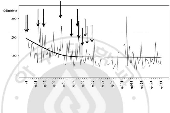
Fig.1. A. laparoscopic resection for endophytic submucosal resection using eversion technique. B. Laparoscopic resection for exophytic submucosal tumor using endoscopic linear staplers. ...4 Fig. 2. Change in operation time, according to the surgeon’s experience in laparoscopic
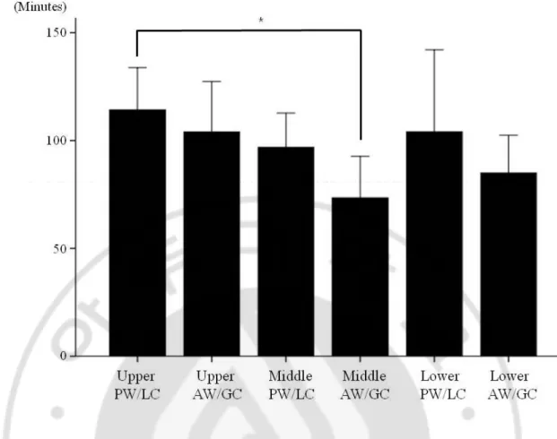
surgery for gastric submucosal tumors. Arrow shows the occurrence of postoperative complications. ...10 Fig. 3. Operation time according to tumor locations. There was no significant difference in operation time among the locations, by one-way ANOVA test. However, operation times for gastric submucosal tumor located in the posterior wall or lesser curvature of upper portion of the stomach was significantly longer than it was for those in the anterior wall or greater curvature of the stomach. * means P<0.05 in post-hoc test (Duncan method). PW posterior wall; LC lesser curvature; AW anterior wall; GC greater curvature. ...11
IV
Table 1. Clinicopathologic and surgical results of 141 enrolled patients…...6 Table 2. Comparison of gastrectomy according to the clinicopathologic features...7 Table 3. Clinical features of patients who were undergone laparoscopic gastrectomy for
gastric submucosal tumor...9 Table 4. Characteristics of patients who had complications...12 Table 5. Comparison of risk of postoperative complications according to the clinicopathologic features...12
1
I. INTRODUCTION
A submucosal tumor (SMT) in the gastrointestinal tract describes an epithelial or non-epithelial mesenchymal tumor, accounting for 1% or less of all gastrointestinal tumors.(Kim et al., 2001) The stomach is the most common site for SMTs. The rate of detection of asymptomatic gastric SMTs has increased in Asian countries including Japan and Korea because of the increasing use of screening gastrofiberscopy for detection of gastric cancer.(Iwahashi et al., 2006)
The first option for management of a gastric SMT is surgical resection, due to the limited preoperative pathologic diagnosis and the high possibility of malignancy.(Berindoague et al., 2006) The first option for management of a gastric SMT is surgical resection, due to the limited preoperative pathologic diagnosis and the high possibility of malignancy.(Matthews et al., 2002) Therefore, wedge resection of the stomach is possible and often results in complete resection of the gastric SMT without subtotal or total gastrectomy. This is fortunate as the latter procedures have a significant impact on the patient’s postoperative quality of life.(Roukos, 1999)
In recent years, laparoscopic resection of gastric SMTs has been regarded as the most appropriate curative approach. Previously, several reports had suggested limiting the indications for the laparoscopic approach to gastric SMTs of 2 cm in diameter, or smaller.(Heinrich and Corless, 2005; Demetri et al., 2007) Since several comparative studies have described the efficacy of laparoscopic surgery compared to open surgery for gastric SMTs,(Matthews et al., 2002; Nishimura et al., 2007; Karakousis et al., 2011) the laparoscopic approach has been applied to most types of resectable gastric SMTs.(Novitsky et al., 2006; De Vogelaere et al., 2012) However, in these reports, the number of enrolled patients was too small to be conclusive. In addition, they did not consider the features of SMTs requiring subtotal or total gastrectomy. Therefore, a clinical study involving a large series of patients undergoing laparoscopic surgery for gastric SMT can be of significant value and aid in the determination of the safety and effectiveness of the procedure across a broad array of SMTs.
Here, we analyzed prospectively collected data concerning laparoscopic resections of gastric SMTs within a single center. We aimed to evaluate the feasibility of this procedure
2
3
II. PATIENTS AND METHODS
Between April 2003 and June 2011, 141 consecutive patients with gastric SMTs underwent laparoscopic surgery in our institution. Indications for surgical treatment of gastric SMTs included, a tumor with a diameter greater than 2 cm; a fine needle aspiration-confirmed gastrointestinal stromal tumor (GIST) of any size; and a clinical GIST suggested by computed tomography (CT) or endoscopic ultrasonography. Also we resected the mass which cause symptoms or patients want resection unless the tumor is smaller than 2 cm.
Initially, we applied laparoscopic surgery to gastric SMT of below 5 cm size. However, indication for laparoscopic surgery was extended to 8cm size after surgeon performed the 70th laparoscopic surgery for gastric SMT.
Data concerning patient clinicopathologic features, surgical procedures, pathology reports, and follow-up results were retrospectively collected from the medical records of all patients. In addition, data regarding surgical parameters, including operation times and postoperative complications, were reviewed.
Surgical Procedures
Two surgeons who had a combined experience of more than 100 laparoscopic surgeries for gastric tumors, including adenocarcinomas, performed all of the surgical procedures. Laparoscopic surgery was performed under general endotracheal anesthesia. A 10-mm trocar for the laparoscope was inserted into the infra-umbilical area, and a 12 mm trocar for a flexible linear stapler was inserted, along with 2 or 3 additional trocars, into the upper abdomen after CO2 pneumoperitoneum was established. Intra-abdominal pressure was maintained at 10–12 mmHg.
Each abdominal cavity was fully explored and the location of the exophytic tumors was easily identified. For exophytic tumors located in posterior wall or lesser curvature of the stomach, the lesser or greater omental fat was dissected to expose the mass. Small endophytic tumors (<2 cm in size) were located using intraoperative sonography or gastrofiberscopy. To avoid gastric deformity after resection, six endophytic tumors of intermediate size (2–5 cm) which was not detected at the serosa surface of the stomach were
4
resected by eversion, as suggested by Hyung et al.(Hyung et al., 2005) For resection of tumors located in the posterior wall of the stomach, anterior gastrotomy was performed (Fig. 1A.). After tumors were identified, a flexible linear stapler was inserted through the gastrostomy site and the stapling was performed. Most tumors were resected by wedge resection using 2 or 3 staplers (Fig. 1B.). Several intracorporeal sutures were made to reinforce the resection lines to reduce the risk of postoperative bleeding. Laparoscopic gastrectomy was performed for several SMTs located at the gastroesophageal junction or the prepyloric area which can cause narrowing or obstruction of stomach after resection. Anastomoses were performed extracorporeally through a 4–5 cm incision into the epigastric area. Most of the tumors were removed through the trocar site, using a laparoscopic bag. For large tumors (>5 cm), the trocar site was extended 3–4 cm to extract the tumor from the abdominal cavity.
During operation, tumors were carefully manipulated in order to prevent the tumor rupture. To extract from abdominal cavity, resected specimen was inserted into the laparoscopic bag, and the pull it out without contact of tumor with wound.
Fig.1 A. laparoscopic resection for endophytic submucosal resection using eversion technique. B. Laparoscopic resection for exophytic submucosal tumor using endoscopic linear staplers.
5
All patients who underwent gastric wedge resection were managed with common clinical pathway. They started sips of water on the second postoperative day, and began on a soft diet on the fourth postoperative day, if they were available. In case of patients who underwent gastrectomy, sips of water was supplied on the third postoperative day, and a soft diet on the fifths postoperative day. All patients were discharged once they exhibited at least 2 days without specific complaints and had normal clinical status and physical examination after starting soft diet. We decided that postoperative morbidity was occurred when the patient required additional management due to unusual events.
Patient follow-up
Only patients diagnosed with GIST were recommended for follow-up every 6 months to 1 year. Abdominal CT and gastrofiberscope examinations were performed to evaluate tumor recurrence. Recurrence was diagnosed by further imaging studies or explorative laparotomy. Statistical analysis
Statistical analyses were performed using the Statistical Package for the Social Sciences version 15.0 (SPSS, IBM Corporation, Armonk, NY, USA). Differences in the numbers of major gastrectomies and complication rates were evaluated relative to clinicosurgical features using the chi-squared test. Operation time, according to the tumor location, was compared using a one-way analysis of variance (ANOVA) test, and a post-hoc test using the Duncan method was performed to make specific comparisons between locations. A P value of less than 0.05 was considered to be statistically significant.
6
III. RESULTS
Laparoscopic surgeries for SMT resections were performed on 141 patients without open conversion. Mean age and body mass index of the patients was 53.9±12.4 years old and 24.5±4.3 kg/m2 (mean±S.D.), respectively. Other clinicopathologic factors are listed in Table 1. The most common location of the tumors was the upper third of the stomach (70 cases) followed by the middle (41 cases) and lower thirds (30 cases). More than 90% of masses were larger than 2 cm in diameter, and 41 patients (29.1%) had tumors larger than 5 cm in diameter. Maximum size of tumors was 9 cm and it was resected by wedge resection. Endophytic masses were found in 61 cases and exophytic masses in 45 cases. GIST was the most common pathologic finding (63.8%); other diagnoses included leiomyoma (14.2%), schwannoma (11%), ectopic pancreas (7%), and polyps (7%).
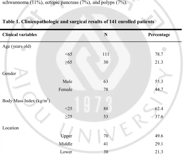
Table 1. Clinicopathologic and surgical results of 141 enrolled patients
Clinical variables N Percentage
Age (years old)
<65 ≥65 111 30 78.7 21.3 Gender Male Female 63 78 55.3 44.7 Body Mass Index (kg/m2)
<25 ≥25 88 53 62.4 37.6 Location Upper Middle Lower 70 41 30 49.6 29.1 21.3
7
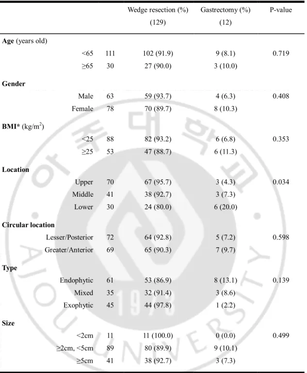
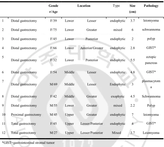
Circular location Lesser/Posterior Greater/Anterior 72 69 51.1 48.9 Type Endophytic Mixed Exophytic 61 35 45 43.3 24.8 31.9 Size <2cm ≥2cm, <5cm ≥5cm 11 89 41 7.8 63.1 29.1 Surgical procedure Wedge resection Distal gastrectomy Proximal gastrectomy Total gastrectomy 129 9 1 2 90.8 7.1 0.7 1.4Among the 141 patients, 129 underwent laparoscopic wedge resections and 12 underwent laparoscopic major gastrectomy. Nine SMTs, located in the prepyloric antrum, were treated by distal gastrectomy, and 3 gastroesophageal junction tumors were treated by either proximal gastrectomy or total gastrectomy. The characteristics of the tumors resected by wedge resection were compared to those resected by major gastrectomy, by age, sex, body mass index, and tumor size; statistically significant correlations were not observed, but gastric SMTs located in the lower third of the stomach were more likely to be removed by major gastrectomy (P = 0.034) (Table 2.). Tumor rupture or spillage did not occur during any of the procedures. The pathological and surgical features associated with each type of surgery are listed in Table 3.
8
Table 2. Comparison of gastrectomy according to the clinicopathologic features
Wedge resection (%) (129)
Gastrectomy (%) (12)
P-value
Age (years old)
<65 ≥65 111 30 102 (91.9) 27 (90.0) 9 (8.1) 3 (10.0) 0.719 Gender Male Female 63 78 59 (93.7) 70 (89.7) 4 (6.3) 8 (10.3) 0.408 BMI* (kg/m2) <25 ≥25 88 53 82 (93.2) 47 (88.7) 6 (6.8) 6 (11.3) 0.353 Location Upper Middle Lower 70 41 30 67 (95.7) 38 (92.7) 24 (80.0) 3 (4.3) 3 (7.3) 6 (20.0) 0.034 Circular location Lesser/Posterior Greater/Anterior 72 69 64 (92.8) 65 (90.3) 5 (7.2) 7 (9.7) 0.598 Type Endophytic Mixed Exophytic 61 35 45 53 (86.9) 32 (91.4) 44 (97.8) 8 (13.1) 3 (8.6) 1 (2.2) 0.139 Size <2cm ≥2cm, <5cm ≥5cm 11 89 41 11 (100.0) 80 (89.9) 38 (92.7) 0 (0.0) 9 (10.1) 3 (7.3) 0.499
9
Table 3. Clinical features of patients who were undergone laparoscopic gastrectmy for gastric submucosal tumor
Gende
r/Age
Location Type Size (cm)
Pathology
1 Distal gastrectomy F/39 Lower Lesser endophytic 3.7 leiomyoma
2 Distal gastrectomy F/75 Lower Greater mixed 6 schwannoma
3 Distal gastrectomy F/45 Lower Posterior endophytic 2 polyp
4 Distal gastrectomy F/66 Lower Anterior/Greater endophytic 2.8 GIST*
5
Distal gastrectomy F/32 Lower Posterior endophytic 5.5 ectopic
pancreas
6 Distal gastrectomy F/54 Middle Lesser endophytic 4.8 GIST*
7
Distal gastrectomy M/69 Middle Lesser Endophytic 3 plasmacytom
a
8 Distal gastrectomy F/42 Middle Greater exophytic 4.5 Schwannoma
9 Distal gastrectomy M/55 Lower Greater mixed 2.2 Polyp
10 Proximal gastrectomy M/45 Upper Greater endophytic 7 leiomyoma
11 Total gastrectomy F/45 Upper Lesser/Posterior endophytic 4 GIST*
12 Total gastrectomy M/27 Upper Lesser/Posterior Mixed 3.7 Leiomyoma
*GIST=gastrointestinal stromal tumor
The mean operation time was 102 minutes, and it was gradually reduced to about 70 minutes, after 30 cases, where it stabilized. However, the surgical time did vary on a case-by-case basis (Fig. 2.). A one-way ANOVA test indicated that the operation times did not differ significantly relative to the tumor location (P = 0.116). The surgical time for resection of gastric SMTs located in the posterior wall of the lesser curvature of the stomach were relatively longer than they were for tumors of the anterior wall or greater curvature of the stomach (Fig. 3.).
10
Fig. 2. Change in operation time, according to the surgeon’s experience in laparoscopic surgery for gastric submucosal tumors. Arrow shows the occurrence of postoperative complications.
11
Fig. 3. Operation time according to tumor locations. There was no significant difference
in operation time among the locations, by one-way ANOVA test. However, operation times for gastric submucosal tumor located in the posterior wall or lesser curvature of upper portion of the stomach was significantly longer than it was for those in the anterior wall or greater curvature of the stomach. * means P<0.05 in post-hoc test (Duncan method). PW posterior wall; LC lesser curvature; AW anterior wall; GC greater curvature.
The mean time to gas out after surgery was 3.3±2.3 days, and the mean length of hospital stay was 7.1±3.4 days. The discharge of forty seven patients (33.3%) who should be managed for postoperative complications and want it to be postponed was behind the planned schedule.
Operative morbidity was 8.5% (12/141), including 2 cases of intra-abdominal bleeding, 1 case of marginal ulcer bleeding, and 1 case of leakage of resection line. Re-operation was
12
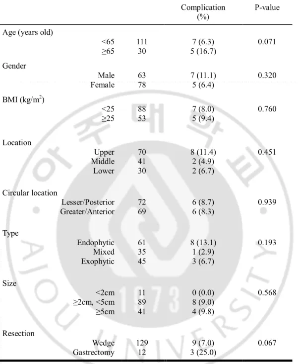
necessary only in the 2 cases of bleeding and leakage. There were no mortality cases (Table 4.). None among age gender, BMI, location, growth type, size, and type of resection were found to have significant influence on post-gastrectomy complications. (Table 5), and there were no complications observed after the 70th patient was operated on.
Of the 90 patients diagnosed with GIST, we selected 52 patients who were followed up more than 12 months. At a mean follow-up of 31 months (range, 12.3 to 56.5 months, median: 17.6 months), recurrence was observed in 2 (1.4%) patients, and no local recurrence was observed. Both of these patients had high-risk GIST; 1 had a tumor that was 5.4 cm in diameter and more than 10 mitoses per 50 high-powered fields were observed; the other case involved a tumor of 7.5 cm in diameter and more than 15 mitoses per 50 high-powered fields. One case was detected liver metastasis at postoperative 16 months, and the other case peritoneal seeding at postoperative 18 months.
13
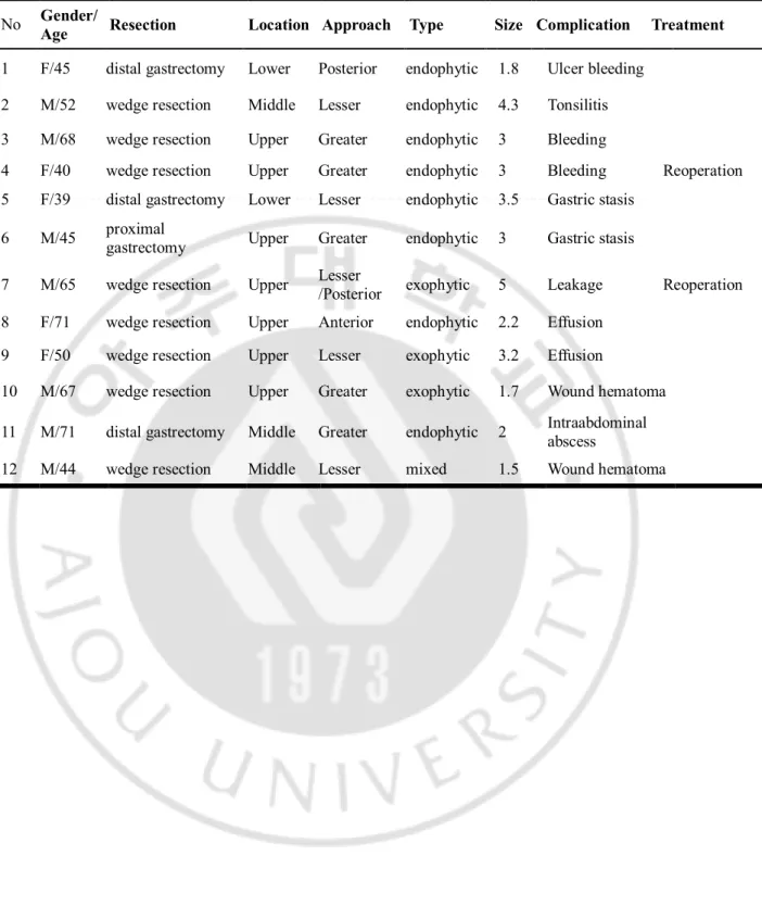
Table 4. Characteristics of patients who had complications
No Gender/ Age Resection Location Approach Type Size Complication Treatment
1 F/45 distal gastrectomy Lower Posterior endophytic 1.8 Ulcer bleeding 2 M/52 wedge resection Middle Lesser endophytic 4.3 Tonsilitis 3 M/68 wedge resection Upper Greater endophytic 3 Bleeding
4 F/40 wedge resection Upper Greater endophytic 3 Bleeding Reoperation 5 F/39 distal gastrectomy Lower Lesser endophytic 3.5 Gastric stasis
6 M/45 proximal gastrectomy Upper Greater endophytic 3 Gastric stasis
7 M/65 wedge resection Upper Lesser /Posterior exophytic 5 Leakage Reoperation 8 F/71 wedge resection Upper Anterior endophytic 2.2 Effusion
9 F/50 wedge resection Upper Lesser exophytic 3.2 Effusion
10 M/67 wedge resection Upper Greater exophytic 1.7 Wound hematoma 11 M/71 distal gastrectomy Middle Greater endophytic 2 Intraabdominal abscess 12 M/44 wedge resection Middle Lesser mixed 1.5 Wound hematoma
14
Table 5. Comparison of risk of postoperative complications according to the clinicopathologic features
Complication (%)
P-value Age (years old)
<65 ≥65 111 30 7 (6.3) 5 (16.7) 0.071 Gender Male Female 63 78 7 (11.1) 5 (6.4) 0.320 BMI (kg/m2) <25 ≥25 88 53 7 (8.0) 5 (9.4) 0.760 Location Upper Middle Lower 70 41 30 8 (11.4) 2 (4.9) 2 (6.7) 0.451 Circular location Lesser/Posterior Greater/Anterior 72 69 6 (8.7) 6 (8.3) 0.939 Type Endophytic Mixed Exophytic 61 35 45 8 (13.1) 1 (2.9) 3 (6.7) 0.193 Size <2cm ≥2cm, <5cm ≥5cm 11 89 41 0 (0.0) 8 (9.0) 4 (9.8) 0.568 Resection Wedge Gastrectomy 129 12 9 (7.0) 3 (25.0) 0.067 BMI=body mass index
15
IV. DISCUSSION
To date, many researchers have reported the efficacy of laparoscopic surgery compared to open surgery for gastric SMTs.(Matthews et al., 2002; Ishikawa et al., 2006; Nishimura et al., 2007; Karakousis et al., 2011) Laparoscopic surgery for gastric SMTs has been shown to yield patient benefits, including enhancing their postoperative recovery.(Cheng et al., 1999; Shimizu et al., 2002) This study reports the evaluation of the largest number of patients who underwent laparoscopic surgery for gastric SMTs at a single center. Most of tumors were resected by laparoscopic wedge resection. However, tumors located at the prepyloric junction showed the highest proportion of distal gastrectomies compared to tumors at other locations. The surgical time was variable in spite of sufficient surgeon’s experience, but there were no complications after 70 cases. Results of the long-term follow-up showed that laparoscopic surgery is feasible for gastric SMTs.
Although laparoscopic resection has become the treatment of choice for gastric SMTs, not all SMTs are resected using this technique. Some authors have limited the indication of laparoscopic surgery to only those patients who have a gastric SMT that is less than 5 cm in diameter.(Shimizu et al., 2002; Ishikawa et al., 2006; Otani et al., 2006) This tumor size limitation might reflect the technical difficulty of laparoscopic minimal resection. In the current series, laparoscopic surgery was applied to all tumors below 5 cm in size, but the limitation was increased to 8cm after the 70th surgery in our institution. As a result, 41 cases (29.0%) involved tumors ≥ 5 cm in diameter. All of these laparoscopic surgeries were successfully performed, without open conversion, presumably because of proficiency of the surgeon in the laparoscopic technique. It is possible for surgeons to perform total or subtotal laparoscopic gastrectomy for SMTs that are impossible to resect by wedge resection. Therefore, the indication for laparoscopic surgery of gastric SMTs below 8cm might have no limitations with respect to tumor location.
Although the surgical procedures can vary according to tumor size and location, most tumors can be resected by wedge resection. It is important to resect gastric SMTs by wedge resection, if possible. Major gastrectomy and reconstruction leads to a defect in gastrointestinal function, such as dumping syndrome and stasis, and can negatively affect the
16
patient’s quality of life. Because resection of gastric SMTs does not require lymph node dissection, it is possible to resect the tumor while preserving most of the stomach. However, stapler resection of endophytic SMTs can lead to stomach deformities because an excessive amount of normal stomach around the tumor needs to be removed due to the extra-luminal approach. Through the use of the eversion method suggested by Hyung et al.(Hyung et al., 2005) , most endophytic SMTs located even in the lower and upper portions of the stomach can be treated by wedge resection. However, the rate of gastrectomy for SMTs in the lower portion of the stomach increased to 20% in this study.
Previous reports have not documented the development of strictures after wedge resection of the stomach near the pylorus, but several authors have reported strictures after endosopic resection.(Tsunada et al., 2008; Iizuka et al., 2010) Such strictures can occur after surgical resection of SMTs near the pyloric ring. Therefore, distal gastrectomy was employed if the resection margin was too close to the pylorus, in order to avoid the possible formation of a postoperative stricture around the pyloric ring.
Several reports have described the tail-road approach for resection of SMTs near the esophagogastric (EG) junction.(Song et al., 2007; Hwang et al., 2009) Proximal or total gastrectomies of these SMTs are associated with increased postoperative complications, including reflux esophagitis. Most surgeons consider that minimal resection, avoiding major gastrectomy, is required for the removal of SMTs near the EG junction. Several approaches to such tumors can be applied to allow minimal resection. For endophytic lesions in the posterior stomach wall, transgastric resection is the treatment of choice. In this method, the surgeon can visualize the exact location of the tumor and confirm its margins.(Watson et al., 1996; Ibrahim et al., 1997) Exophytic SMTs located on the lesser curvature of the stomach or on its posterior wall can be removed completely after dissection of the gastrocolic or gastrohepatic ligaments to identify the tumor margin. For this reason, the surgical time for SMTs in this region were longer than for those conducted on tumors located in other regions of the stomach, in the present case series. However, most SMTs in this region were resected by wedge resection, except for 3 cases of large tumors where malignancy was expected in the preoperative study. In these instances, major gastrectomy was inevitable. One of these cases was later diagnosed as having a 3.7 cm leiomyoma. If an exact diagnosis, by fine needle biopsy, had been available earlier, a minimal resection, like enucleation, could have
17
been performed. Therefore, thorough preoperative investigations are required to obtain a correct diagnosis and for the selection of appropriate techniques.
Present study showed fair results with regard to the safety of the procedure. There were no mortalities, and no complications were observed after the 70th case. Laparoscopic surgery for gastric cancer has become popular in Korea, because of the high proportion of early gastric cancers. First laparoscopic surgery for gastric SMTs was started after they performed the first laparoscopic gastrectomy for early gastric cancers. Now, they performed over 100 laparoscopic surgeries for gastric cancers a year. Because the laparoscopic gastrectomy for gastric cancer requires the complicated procedure, laparoscopic resection for SMTs is assumed to have become a safe method. The variation of operative time should be considered according to complexity of procedures for tumors that are located in the lesser curvature and posterior wall of the stomach or closed to esophagogastric junction or pylorus.
Two of the 52 patients diagnosed with GIST demonstrated a recurrence of their cancer within 12 months of surgery. In both these cases, the tumors were greater than 5 cm in diameter. Although the number of cases is insufficient to be conclusive, large tumors (≥5 cm) appeared to be more likely to recur than did the smaller ones (<5 cm). Previous clinical studies on patients who underwent open laparotomy for GIST also indicated that tumor sizes greater than 5 cm in diameter were prognostic of poor survival.(Ng et al., 1992; DeMatteo et al., 2000; Pierie et al., 2001) We assumed that the laparoscopic surgery for tumors with greater than 5cm did not affect on the recurrence of them. However, prospective clinical study will be required for confirmative results about it.
18
V. CONCLUSION
In conclusion, we confirmed that laparoscopic surgery for gastric SMTs is technically safe and oncologically feasible. Laparoscopic resection could be a useful and promising procedure for any kind of gastric SMT if size is below 8cm. However, wedge resection was considered according to the location and characteristics of tumor.
19
REFERENCES
1. Berindoague R, Targarona EM, Feliu X, Artigas V, Balague C, Aldeano A, Lahoud A, Navines J, Fernandez-Sallent E, Trias M: Laparoscopic resection of clinically suspected gastric stromal tumors. Surg Innov 13: 231-237, 2006
2. Cheng HL, Lee WJ, Lai IR, Yuan RH, Yu SC: Laparoscopic wedge resection of benign gastric tumor. Hepatogastroenterology 46: 2100-2104, 1999
3. De Vogelaere K, Van Loo I, Peters O, Hoorens A, Haentjens P, Delvaux G: Laparoscopic resection of gastric gastrointestinal stromal tumors (GIST) is safe and effective, irrespective of tumor size. Surg Endosc 26: 2339-2345, 2012
4. DeMatteo RP, Lewis JJ, Leung D, Mudan SS, Woodruff JM, Brennan MF: Two hundred gastrointestinal stromal tumors: recurrence patterns and prognostic factors for survival. Ann Surg 231: 51-58, 2000
5. Demetri GD, Benjamin RS, Blanke CD, Blay JY, Casali P, Choi H, Corless CL, Debiec-Rychter M, DeMatteo RP, Ettinger DS, Fisher GA, Fletcher CD, Gronchi A, Hohenberger P, Hughes M, Joensuu H, Judson I, Le Cesne A, Maki RG, Morse M, Pappo AS, Pisters PW, Raut CP, Reichardt P, Tyler DS, Van den Abbeele AD, von Mehren M, Wayne JD, Zalcberg J: NCCN Task Force report: management of patients with gastrointestinal stromal tumor (GIST)--update of the NCCN clinical practice guidelines. J Natl Compr Canc Netw 5 Suppl 2: S1-29; quiz S30, 2007 6. Heinrich MC, Corless CL: Gastric GI stromal tumors (GISTs): the role of surgery in
the era of targeted therapy. J Surg Oncol 90: 195-207; discussion 207, 2005
7. Hwang SH, Park do J, Kim YH, Lee KH, Lee HS, Kim HH, Lee HJ, Yang HK, Lee KU: Laparoscopic surgery for submucosal tumors located at the esophagogastric junction and the prepylorus. Surg Endosc 23: 1980-1987, 2009
8. Hyung WJ, Lim JS, Cheong JH, Kim J, Choi SH, Noh SH: Laparoscopic resection of a huge intraluminal gastric submucosal tumor located in the anterior wall: eversion method. J Surg Oncol 89: 95-98, 2005
9. Ibrahim IM, Silvestri F, Zingler B: Laparoscopic resection of posterior gastric leiomyoma. Surg Endosc 11: 277-279, 1997
20
Stricture after endoscopic submucosal dissection for early gastric cancers and adenomas. Dig Endosc 22: 282-288, 2010
11. Ishikawa K, Inomata M, Etoh T, Shiromizu A, Shiraishi N, Arita T, Kitano S: Long-term outcome of laparoscopic wedge resection for gastric submucosal tumor compared with open wedge resection. Surg Laparosc Endosc Percutan Tech 16: 82-85, 2006
12. Iwahashi M, Takifuji K, Ojima T, Nakamura M, Nakamori M, Nakatani Y, Ueda K, Ishida K, Naka T, Ono K, Yamaue H: Surgical management of small gastrointestinal stromal tumors of the stomach. World J Surg 30: 28-35, 2006
13. Karakousis GC, Singer S, Zheng J, Gonen M, Coit D, DeMatteo RP, Strong VE: Laparoscopic versus open gastric resections for primary gastrointestinal stromal tumors (GISTs): a size-matched comparison. Ann Surg Oncol 18: 1599-1605, 2011 14. Kim CJ, Day S, Yeh KA: Gastrointestinal stromal tumors: analysis of clinical and
pathologic factors. Am Surg 67: 135-137, 2001
15. Matthews BD, Walsh RM, Kercher KW, Sing RF, Pratt BL, Answini GA, Heniford BT: Laparoscopic vs open resection of gastric stromal tumors. Surg Endosc 16: 803-807, 2002
16. Ng EH, Pollock RE, Romsdahl MM: Prognostic implications of patterns of failure for gastrointestinal leiomyosarcomas. Cancer 69: 1334-1341, 1992
17. Nishimura J, Nakajima K, Omori T, Takahashi T, Nishitani A, Ito T, Nishida T: Surgical strategy for gastric gastrointestinal stromal tumors: laparoscopic vs. open resection. Surg Endosc 21: 875-878, 2007
18. Novitsky YW, Kercher KW, Sing RF, Heniford BT: Long-term outcomes of laparoscopic resection of gastric gastrointestinal stromal tumors. Ann Surg 243: 738-745; discussion 745-737, 2006
19. Otani Y, Furukawa T, Yoshida M, Saikawa Y, Wada N, Ueda M, Kubota T, Mukai M, Kameyama K, Sugino Y, Kumai K, Kitajima M: Operative indications for relatively small (2-5 cm) gastrointestinal stromal tumor of the stomach based on analysis of 60 operated cases. Surgery 139: 484-492, 2006
20. Pierie JP, Choudry U, Muzikansky A, Yeap BY, Souba WW, Ott MJ: The effect of surgery and grade on outcome of gastrointestinal stromal tumors. Arch Surg 136:
21
383-389, 2001
21. Roukos DH: Current advances and changes in treatment strategy may improve survival and quality of life in patients with potentially curable gastric cancer. Ann
Surg Oncol 6: 46-56, 1999
22. Shimizu S, Noshiro H, Nagai E, Uchiyama A, Mizumoto K, Tanaka M: Laparoscopic wedge resection of gastric submucosal tumors. Dig Surg 19: 169-173, 2002
23. Song KY, Kim SN, Park CH: Tailored-approach of laparoscopic wedge resection for treatment of submucosal tumor near the esophagogastric junction. Surg Endosc 21: 2272-2276, 2007
24. Tsunada S, Ogata S, Mannen K, Arima S, Sakata Y, Shiraishi R, Shimoda R, Ootani H, Yamaguchi K, Fujise T, Sakata H, Iwakiri R, Fujimoto K: Case series of endoscopic balloon dilation to treat a stricture caused by circumferential resection of the gastric antrum by endoscopic submucosal dissection. Gastrointest Endosc 67: 979-983, 2008
25. Watson DI, Game PA, Devitt PG: Laparoscopic resection of benign tumors of the posterior gastric wall. Surg Endosc 10: 540-541, 1996
-국문요약-
위 점막하 종양의 복강경적 절제: 단일기관 141례의 분석
아주대학교 대학원 의학과 유 기 상 (지도교수: 한 상 욱) 위 점막하 종양의 치료는 수술적 절제이다. 최근 수술 기술의 발전은 대 부분의 종류의 절제 가능한 위 점막하 종양의 치료에 복강경적 절제를 널리 적 용하는데 이바지 하였다. 이 연구는 단일 기관에서의 위 점막하 종양의 복강경적 절제의 성적을 분석해 그 효용성을 확인하는데 있다. 2003년 4월부터 2011년 6월까지 아주대학교 병원에서 시행한 총 141례의 위 점막하 종양에 대한 복강경적 절제술을 받은 환자를 대상으로 하였다. 이들에 대한 인구통계 및 종양의 특성, 수술적 결과 및 종양학적 결과에 대해 분석하였 다. 위장관 기질성 종양이 가장 많은 조직학적 소견이었으며(90례) 대부분 위의 상부 1/3에 위치하고 있었다(70례). 위 쐐기 절제술을 128명의 환자에서 시행하였 으며 13명의 환자에 대해서는 위 절제술을 시행하였다. 평균 수술 시간은 102분 이었으며 30례 이후의 수술에 대해 대략 70분 정도로 안정화 되었다. 위의 후벽 혹은 상부 1/3의 소만부에 위치한 종양의 수술 시간이 다른 곳에 위치한 종양의 수술시간에 비해 긴 것으로 확인되었다. 2례의 복강내 출혈, 1례의 궤양성 출혈, 1 례의 문합부 유출을 포함한 12례의 술후 합병증이 발생하였다. 그러나 70례 이후 의 수술에서는 합병증이 발생하지 않았다. 추적관찰 기간 중 2례에서 위장관 기 질성 종양의 재발이 확인되었다. 복강경하 위 점막하 종양의 절제술은 안전하고 효용성이 있는 것으로 판 단되며 특히 술자의 경험 및 기술이 안정화되었을 때는 더욱 그러한 것으로 판 단된다. 복강경적 절제술은 어느 종류의 위 점막하 종양에 상관없이 유용한 것으로 판단된다.
핵심어: 점막하 종양, 복강경, 위종양