pISSN 1598-298X / eISSN 2384-0749 J Vet Clin 38(2) : 82-84 (2021)
https://doi.org/10.17555/jvc.2021.04.38.2.82
82
Splenic Mast Cell Tumors in Two Cats
Ji-Youl Jung*, Nak-Hyoung Kim*, So-Jeong Yim*, Kyung-Hwa Hong**, Ja-Sil Park*** and Jae-Hoon Kim*1
*College of Veterinary Medicine and Veterinary Medical Research Institute, Jeju National University, Jeju 63243, Korea **Anyang Animal Medical Center, Anyang 14006, Korea
***Dasom Feline Medical Center, Busan 48415, Korea
(Received: January 14, 2021 / Revised: March 09, 2021 / Accepted: March 24, 2021)
Abstract : Two 11-year-old cats, female Korean shorthair cat and male Siamese cat, with abdominal distention were presented to the local animal hospitals. Radiographic and ultrasonographic examinations revealed moderate to severe splenomegaly in both cats. In Korean shorthair cat, multiple masses were also existed on the anal and facial skin. Surgically excised whole spleens of two cats were requested for histopathologic examination. Histopathologically, numerous neoplastic round cells with cytoplasmic fine granules were widely infiltrated in the splenic parenchyma. The cytoplasmic granules were metachromatic on toluidine blue staining. These splenic masses were diagnosed as splenic mast cell tumors. Among them, Korean shorthair cat was remained healthy for at least 1 year after splenectomy. Because of no visiting of owner, we were only able to know the information for Siamese cat until 10 months after the splenectomy. To our best knowledge, this is the first detail case reports for splenic mast cell tumors in cats in Korea. Key words : cat, histopathology, mast cell tumor, spleen, splenectomy.
Introduction
Mast cell tumors (MCTs) are the third most common type of neoplasia in cats, after lymphoid malignancies and mammary tumors (1). MCTs in cats are generally classified into three cat-egories based on the involved organ: cutaneous and visceral such as splenic and intestinal MCTs, although multiple organ systems may also be involved simultaneously. Splenic MCT is one of the most common splenic tumors in cats, representing 15-26% of all splenic neoplasia (4,13). Cutaneous lesions are typically benign, but splenic and intestinal lesions are often associated with a poor prognosis because of the increased invasiveness and frequency of local and distant metastasis associated with these tumor types (1). Cats with the visceral MCTs may have signs of systemic illness including lethargy, anorexia, weight loss, and intermittent vomiting (1,2).
Splenectomy has been advocated as the primary therapy for splenic MCTs, with previously published individual sur-vival times ranging from 8 to 38 months (7,14). The efficacy of adjuvant chemotherapy and steroids is still largely unknown. Although splenic MCTs were common in feline popula-tions of other countries, there was no case report and avail-able data for this tumor in Korea. This report describes the histopathologic and immunohistochemical features in two cases of splenic MCTs in cats in Korea.
Cases
An 11-year-old neutered female Korean shorthair cat (case
1) was presented to a local animal hospital with various small masses on the anal and facial skin and with abdominal distention. The initial purpose of the visit was related to the cutaneous symptoms. Hematological analysis revealed throm-bocytopenia (102 K/L, reference range 150-500 K/L). Bio-chemical analysis showed normal results. Fine needle aspiration (FNA) tests were performed to the skin mass around anus and spleen.
An 11-year-old neutered male Siamese cat (case 2) was presented to other local animal hospital with anorexia, spo-radic vomiting, and abdominal distention. Hematological anal-ysis revealed polycythemia (14 M/L, reference range 5-11 M/L). Biochemical profiling indicated an increase in liver enzyme (ALT 306 U/L, reference range 12-118 U/L).
On radiographic and ultrasonographic examinations,
sple-1Corresponding author.
Splenic Mast Cell Tumors in Two Cats 83
nomegaly was observed in both cats (Fig 1). Because the splenic neoplasia was suspected in these cases, the entire spleens of two cats were surgically excised. Grossly, the excised spleens showed severe swelling with round periph-eral edges, especially in Korean shorthair cat (Fig 2A). The spleen was firm and dark red to gray color. In Siamese cat, multiple pale to tan colored abnormal round nodular masses were observed in the splenic parenchyma (Fig 2B). Accord-ing to Wright-Giemsa stainAccord-ing for FNA slides from the anal skin and the spleen, characteristic monomorphic population of round cells with abundant cytoplasmic metachromatic gran-ules that were consistent with mast cells were clearly demon-strated. Surgically excised spleen samples were immediately fixed in 10% neutral buffered formalin and submitted to the Veterinary Pathology Laboratory in Jeju National University for routine pathologic examination. The samples were pro-cessed and stained with hematoxylin and eosin (H&E), and toluidine blue stain for light microscopy examination. To assess KIT expression, immunohistochemistry (IHC) for CD117 (rabbit polyclonal antibody, 1:500, Dako, Denmark) was also performed.
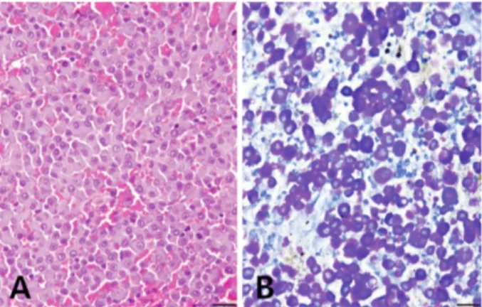
Histopathologic examination revealed diffusely distributed numerous round tumor cells throughout the splenic paren-chyma in case 1. These tumor cells were round to polyhedral in shape with a finely granulated cytoplasm (Fig 3A). Nuclei were large, round, vesicular, and hyperchromatic with margi-nated chromatin, and had prominent nucleoli. The mitotic count was very low. On the other hand, multifocal nodular masses were observed in the spleen of case 2. Numerous round cells were widely distributed in the nodular masses and occasionally in the normal splenic red pulp. The mor-phology of the tumor cells was similar to that of case 1. Tolu-idine blue stain demonstrated metachromatic cytoplasmic
granules of tumor cells in both cats (Fig 3B). However, the neoplastic mast cells in the spleen of two cats were all nega-tive for IHC staining using CD117 antibody.
Based on the clinical and gross findings, histopathologic examination, and IHC results, two cats were diagnosed with splenic MCTs.
The cats did not receive any specific treatment for splenic tumors except splenectomy. Korean shorthair cat (case 1) was alive and healthy for 1 year after surgery. Because of no visiting of owner, we were only able to know the informa-tion for Siamese cat (case 2) until 10 months after the sple-nectomy.
Discussion
Splenic MCT is predominantly a disease of older cats with no breed or sex predilection. Although the sample size was very low in this study, the age of both cats was 11 years. This is very similar with previously reported data on the onset age of MCTs (3,6). In agreement with previous studies (3,6), male Siamese cat was presented with nonspecific gastrointes-tinal signs such as anorexia and sporadic vomiting. In con-trast, female Korean shorthair cat had two concurrent MCTs in spleen and anal skin. Simultaneous occurrences of cutane-ous tumors have been identified in 3-23% of cats with splenic MCT (8,12) This phenomenon is not well under-stood until today, and there is no way to distinguish between a primary splenic MCT and a secondary metastatic splenic MCT originated from the primary skin tumor. The male Sia-mese cat did not show any involvement in regional lymph nodes, liver, and intestine.
Splenomegaly is the most common clinical feature in cats with visceral MCTs under the physical examination and diag-nostic imaging methods. However, various other splenic conditions can also be present with moderate to severe sple-nomegaly. The feline spleen with MCT may appear mottled, nodular or irregular, and enlarged. According to the previous large scaled study of sonographic findings in 101 cats (4), 25 (83%) of 30 cats with lymphosarcoma and 25 (93%) of 27 cats with MCT had splenomegaly, respectively. Even 11 Fig 2. Gross findings. (A) Korean shorthair cat. Note the
mark-edly enlarged spleen with round edges. (B) Siamese cat. Multiple pale to tan colored abnormal round nodular masses (arrows) were observed in the formalin-fixed splenic parenchyma.
Fig 3. Histopathologic findings. (A) Neoplastic cells were round to polyhedral in shape with finely granulated cytoplasm in Korean shorthair cat (H&E, Bar = 20 m). (B) The cytoplasmic granules of tumor cells were metachromatic in Siamese cat (Toluidine blue, Bar = 20 m).
84 Ji-Youl Jung, Nak-Hyoung Kim, So-Jeong Yim, Kyung-Hwa Hong, Ja-Sil Park and Jae-Hoon Kim
(41%) of 27 cats with extramedullary hematopoiesis and/or lymphoid hyperplasia also had splenomegaly. Hence, there are no imaging methods to distinguish the characteristic find-ings of feline splenic MCT from other spleen diseases (4). Therefore, the differential diagnosis of splenic tumors can be made by the cytological evaluation of FNA or the histopatho-logic examination for biopsy sample of the mass. For splenic tumors, the ultrasonographic appearance of the affected organ can be useful for guiding FNA target site for cytology (4). In this study, both cats showed splenomegaly with variable degree. Splenic MCTs were easily diagnosed in both cats by histopathologic examination from the surgically excised spleens. C-kit proto-oncogene product (KIT, CD117) is a receptor tyrosine kinase for stem cell factor produced by a number of cells including mast cells (11). Tyrosine kinases are cell membrane-bound growth factor receptors that, when mutated, can result in uncontrolled cellular proliferation (5). In canine cutaneous MCTs, c-kit mutation or KIT protein overexpression is a sig-nificant prognostic marker for MCTs or systemic mastocyto-sis and are considered good targets for treatment with kinase inhibitors (11,15). However, the correlation between KIT expression and tumor proliferative activity or disease out-come has been found to be very low in feline MCTs, espe-cially cutaneous MCTs (11). In addition, the KIT expression in feline splenic MCTs was lower than that of cutaneous MCTs (9). No c-kit mutations were identified in feline splenic MCT specimens in a previous study (2). This result indi-cated that receptor tyrosine kinase inhibitor therapy for splenic MCTs may not be benefit for the treatment in cats. In accordance with previous reports, no KIT protein was detected in two feline spleen samples of present study.
Treatment methods for feline MCTs include histamine blockade, surgery including splenectomy, chemotherapy, and receptor tyrosine kinase inhibition (5). Among them, several studies indicated that surgery was the best choice of treat-ment due to relatively long survival times for splenectomy alone (7,13). In a more recent study, two cat groups with splenic MCTs that underwent splenectomy with/without che-motherapy had prolonged survival times than cats in other group received chemotherapy alone (3). Regardless, splenec-tomy of cats with splenic MCTs has been associated with median survival times of 12-19 months (range: 0-64 months) (6,7,10,12). In this study, splenectomy without additional chemotherapy was performed in both cats at the time of con-firming splenomegaly using sonographic examination. After the surgery, the female Korean shorthair cat and the male Siamese cat were still alive and healthy for 1 year and 10 months, respectively.
Conclusion
Because of the small sample size in this study, we could not find any significant prognostic factors for the survival of cats. However, we believe that the result of our study would be helpful to make an accurate diagnosis for radiographic and histopathological examinations of feline splenic MCTs.
Acknowledgment
This research was supported by the 2020 scientific promo-tion program funded by Jeju Napromo-tional University.
Conflict of Interest
No conflicts of interest have been declared.
References
1. Carpenter JL, Andrews LK, Holzworth J. Tumors and tumor-like lesions. In: Disease of the Cat: Medicine and surgery. Philadelphia: WB Saunders. 1987: 406-583.
2. Dank G, Chien MB, London CA. Activating mutations in the catalytic or juxtamembrane domain of c-kit in splenic mast cell tumors of cats. Am J Vet Res 2002; 63: 1129-1133. 3. Evans BJ, O’Brien D, Allstadt SD, Gregor TP, Sorenmo KU.
Treatment outcomes and prognostic factors of feline splenic mast cell tumors: A multi-institutional retrospective study of 64 cases. Vet Comp Oncol 2018; 16: 20-27.
4. Hanson JA, Papageorges M, Girard E, Menard M, Hebert P. Ultrasonographic appearance of splenic disease in 101 cats. Vet Radiol Ultrasound 2001; 42: 441-445.
5. Henry C, Herrera C. Mast cell tumors in cats, clinical update and possible new treatment avenues. J Feline Med Surg 2013; 15: 41-47.
6. Kraus KA, Clifford CA, Davis GJ, Kiefer KM, Drobatz KJ. Outcome and prognostic indicators in cats undergoing splenectomy for splenic mast cell tumors. J Am Anim Hosp Assoc 2015; 51: 231-238.
7. Liska WB, MacEwen EG, Zaki FA, Garvey M. Feline systemic mastocytosis: A review and results of splenectomy in seven cases. J Am Anim Hosp Assoc 1979; 15: 589-597.
8. Litster AL, Sorenmo KU. Characterisation of the signalment, clinical and survival characteristics of 41 cats with mast cell neoplasia. J Feline Med Surg 2006; 8: 177-183.
9. Mallett CL, Northrup NC, Saba CF, Rodriguez CO, Rassnick KM, Gieger TL, Childress MO, Howerth EW. Immunohisto-chemical characterization of feline mast cell tumors. Vet Pathol 2013; 50: 106-109.
10. Meuten DJ. Mast cell tumors. In: Tumors in Domestic Animals, 5th ed. Ames: Wiley Blackwell. 2017: 176-202.
11. Sabattini S, Bettini G. Prognostic value of histologic and immunohistochemical features in feline cutaneous mast cell tumors. Vet Pathol 2010; 47: 643-653.
12. Skeldon NCA, Gerber KL, Wilson RJ, Cunnington SJ. Masto-cytaemia in cats: prevalence, detection and quantification methods, haematological associations and potential implications in 30 cats with mast cell tumors. J Feline Med Surg 2010; 12: 960-966.
13. Spangler WL, Culbertson MR. Prevalence and type of splenic diseases in cats: 455 cases (1985-1991). J Am Vet Med Assoc 1992; 201: 773-776.
14. Thamm DH, Vail DM. Mast cell tumors. In: Small animal clinical oncology, 3rd ed. Philadelphia: Saunders. 2007:
402-424.
15. Webster JD, Yuzbasiyan-Gurkan V, Kaneene JB, Miller R, Resau JH, Kiupel M. The role of c-KIT in tumorigenesis: evaluation in canine cutaneous mast cell tumors. Neoplasia 2006; 8: 104-111.