The causes of restrictive respiratory disease in patients with stroke are rib cage movement limita-tions and core muscle weakness.1,2
Breathing in patients with stroke decreases the maximum amount of spontaneous ventilation due to damage to the cortical-transverse membrane nerves and deterioration of the diaphragm.3,4The diaphragm
is the main respiratory muscle responsible for 70% of one breath. In patients with stroke with weakened muscles, the respiratory function is reduced by approximately half.5,6
To increase the respiratory function of these patients, diaphragmatic breathing exercises have been implemented. The diaphragmatic breathing exercise is the easiest intervention in the early stages of respiratory rehabilitation. For the diaphragm to contract normally, not only the anterior protrusion of the abdomen but also the expansion of the lower rib cage is required. Maitland joint mobilization is a treatment technique that applies passive movement divided by the therapist’s hand in five grades.7 The
more chronic the patient with stroke, the more soft tissue builds up, which leads to movement limita-tions.8
Effects of Rib Cage Joint Mobilization Combined with
Diaphragmatic Breathing Exercise on the Pulmonary Function
and Chest Circumference in Patients with Stroke
INTRODUCTION
Background: Patients with stroke have core muscle weakness and limited rib cage movement, resulting in restrictive lung disease.
Objectives: To examine the comparison of effects of rib cage joint mobilization combined with diaphragmatic breathing exercise and diaphragmatic breathing exercise on the pulmonary function and chest circumference in patients with stroke.
Design: A cluster randomized controlled trial.
Methods: Twenty-four patients were randomly assigned to an experimental group (rib cage joint mobilization combined with diaphragmatic breathing exercise group) and control group (diaphragmatic breathing exercise group). Patients in the experimental group underwent rib cage joint mobilization for 15 min and diaphragmatic breathing exercise for 15 min. The control group underwent diaphragmatic breathing exercise for 30 min. Both groups under-went exercise thrice a week for 4 weeks. The pulmonary function and chest circumference were measured using the MicroLab spirometer and a tape measure, respectively.
Results: After the intervention, the pulmonary function and chest circumference significantly improved in both groups. These improvements were significantly higher in the experimental group than those in the control group.
Conclusion: Rib cage joint mobilization combined with diaphragmatic breathing exercise improves pulmonary function and chest circumference in patients with stroke.
Keywords: Diaphragmatic breathing exercise; Rib cage; Joint mobilization; Respiratory function; Stroke
Ayeon Kim, PT, MSa, Youngwha
Song, Prof., PhDb, Geurin Hong, PT,
MSa, Dajeong Kim, PT, MSa, Soonhee
Kim, Prof., PhDa
aDepartment of Physical Therapy, Yongin
University, Yongin, Republic of Korea
bDepartment of Physical Therapy, Dongnam
Health University, Suwon, Republic of Korea
Received : 18 April 2020 Revised : 02 June 2020 Accepted : 10 June 2020
Address for correspondence Soonhee Kim, Prof., PhD
Department of Physical Therapy, Yongin University 134, Yongindaehak-ro, Cheoin-gu, Yongin-si, Gyeonggi-do, Republic of Korea
Tel: 82-10-3844-9972 E-mail: [email protected]
In particular, inhalation limitation occurs due to the rib cage construction,1 and joint mobilization applied
to the rib cage increases the respiratory function and muscle activity and tension in patients with stroke and improve functional activity.9As described earlier,
joint mobilization applied to patients with stroke has been known as an intervention method for improving the respiratory function.10 However, to date, most
studies have compared diaphragmatic respiration and joint mobilization. In this study, we investigated the effects of diaphragmatic breathing exercise and diaphragmatic breathing exercise combined with rib cage joint mobilization on the pulmonary function and chest circumference.
The study was an assessor-blinded and Cluster randomized trial. A total of 24 adult patients who were admitted to the B hospital participated in this study. These patients were hospitalized for stroke under the diagnosis of a rehabilitation medicine doc-tor at a nursing home hospital in Gyeonggi-do. The intervention period was conducted thrice a week for 30 min for four weeks. The inclusion criteria of the subjects were as follows: those who had been diag-nosed with stroke for 6 months or more, had 10% less forced vital capacity (FVC) than the normal predicted value, and had a Korean-Mini Mental State Examination (K-MMSE) score of 24 or more. Those who had past or present lung-related damage and those with orthopedic disease in the rib cage, unsta-ble angina, congestive heart failure, peripheral artery disease, and depression were excluded from this study. This study was approved by the Institutional Review Board of Yongin University (2-1040966-AB-N-01-20-1910-HSR-156-6). Before the initial eval-uation, all subjects provided written informed con-sent.
Three physical therapists, including researchers, with >3 years of experience in neurological physical therapy participated in the intervention. The diaphragmatic breathing exercise exerted force resistance upon inhalation in the supine position and induced contraction of the rectus abdominis during exhalation.11 The diaphragmatic breathing exercise
combined with rib cage joint mobilization was
per-formed by turning the head from the prone position toward the comfortable side, followed by the center of the posterior–anterior mobilization and transverse mobilization. Each exercise was performed thrice a week for a total of 4 weeks (12 times).12In the case of
diaphragmatic breathing exercise, the researcher applied resistance to the diaphragm during inhalation and stretched to the diaphragm muscle during exha-lation. The subjects were instructed to relax, and both hands of the researchers were placed on the rectus muscle under the rib cartilage; then, the subjects were instructed to deeply and slowly inhale through the nose.11,13 Rib cage joint mobilization in the center
of the thoracic vertebrae felt the end feeling of each segment in the spine and recorded the joints with low mobility. The researcher put their thumb on one hand on a spinous process recorded as having low mobility and supported the hand with the other thumb. In transverse joint mobilization, both sides of the area where low mobility was recorded were compared and selected, and then, the thumb was placed in the transverse direction.7 Joint mobilization was applied
in grade 3 according to the Maitland classification, and the intervention time was 4 sets, 5 minutes per set, and the rest time was 1 minute, for a total of 23 minutes.
Pulmonary function
All assessments were performed by an physical therapist unrelated to this study. The evaluator was thoroughly familiar with the evaluation method and blinded to the study group prior to the evaluation. A lung spirometer was used to evaluate the respiratory function in this study.14 Pulmonary function
meas-urements were conducted in accordance with the pulmonary function test guidelines. The subjects sat on a chair with a backrest, looked at the front, and exhaled air into the spirometer after the maximum possible exhalation (to maintain exhalation for 6 s). The measurement variables were the peak expiratory flow (PEF), amount of effort exhalation, and forced expiratory volume in 1 s (FEV1). All items were measured three times, and the average value was recorded.15
Chest circumference
To measure the ability to expand around the chest, a spring tape measure was used to measure the upper and lower chest circumferences. To measure the upper chest circumference, markers were used to mark the space between the third rib along the center
SUBJECTS AND METHODS
Subjects
Outcome Measures
line of the clavicle and the fifth spine. The lower chest circumference was marked on the tip of the spin-ous process and the spinspin-ous spine of the tenth spine.15
All statistical data were analyzed using SPSS 21.0 software (IBM Corp., Armonk, NY, USA). Both the experimental and control groups satisfied the normal distribution. Thus, parametric tests were used. The homogeneity test between the two groups was per-formed using an independent t-test. The independent sample t-test and chi-square test were used for the general characteristics of the subjects. A paired t-test was performed for the effects of exercise. The with-in-subject factor was time (before and after the test), whereas the between-subject factor was group-by-time (experimental and control groups). When signif-icant differences were the interactions or main effects (group-by-time), t-test was used. The statistical significance level was set at α=.05.
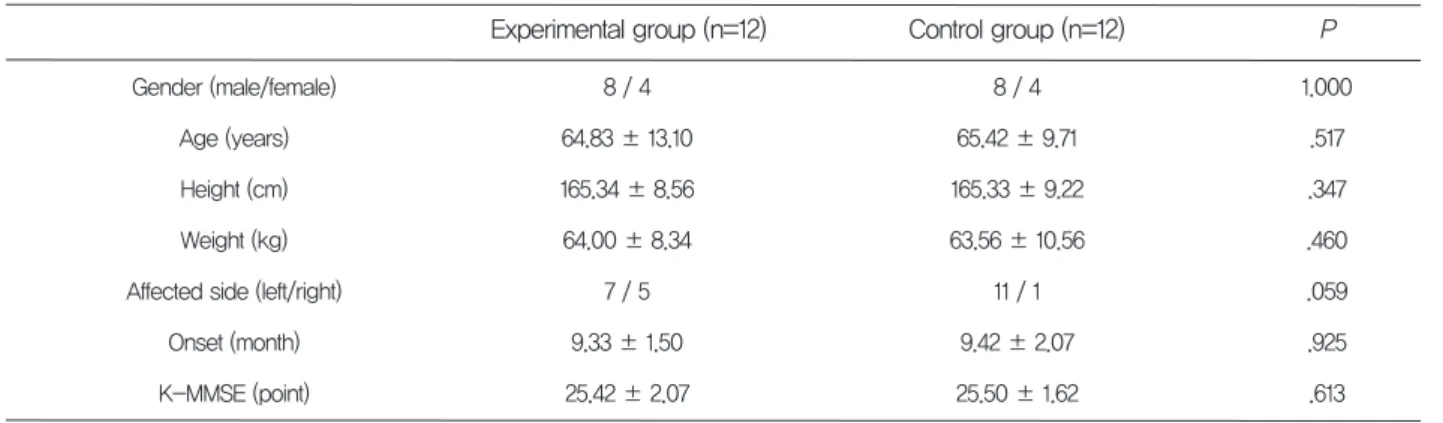
The general characteristics of the subjects are shown in Table 1. The gender of the experimental group was 8 men and 4 women, the average age was 64.83±13.10 years, the average height was 165.34±8.34 cm, and the average weight Was 64.00±8.34 kg, the site of the paralysis was 7 right hemiplegic, 5 left hemiplegic, the average onset peri-od was 9.33±1.50 months, and the average K-MMSE score was 25.42±2.07 points. The gender of the control group was 8 men and 4 women, the aver-age aver-age was 65.42±9.71 years, the averaver-age height was 165.33±9.22 cm, and the average weight was 63.56±10.56 kg, the site of the paralysis was 11 right hemiplegic, 1 left hemiplegic, the average onset peri-od was 9.42±2.07 months, and the average K-MMSE score was 25.50±1.62 points.
Data and Statistical Analysis
General characteristics of the subjects
*P<.05
K-MMSE: Korean version of Mini-Mental State Examination
(Mean±SD)
Gender (male/female) Age (years) Height (cm) Weight (kg) Affected side (left/right)
Onset (month) K-MMSE (point) Control group (n=12) 8 / 4 65.42 ± 9.71 165.33 ± 9.22 63.56 ± 10.56 11 / 1 9.42 ± 2.07 25.50 ± 1.62 Experimental group (n=12) 8 / 4 64.83 ± 13.10 165.34 ± 8.56 64.00 ± 8.34 7 / 5 9.33 ± 1.50 25.42 ± 2.07 P 1.000 .517 .347 .460 .059 .925 .613 Table 1. General characteristics
*P<.05
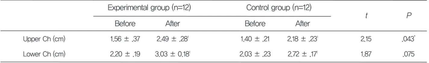
1There was a significant difference between before and after the test (P<.05) *The experimental group improved more than the control group
FEV1: Forced expiratory volume in 1 s FVC: Forced vital capacity
PEF: Peak expiratory flow
Experimental group: Rib cage joint mobilization combined with diaphragmatic breathing exercise group Control group: Diaphragmatic breathing exercise group
(Mean±SD) FEV1 (ℓ) FVC (ℓ) PEF (ℓ/min) Control group (n=12) Experimental group (n=12) Before After 2.34 ± .62 2.74 ± .57 245.58 ± 80.89 2.70 ± .551 3.11 ± .561 328.91 ± 76.08 Before After 2.18 ± .68 2.63 ± .55 224.75 ± 90.81 2.29 ± .701 2.89 ± .711 249.50 ± 96.061 .013* .231 .009* P -2.698 -1.232 -2.870 t
Table 2. Comparison of the pulmonary function before and after
Changes in the pulmonary function and chest circumference between the two groups
After four weeks of intervention, all groups experi-enced significant increases in the pulmonary function and chest circumference. The pulmonary function and chest circumference were significantly different between the two groups (Tables 2 and 3).
This study confirmed the changes in Maitland joint mobilization and diaphragmatic breathing exercises applied to the pulmonary function and chest circum-ference in patients with stroke with reduced respira-tory function. As a result of the study, both experi-mental and control groups showed significant differ-ences between the pulmonary function and chest cir-cumference before and after intervention.
Stroke is a disease that causes irreversible damage to the central nervous system due to bleeding and infarction in the cerebral blood vessels.16Patients with
stroke develop respiratory failure due to damage to the nervous system.17 Breathing in these patients
generally results in weakened respiratory muscles and reduced pulmonary volume, and reduced diaphragmatic motion reduces the effectiveness of coughing. In addition, in patients with stroke, asym-metric chest pump movements, deterioration of the upper chest volume, and compensatory movements to expand the rib cages in the lower abdomen upon inspiration were associated with rib cage move-ments.18 Recently, the respiratory muscle training
increased chest circumference, reduced asymmetric chest circumference, and increased swelling through the inhalation trainer improved the quality of life of patients with stroke. The diaphragmatic breathing
exercise is a method of contracting the diaphragm muscle in the state where the upper rib cage is relaxed as much as possible to effectively contract the diaphragm.11 In a recent study, diaphragmatic
breathing exercise was applied to patients with stroke in combination with various methods. Increasing diaphragm movement has been introduced as an effective method for improving the respiratory func-tion in these patients. Because the diaphragm muscle is attached to the chest wall, the limit on diaphragm contraction decreases as the mobility of the rib cage increases.19Joint mobilization was used to reorder the
joints of patients with stroke.7 It is a manual
treat-ment method to control pain and increase the range of joint movement and is graded in a total of 5 steps to induce recovery between the soft tissues or joints that are constructed by stretching and sliding them. Sutbeyaz et al.10showed that diaphragmatic breathing
exercise showed a significant difference in the respi-ratory function tests in patients with stroke. Lee20
found that the diaphragmatic breathing exercise was also significant for the pulmonary function tests and chest circumference. Repeated diaphragmatic breathing exercises helped to improve posture by relaxing the rigid rib cage and reducing the kyphosis of the spine.21 This is considered to be the result of
the positive effects on the activity of the respiratory muscles necessary for the respiratory function, ability to expand the rib cage, and improvement of the res-piratory function, consistent with the results of this study. Park and Park22 showed that Maitland joint
mobilization applied to patients with chronic stroke was effective in the pulmonary function and chest circumference. Fugl-Meyer et al.1 reported that the
limit of inspiration is due to the formation of the breast cage. Kriel23 suggested that rib cage joint
mobilization increases the volume of the rib cage and increases inspiration. Rib cage flexibility and muscle
*P<.05
1There was a significant difference between before and after the test (P<.05) *The experimental group improved more than the control group
Upper Ch: Upper chest circumference Lower Ch: Lower chest circumference
Experimental group: Rib cage joint mobilization combined with diaphragmatic breathing exercise group Control group: Diaphragmatic breathing exercise group
(Mean±SD) Upper Ch (cm) Lower Ch (cm) Control group (n=12) Experimental group (n=12) Before After 1.56 ± .37 2.20 ± .19 2.49 ± .281 3.03 ± 0.181 Before After 1.40 ± .21 2.03 ± .23 2.18 ± .231 2.72 ± .171 .043* .075 P 2.15 1.87 t
Table 3. Comparison of the chest circumference before and after
relaxation increased the muscle function and decreased the breathing effort. The activation of the parasympathetic nerves and suppression of the sym-pathetic nerves have created an environment of increased rib cage mobility and diaphragm contrac-tion. Therefore, it was confirmed through this study that the diaphragmatic breathing exercise combined with rib joint mobilization was more significant for the pulmonary function and chest circumference. Since this study included patients with stroke for more than 6 months after the onset and we studied some stroke patients, future studies require various additional evaluation methods. It would be necessary to compare the differences between the methods.
In conclusion, rib cage joint mobilization with diaphragmatic breathing exercise improved FEV1, FVC, PEF, and chest circumference in patients with stroke. In addition, rib cage joint mobilization com-bined with diaphragmatic breathing exercise was more effective for FEV1, PEF, and upper chest cir-cumference than only diaphragmatic breathing exer-cise.
The author declares that there are no conflicts of interest.
CONCLUSION
4. 5. 6. 7. 8. 9. 10. 11. 12. 13. 14. 15.Teixeira-Salmela LF, Parreira VF, Britto RR, et al. Respiratory pressures and thoracoabdominal motion in community-dwelling chronic stroke survivors. Arch Phys Med Rehabil. 2005;86(10): 1974-1978.
Khedr EM, El Shinawy O, Khedr T, et al. Assessment of corticodiaphragmatic pathway and pulmonary function in acute ischemic stroke patients. Eur J Neurol. 2000;7(3):323-330. Roffe C, Sills S, Pountain SJ, et al. A randomized controlled trial of the effect of fixed-dose routine nocturnal oxygen supplementation on oxygen saturation in patients with acute stroke. J Stroke Cerebrovasc Dis. 2010;19(1):29-35.
Maitland GD, Hengeveld E, Banks K, et al. Maitland's vertebral manipulation (Vol. 1). Philadelphia, PA: Elsevier Butterworth-Heinemann; 2013.
Winstein CJ, Miller JP, Blanton S, et al. Methods for a multisite randomized trial to investigate the effect of constraint-induced movement therapy in improving upper extremity function among adults recovering from a cerebrovascular stroke. Neurorehabil Neural Repair. 2003;17(3):137-152. Park S, Yang D, Kim J, et al. Effects of Neck Stabilizing Exercise Combined with Transcranial Direct Current Stimulation on Muscle Characteristics and Function in Patients with Cervicogenic Headache. Journal of The Korean Society of Integrative Medicine. 2019;7(3):159-169.
Sutbeyaz ST, Koseoglu F, Inan L, et al. Respiratory muscle training improves cardiopul-monary function and exercise tolerance in sub-jects with subacute stroke: A randomized con-trolled trial. Clin Rehabil. 2010;24(3):240-250. Kigin CM. Breathing exercises for the medical patient: The art and the science. Phys Ther. 1990;70(11):700-706.
Jowsey P, Perry J. Sympathetic nervous system effects in the hands following a grade III postero-anterior rotatory mobilisation technique applied to T4: A randomised, placebo-controlled trial. Man Ther. 2010;15(3):248-253.
Kisner C, Colby LA. Therapeutic exercise: Foundations and techniques. 4th. Philadelphia: F.A Davis Company; 2002
Crapo RO. Pulmonary-function testing. N Engl J Med. 1994;331(1):25-30.
Bockenhauer SE, Chen H, Julliard KN, et al. Measuring thoracic excursion: Reliability of the cloth tape measure technique. J Am Osteopath Assoc. 2007;107(5):191-196.
REFERENCES
1.
2.
3.
Fugl-Meyer AR, Linderholm H, Wilson AF. Restrictive ventilatory dysfunction in stroke: Its relation to locomotor function. Scand J Rehabil Med Suppl. 1983;9:118-124.
MacKay-Lyons MJ, Howlett J. Exercise capacity and cardiovascular adaptations to aerobic training early after stroke. Top Stroke Rehabil. 2005;12 (1):31-44.
Cohen E, Mier A, Heywood P, et al. Diaphra-gmatic movement in hemiplegic patients meas-ured by ultrasonography. Thorax. 1994;49(9) :890-895.
16. 17.
18. 19.
Sacco RL, Kasner SE, Broderick JP, et al. An updated definition of stroke for the 21st century. Stroke. 2013;44(7):2064-2089.
de Almeida ICL, Clementino ACCR, Rocha EHT, et al. Effects of hemiplegy on pulmonary function and diaphragmatic dome displacement. Respir Physiol Neurobiol. 2011;178(2):196-201.
Kim K. The effect of chest expansion and pul-monary function of stroke patients after breath-ing exercise. J Kor Phys Ther. 2009;21(3):25-32. Yelvar GDY, Çirak Y, Demir YP, et al. Immediate effect of manual therapy on respiratory functions and inspiratory muscle strength in patients with COPD. Int J Chron Obstruct Pulmon Dis. 2016;11:1353-1357.
20.
21. 22.
23.
Leelarungrayub D. Chest mobilization techniques for improving ventilation and gas exchange in chronic lung disease. INTECH Open Access Publisher; 2012.
Obayashi H, Urabe Y, Yamanaka Y, et al. Effects of respiratory-muscle exercise on spinal curva-ture. J Sport Rehabil. 2012;21(1):63-68.
Park SJ, Park SE. Effect of upper thoracic mobi-lization on cervical alignment in stroke patients with forward head posture: A case study. J Int Acad Phys Ther Res, 2018;9(2):1513-1516.
Kriel A. An investigation into the immediate effect of rib mobilization and sham laser applica-tion on chest wall expansion and lung funcapplica-tion in healthy asymptomatic males: A pilot study (Dissertation). South Africa: Durban Institute of Technology; 2005.