Heat Shock Protein 90 InhibitorDecreases
Collagen Synthesis of Keloid Fibroblasts and
Attenuates the Extracellular Matrix on the
Keloid Spheroid Model.
Jae-Yeon Park
Department of Medicine
Heat Shock Protein 90 Inhibitor Decreases
Collagen Synthesis of Keloid Fibroblasts and
Attenuates the Extracellular Matrix on the
Keloid Spheroid Model.
Directed by Professor Won Jai Lee
The Master's Thesis
submitted to the Department of Medicine,
the Graduate School of Yonsei University
in partial fulfillment of the requirements for the degree
of Master of Medical Science
Park Jae-Yeon
ACKNOWLEDGEMENTS
I would like to express the deepest appreciation to my thesis supervisor
Professor Won Jai Lee. Without his guidance and persistent help this
dissertation would not have been possible. His keen observation and sound
judgment in selecting, laying out and providing feedbacks enabled to discover
the better part of myself.
I would like to thank my committee members, Professor Dai Hyun Lew and
Professor Ju Hee Lee who demonstrated persistent support and guidance
throughout the whole research.
In addition, this dissertation was also the product of a large measure of
serendipity, fortuitous encounters who have changed the course of my academic
career. Had I not made the encounter with Professor Kwan Chul Tark in 1998 at
an operating room volunteer service for premedical students, I would probably
not have pursued plastic surgery. He enlightened me by showing the various
new fields in plastic surgery in the operating room and added fuel to my
enthusiasm. He is both my mentor and master.
I would like to thank my parents in continuously guiding and supporting me
both for my academic and career life as well as bring me up and shaping me as
who I am today.
<TABLE OF CONTENTS>
ABSTRACT ··· 1
I. INTRODUCTION ··· 3
II. MATERIALS AND METHODS ··· 5
1. Human dermal fibroblast and keloid-derived fibroblast cells ··· 5
2. qRT-PCR analysis of expression levels of type I and III collagen ··· 5
A. Western blotting analysis for Akt and Smad 2/3 complex ··· 5
3. Preparation and 17AA treatment of keloid spheroids ··· 6
4. Immunohistochemistry (IHC) ··· 6
5. Statistics ··· 7
III. RESULTS ··· 8
1. Hsp 90 expression was increasedin keloid tissues
compared with adjacent normal tissues ··· 8
2. 17AAG down-regulates mRNA expression
of type I collagwn in KTs ··· 10
3. Suppression of Smad 2/3 complex expression in KFs
under 17AAGtreatement ··· 11
4. Collagen deposition and intensity were decreased on the
keloid spheroid which treated with 17 AAG ··· 12
5. 17 AAG desreases the expression of type I, III collagen,
elastin, and fibronectin in keloid spheroids ··· 14
IV. DISCUSSION ··· 19
V. CONCLUSION ··· 21
REFERENCES ··· 22
LIST OF FIGURES
Figure 1. Histologic analysis of keloid tissue.
··· 9
Figure 2. Quantitative analysis of mRNA expression of type
I and III collagen.
··· 10
Figure 3. Immunoblotanalysis of Smad 2/3.
complex expression in KFs. ··· 11
Figure 4. Masson’s trichrome staining of the keloid spheroid.
··· 12,13
Figure 5. Immunohistochemical staining of keloid spheroid sections
for typeI collagen from 17AAG-treated keloid tissues. ··· 15
Figure 6. Immunohistochemical staining of keloid spheroid sections for
typeIII collagen from 17AAG-treated keloid tissues. ··· 16
Figure 7. Immunohistochemical staining of keloid spheroid sections for
elastin from 17AAG-treated keloid tissues. ··· 17
Figure 8. Immunohistochemical staining of keloid spheroid sections for
1
ABSTRACT
Heat Shock Protein 90 InhibitorDecreases Collagen Synthesis of Keloid Fibroblasts and
Attenuates the Extracellular Matrix on the Keloid Spheroid Model.
Jae-Yeon Park
Department of Medicine
The Graduate School, Yonsei University
(Directed by Professor Won Jai Lee)
The 90-kDa heat-shock protein (Hsp90) is very abundant cytosolicproteins representing more than 1% of total proteins, andthis is induced in response to a widevariety of physiological and environmental stress. Interestingly, inhibition ofHsp90 by 17-allylamino-demethoxy-geldanamycin (17AAG), which is a geldanamycin analog that specifically inhibits the ATPase activity of Hsp90, compromises TGFβ-mediated transcriptionalresponses by enhancing TGF- β receptor I and II degradation, thus preventingSmad2/3 activation.Keloid scars are pathologic proliferations of the dermal skin layer resulting from excessive collagen deposition.Also, TGF-β is one of the most well-studied growth factors and seems to play the main role in the pathophysiology of keloids. Thus, we examined whether Hsp90 might regulate TGF-β signaling in pathogenesis and treatment of keloids and investigated the expression of Hsp90 in keloid tissue and normal tissue by immunohistochemistry (IHC).
Hsp 90 protein expression was examined byimmunohistochemistry in keloid tissues. HDFs and KFs were treated with a various amount (5, 10, 20 μM) of 17AAG and mRNA levels of collagen type I and III were assessed by real time RT-PCR. The effect of 17AAG onprotein expression of kinase B (Akt)and Smad 2/3 complex was evaluated by Western blot assay.Additionally, the expression levels of major extracellular matrix (ECM)were investigated by Masson-Trichrome stain and immunohistochemistry in keloid spheroids treated with a various amount of 17AAG.
Overexpression of Hsp90 in human keloid tissuesamplescompared to the adjascent normal tissue. Also,Hsp 90 inhibitor such as 17AAG decreased the mRNA expression of type I collagen and Smad-2/3 complexin KFs. Masson’s trichrome staining of keloid sections revealed that collagen deposition was decreasedin spheroids treated with 17AAG. In addition, dense and coarse collagen bundles were replaced by thin and shallow collagen
2
bundles. Immunohistochemical analysis using keloid spheroid showed that expressions of ECM proteinssuch as collagen I, III, fibronectin, and elastin were markedly decreased in keloid spheroidstreated with 17AAG.
These results suggest that the antifibrotic effect ofHsp90 inhibitor such as 17AAG may have therapeutic effects on keloids.
---
Key words : keloid, Hsp 90, 17AAG, Collagen, keloid spheroid
3
Heat Shock Protein 90 InhibitorDecreases Collagen Synthesis of Keloid Fibroblasts and
Attenuates the Extracellular Matrix on the Keloid Spheroid Model.
Jae-Yeon Park
Department of Medicine
The Graduate School, Yonsei University
(Directed by Professor Won Jai Lee)
I. INTRODUCTION
Keloid is pathologic proliferations of the dermal layer of the skin and results from a pathological wound healing response, resulting in excessive synthesis of extracellular matrix (ECM) components and deposition.However, treatment of keloid scar is extremely difficult because of highrecurrence of keloid after surgery and spread beyond the keloid margin. Therefore, although many strategies are available for keloid scars, none are fully effective1-3.Traditionally, abnormal fibroblasts have been considered to be the cause of the
abnormal scarring that occurs with keloids, hypertrophic scars, and pathologic organ fibrosis.During abnormal dermal fibrosis or recurrence after keloid surgery,fibroblasts are activated and acquire a myofibroblast-likephenotype that is characterized by increased proliferationand extracellular matrix synthesis4-5. Therefore,
suppression of KFs proliferation and activation has been proposed astherapeutic strategies for the treatment and prevention of keloids.TGF-β is a key regulatory growth factor ofextracellular matrix (ECM) assembly and remodelingthatTGF-β/Smad signaling plays a central role in keloidpathogenesis1, 6-7. Therefore, modulating of
TGF-β synthesis or activity represents a potential and logical approach to treat the hypertrophic scar or keloid. The 90-kDa heat-shock protein (Hsp90) is very abundant cytosolicproteins representing more than 1% of total proteins, andthis is induced in response to a widevariety of physiological and environmental stress8. Hsp 90
functions by facilitating protein folding and stabilization and has been shown to form complexes with many client proteins that are important for cell growth, survival, and differentiation8-11. Also, Hsp90 is also implied in
the balance of apoptosis versuscell survival after induction of stress Hsp90 regulates the activity and stability of manytranscription factors and kinases implicated in apoptosis,12 such as NF-κB, p53, Akt, Raf-1 and JNK.8, 11, 13-14 The small-molecule 17-allylamino-demethoxy-geldanamycin (17AAG) is a geldanamycin analog that
specifically inhibits the ATPase activity of Hsp90.9-10, 12 Interestingly, inhibition of Hsp90 by 17AAG
4
degradation in a Smurf2 ubiquitin E3 ligase-dependent manner, thus preventing Smad2/3 activation.13, 15 TGF- β
receptor I and II specifically interact with Hsp90 and are clients of this cellular chaperone.13
Recently, the overexpression of Hsps indicates that both a proliferative (hsp70) and a collagen synthesis (hsp47, hsp27) component are present in keloid tissue.16 Hsp47 has been reported to be upregulated in keloid fibroblasts
andcould induce excessive collagen accumulation by enhancing synthesis and secretion of collagen.17 Also, we
demonstrated that Hsp70 is overexpressed in keloid fibroblasts and tissue and this overexpression of Hsp70 may be involved in the pathogenesis of keloids.18 However, the clinical significance of Hsp90 inhibitors such as
17AAG in disease models with aberrant TGF-b responses such as keloid and hypertrophic scar remains to be determined.
Here, we hypothesed whether Hsp90 might regulate TGF-β signaling in pathogenesis and treatment of keloids and investigated the expression of Hsp90 in keloid tissue and normal tissue by immunohistochemistry (IHC). Based on this findings, we have treated hsp 90 inhibitor like 17AAG on the keloid fibroblasts (KFs) to examine the therapeutic potential of 17AAG for treating keloid and hypertrophic scar. Additionally, the expression levels of extracellular matrix (ECM) such as type I and III collagen, fibronectin, and elastin were investigated by immunohistochemistry in keloid spheroids19 treated with 17 AAG.
5
II. MATERIALS AND METHODS
1. Human dermal fibroblast and keloid-derived fibroblast cells
Human normal dermal fibroblasts (HDFs) and keloid fibroblasts (KFs) were obtained from the ATCC (American Type Culture Collection, Manassas, VA). Cells were cultured in Dulbecco’s Modified Eagle’s Medium (DMEM; GIBCO, Grand Island, NY) supplemented with 10% heat-inactivated fetal bovine serum (FBS), penicillin (100 UmL-1), streptomycin (100 μgmL-1).The culture medium was changed in 2-3 day
intervals. KFs cells was exposed to a various amounts (2, 5, 10μM) of 17-(allylamino)-17-demethoxygeldanamycin (17AAG) (Sigma, Saint Louis, Mo, USA) for 48h, incubated in CO2 incubator at
37 °C.
2. qRT-PCR analysis of expression levels of typeI and III collagen
KFs (5×10⁵ cells) were treated with various amounts of (17AAG) and at 2 days post-treeatment, total RNA was
prepared with TRIzol® reagent (Gibco BRL, Grand Island, NY), and complementary DNA was prepared from
0.5 μg total RNA by random priming using a first-strand cDNA synthesis kit (Promega Corp., Madison, WI), under the following conditions: 95°C for 5 min, 37°C for 2 hr, and 75°C for 15 min. Taqman® primer/probe kits
[assay ID: Hs00164004_m1 (collagen typeI) and Hs00164103_m1 (collagen typeIII)] were used to analyze mRNA expression levels with an ABI Prism® 7500 HT Sequence Detection System (primer kits and instrument
from Applied Biosystems, Foster City, CA). Target mRNA levels were measured relative to an internal glyceraldehyde-3-phosphate dehydrogenase (GAPDH) control (assay ID: Hs99999905_m1, Applied Biosystems). For cDNA amplification, AmpliTaqGold® DNA polymerase (Applied Biosystems) was activated
by 10 min incubation at 95°C; this was followed by 40 cycles of 15 sec at 95°C and 1 min at 60°C for each cycle. To measure cDNA levels, the threshold cycle at which fluorescence was first detected above baseline was determined, and a standard curve was drawn between starting nucleic acid concentrations and the threshold cycle. Target mRNA expression levels were normalized to GAPDH levels, and relative quantization was expressed as fold-induction compared with control conditions in each cell type.
A. Western blotting analysis for Akt and Smad 2/3 complex.
KFs were grown to 70% confluence in 100×20 mm cell culture dishes. Cultured KFs were exposed to 17AAG (5μM) for 48 hours.Protein (20μg) wassubjected to 10% SDS-polyacrylamide gel electrophoresis, and then
6
electrophoretically transferredon to a PVDF membrane (Millipore, Billerica, MA, USA). Membrane was blocked with blocking buffer for 1h and then incubated with primary antibodies against Smad2/3 complex (CellSignaling Technology, Beverly, MA) and actin (1:5000, mouse monoclonal; Sigma–Aldrich, St. Louis, MO, USA). Incubate overnight at 4°C. Secondary antibodies against HRP-conjugated rabbit antibody (1:2000, Santa Cruz Biotechnology, INC., Santa Cruz, CA, USA) and mouse antibody (1:2000, Santa Cruz) were then added and incubated for another 2 h at room temperature.After reaction with secondary antibodies, the membrane blot was developed with an electrochemiluminescence(ECL) blotting system (Amersham Pharmacia Biotech Inc, Piscataway, NJ, USA) according to the manufacturer's instructions and the densities of the bands on the developed film were analysed using the “Image J” software.
3. Preparation and 17AAG treatment of keloid spheroids
Keloid tissues were obtained from active-stage keloid patients (n=3), after obtaining informed consent according to a protocol approved by the Yonsei University College of Medicine Institutional Review Board (IRB). All experiments involving humans were performed in adherence to the Declaration of Helsinki tenets. Keloid spheroids were prepared by dissecting keloid central dermal tissue into 2-mm diameter pieces with sterile 21-gauge needles.Explants were plated individually in HydroCell® 12-well plates (designed to prevent
cellattachment; Nunc, Rochester, NY)and cultured in DMEM supplemented with 10% FBS. For treatments of keloid spheroids, 100 μL of culture medium/well was substituted with 100 μL of various amounts of 17AAG suspended in culture medium. After 3 days, 17 AAG treated keloid spheroids were fixed with 10% formalin, paraffin-embedded, and cut into 5-μm-thick sections.
4. Immunohistochemistry (IHC)
Keloid tissues were obtained from active-stage keloid patients (n=4) after having obtained informed consent according to a protocol approved by the Yonsei University College of Medicine Institutional Review Board. All experiments involving humans were performed in adherence to the Helsinki Guidelines.Formaldehyde-fixed tissues were transferred to a paraffin-embedded block, sectioned at 4-μm thicknesses. After deparaffination and rehydration, endogenous peroxidase activity was blocked by 10 min incubation at room temperature with absolute methanol containing 1% H2O2. Sections were treated with HSP90 (1:250, Rabbit polyclonal, Abcam
7
Sensitive™ Polymer-HRP IHC, Bio Genex) for 1 hours at room temperature. The bound complexes were visualized by incubating the tissue sections with 0.05% diaminobenzidine and 0.003% hydrogen peroxide. The sections were counterstained with Harris hematoxylin for nuclei, dehydrated, and mounted. The expression level of HSP90wAS semi-quantitatively analyzed using MetaMorph image analysis software (Molecular Devices, Sunnyvale, CA, USA). Results are expressed as the mean optical density for six different digital images per one sample.
Keloid spheroid sections were incubated at 4°C overnight with primary antibody mouse, anti-collagen typeI (ab6308; Abcam, Ltd., Cambridge, UK), mouse anti-collagen typeIII (C7805; Sigma, St. Louis, MO), mouse anti-elastin (E4013; Sigma), or mouse anti-fibronectin (sc-52331; Santa Cruz Biotechnology, Santa Cruz, CA), and then incubated at room temperature for 20 min with the Dako Envision™ Kit (Dako, Glostrup, Denmark) as secondary antibody. The intensity of fibrosis was measured semi-quantitativelyon the Masson-Trichrome stained sectionsand expression levels of typeI and III collagen, elastin, and fibronectin were semi-quantitatively analyzed using MetaMorph® image analysis software (Universal Image Corp., Buckinghamshire, UK). Results
are expressed as the mean optical density for six different digital images.
5. Statistics
Results are expressed as the mean ± standard error of the mean (SEM). Data were analyzed by a repeated-measures one-way ANOVA. Two sets of independent sample data were compared using a paired t-test;p-values<0.05 was considered indicative of statistically significant differences.
8
III. RESULTS
1. Hsp 90 expression was increasedin keloid tissues compared with adjacent normal tissues.
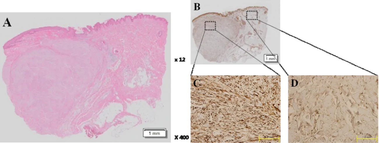
With H&E staining, we found that keloid tissue had a dense and excessive deposition of collagen, which was extended over the clinical keloid margin into the extra-lesionaldermal tissue (Figure 1A). To evaluate the expression pattern of Hsp 90protein in keloid tissue (n=4), immunohistochemistric staining wasassessed (Figure 1B). Compared to extra-lesional normal tissue (Figure 1C), markedly increased immunoactivities for Hsp 90 was noted in central and peripheral keloid region(Figure 1D).The increased expression of Hsp 90 protein was semi-quantitatively measured with MetaMorph® image analysis software and found that the optical density(OD) 20629±2235 in keloid tissue and 4303± 488.1 in normal tissue. In keloid tissue, the expression of Hsp90 increased by4.8 times than normal tissue and this difference was statistically significant (p<0.01, Figure1E).
Figure 1. Histologic analysis of keloid tissue. (A) H&E staing. Under the light microscope (×12), we found that keloid tissue had a dense and excessive deposition of collagen. (B) Immunohistochemical staining of keloids and adjacent normal dermal tissues. We could confirm that the expression of HSP90 immunoactivitiesin keloid tissue (C)was increased than that in adjacent normal tissue (D) using immunohistochemistry.(E) On the semi-quantitative analysis using Metamorph image analysis software, the optical density(OD) was examined 20629 ± 2235 in keloid tissue and 4303 ± 488.1 in normal tissue. In keloid tissue, the expression of HSP90 increased by 4.8 times than normal tissue and this difference was statistically significant (**p<0.01).
2. 17 AAG down-regulates mRNA expression of type I collagen in KFs
In the KFs, mRNA expression of type I and III collagen was examined using qRT–PCR. Quantitative analysis using qRT–PCR indicated that the mRNA levels of type I collagen in the KFs were significantly decreased with various amounts of 17AAG treatment (5, 10, 20 μM) by 70%, 53%, and 61%, respectively (Figure 2A. mean of five repeated experiments, **p<0.01) compared with non-treated KFs, showing that Hsp90 inhibitor such as 17AAG reduces the expression of type I collagen in KFs. However, mRNA expression of type III collagen was not reducedcompared with non-treated KFsand it was slightly increased,but no significance (Figure 2B. mean five repeated experiments). These results suggest that 17AAGattenuates the mRNA levels of type I collagen in KFs, which have upregulated collagen expression.
10
Figure 2. Quantitative analysis of mRNA expression of type I and III collagen. (A) qRT–PCR analysis indicated that typeI collagen mRNA levels in KFs treated withvarious amounts of 17AAG treatment (2, 5, 10 μM, 2days)was significantly decreased (**p<0.01) compared with non-treated cells. (B) However, mRNA expression of type III collagen was not reducedcompared with non-treated KFs and it was slightly increased, but no significance. Relat ive colla gen I m A exp essio RN r * * *
A
B
Relat ive colla gen III mRN Aexp
3. Suppression ofSmad 2/3 complex expression in KFs under 17AAGtreatement
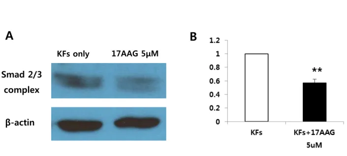
To examine the mechanism by which the 17AAG suppressed collagen mRNA expression, the expression ofSmad 2/3 complex protein was investigated using immunoblot analysis. 17AAGdecreasedthe expression of Smad 2/3 complex protein in KFs compared with non-treated KFs(Figure 3A).The degree of decrease was 37% compared with KFs cell only (**p<0.01, Figure 3B). These results revealed that 17AAG decreased collagen type I mRNA expressionby inhibiting TGFβ-mediated transcriptionalresponses thus preventingSmad2/3 activation. 11
B
β-actin
17AAG 5μMSmad 2/3
complex
KFs onlyA
**
Figure 3. Immunoblotanalysis of Smad 2/3 complex expression in KFs. (A) 17AAG (5 μM)decreasedthe expression of Smad 2/3 complex protein in KFs compared with non-treated KFs. (B) Quantification of immunoblot results expressed as mean±s.e. of four independent experiments. It showed that the suppression of Smad 2/3 complex protein expression was significant in 5µM 17AAG-treated KFs (**p<0.001).
4. Collagen deposition and intensity were decreased on the keloid spheroid which treated with 17 AAG
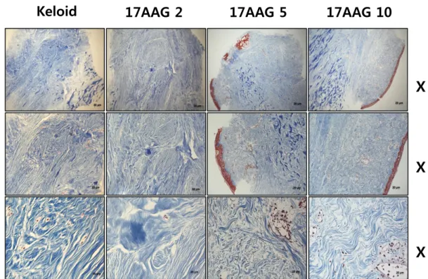
Keloid spheroidsderived from active-stage keloid patients (n=3) were cultured with treatment of various amount of 17 AAG (0, 2, 5, 10 μM). The intensity of fibrosis was measured semi-quantitativelyon the M-T stained sections. With Masson’s trichrome staining, we found that keloid tissue had more dense and excessive deposition of collagen compared with adjacent normaldermal tissue. Also, irregular bundle-shaped collagen arrangement was showed on the keloid spheroid tissue (Figure 4A). After various amounts of 17 AAG (2, 5, 10 μM) treatment, Masson-Trichromestaing showed that collagen deposition and intensity were decreased on the keloid spheroid which treated with 17AAG. Also, dense and coarse collagen bundles were replaced by thin and shallow collagen bundles on the keloid spheroid which treated by various amount of 17AAG (Figure 4B).
12
Normal tissue
X5
X10
X40
A
Keloid tissue
21
V. CONCLUSION
Heat shock protein 90 inhibitor, 17-(allylamino)-17-demethoxygeldanamycin, decreases collagen synthesis of keloid fibroblastsand attenuates the extracellular matrix accumulation, including type I and III collagen, elastin and fibronectin,in the keloid spheroid model. And Smad pathway is involved in these series of changes. These results suggest that the antifibrotic effect ofHsp90 inhibitor such as 17AAG may have therapeutic potentials on keloids.
13
17AAG 2
X
X
X
17AAG 5
17AAG 10
B
Keloid
Figure 4. Masson’s trichrome staining of the keloid spheroid. (A) Comparison between keloid tissue and adjacent normal tissue. The keloid tissue had more dense and excessive deposition of collagen compared with adjacent normal dermal tissue. Also, irregular bundle-shaped collagen arrangement was showed on the keloid spheroid tissue. (B) Effects of 17AAG treatment on the keloid spheroid. Collagen deposition and intensity were decreased on the keloid spheroid which treated with 17AAG. Also, dense and coarse collagen bundles were replaced by thin and shallow collagen bundles.
14
5. 17 AAG decreases the expression of type I, III collagen, elastin, and fibronectin in keloid spheroids.
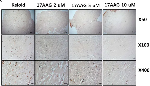
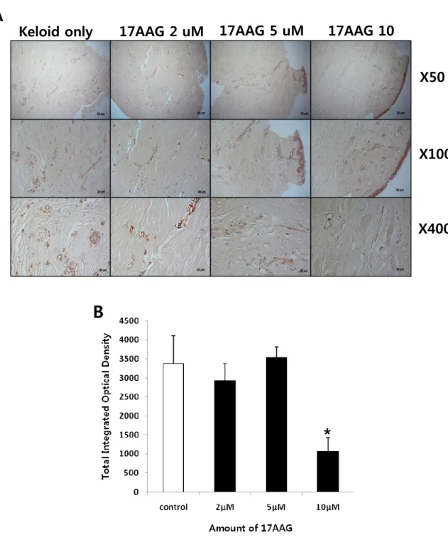
Keloid spheroidsderived from active-stage keloid patients (n=3) were cultured with treatment of various amount of 17 AAG (2, 5, 10 μM). Immunohistochemical staining revealed that the expression of type I collagen, type III collagen, elastin, and fibronectin in keloid spheroids treated with 17AAG was significantly reduced compared with no treated keloid spheroids (Figure 5 - 8)using semi-quantitative measurements and MetaMorph image analysis software.
Immunohistochemical staining revealed that the expression of type I collagen protein was reduced by 2%, 70%, and 89%, respectively with increasing amounts of 17AAG (2, 5, 10 μM)compared with non-treated keloid spheroid. Especially, there is a statistical significance on the 5 and 10 μM 17AAG-treated keloid spheroids (**p<0.01, Figure 5A and B). This result correlated with down-regulation of collagen I mRNA expressionin
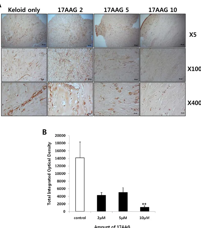
vitro. The expression of typeIII collagen protein was significantly reduced by 68%with 10 μM 17AAG
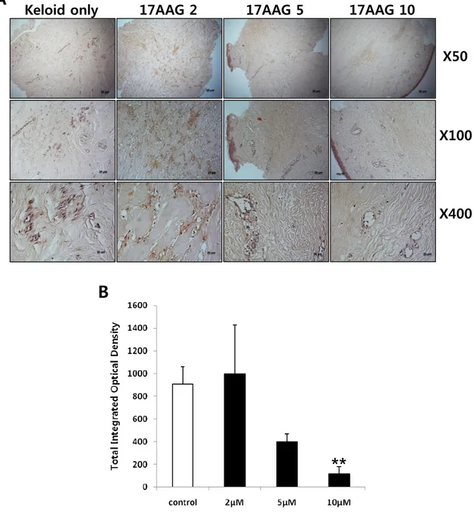
treatment compared with non-treated keloid spheroid (*p<0.05, Figure 6A and B). Similarly results were obtained the expression of elastin and fibronectin. The expressions of elastin and fibronectinwere significantly reduced by 92% and 84%, respectively, with 10 μM 17AAG treatment compared with non-treated keloid spheroid (**p<0.01, Figure 7 and 8). Together, these data strongly suggest that expression of major ECM components such as type I collagen, type III collagen, elastin, and fibronectin was decreased byHsp90 inhibitor like 17AAG.These results suggest that 17AAG has a prominent role in remodeling ECMcomponents.
A
Keloid
17AAG 2 uM
17AAG 5 uM
17AAG
X50
X100
X400
10 uM
15
Figure 5. Immunohistochemical staining of keloid spheroid sections for typeI collagen from 17AAG-treated keloid tissues. (A) Representative light micrographs ofcollagen I immunohistochemistry of spheroid tissues cultured with various amounts of 17AAG (2, 5, 10 μM) for 3days. Original magnification: 50×, 100×, and 400×.(B) Semi-quantitative analysis of panel (A) resultsusing MetaMorph imaging analysis software. The expression of type I collagen protein was reduced by 2%, 70%, and 89%, respectively with increasing amounts of 17AAG (2, 5, 10 μM) compared with non-treated keloid spheroid. Especially, there is a statistical significance on the 5 and 10 μM 17AAG-treated keloid spheroids (**p<0.01).
l
*
*
B
Keloid only
17AAG 2 uM
X50
X100
X400
17AAG 5 uM
17AAG 10
A
B
*
Figure 6. Immunohistochemical staining of keloid spheroid sections for typeIII collagen from 17AAG-treated keloid tissues. (A) Representative light micrographs ofcollagen III immunohistochemistry of spheroid tissues cultured with various amounts of 17AAG (2, 5, 10 μM) for 3days. Original magnification: 50×, 100×, and 400×.(B) Semi-quantitative analysis of panel (A) resultsusing MetaMorph imaging analysis software. The expression of type III collagen protein was reduced by 68% with 10 μM 17AAG treatment compared with non-treated keloid spheroid (*p<0.05).
A
Keloid only
17AAG 2
17AAG 5
M
17AAG 10
17X5
X100
X400
M
Figure 7. Immunohistochemical staining of keloid spheroid sections for elastin from 17AAG-treated keloid tissues. (A) Representative light micrographs ofelastin immunohistochemistry of spheroid tissues cultured with various amounts of 17AAG (2, 5, 10 μM) for 3days. Original magnification: 50×, 100×, and 400×. (B) Semi-quantitative analysis of panel (A) resultsusing MetaMorph imaging analysis software. The expression of typeelastin was reduced by 92% with 10 μM 17AAG treatment compared with non-treated keloid spheroid (*p<0.05).
B
18
X50
X100
X400
G 10
Figure 8. Immunohistochemical staining of keloid spheroid sections for fibronectin from 17AAG-treated keloid tissues. (A) Representative light micrographs offibronectin immunohistochemistry of spheroid tissues cultured with various amounts of 17AAG (2, 5, 10 μM) for 3days. Original magnification: 50×, 100×, and 400×. (B) Semi-quantitative analysis of panel (A) resultsusing MetaMorph imaging analysis software. The expression of fibronectinprotein was reduced by 84% with 10 μM 17AAG treatment compared with non-treated keloid spheroid (*p<0.05).
Keloid only
17AAG 2
17AAG 5
17AA
A
B
19
IV. DISCUSSION
In this study, we have demonstrated that overexpression of Hsp90 in human keloid tissue samplescompared to the adjacent normal tissue and its inhibitor such as 17AAG decreases collagen type I as well as Smad 2/3 complex expression in Keloid fibroblasts. Also, Masson’s trichrome staining of keloid spheroid sections revealed that dense and coarse collagen bundles were replaced by thin and shallow collagen bundles by 17 AAG treatment. Immunohistochemical analysis using keloid spheroid showed that expressions of ECM proteinssuch as collagen I, III, fibronectin, and elastin were markedly decreased in keloid spheroids treated with 17AAG.
Excessive ECM accumulation resulting from an aberrant ECM protein synthesis and degradation is one of the important causes of the hypertrophic scars and keloids. There are several hypotheses as to the cause of keloid formation, including altered growth-factor regulation, immune dysfunction, aberrant collagen turnover, sebum or sebocytes as self-antigens, altered mechanics and altered apoptotic signaling of keloid fibroblasts.3, 20 Among
these, abnormal increases in growth factors and cytokines like TGF-β plays a critical role in the pathogenesis of keloid.Generally, TGF-β is one of the well-studied growth factors and seems to play the main role in the pathophysiology of keloids.1, 6-7 Therefore, inhibiting TGF-β1-dependent signaling either by TGF-βI/TGF-βII
receptor neutralizing antibodies, truncated receptor, antisense oligonucleotides, and Smad2-/Smad3-specific siRNAs decrease procollagen gene expression and inhibit fibrosis progression.3, 7, 21-24
In this study, both type I and III collagen were reduced in keloid spheroid after treatment of 17-AAG. There is a report that the ratio of type I/III collagen of keloids is significantly elevated compared to that of normal scars.25 Interestingly, in keloid spheroid section, the amount of type I collagen is more reduced compared to that
of type III collagen. Therefore, we can guess that treatment of 17-AAG on keloid spheroid can change the ratio of type I/III collagen more similarly to the ratio of normal scar.
TGF-β is known to increase tropoelastin mRNA abundance and elastin formation.26-27 There was also a report
that elastin mRNA can be stabilized by TGF-β pathway.28 Moreover, TGF-β1 can limit elastin degradation by
decreasing levels and activity of matrix metalloproteinase (MMP)-2 and MMP-9.29 Thus, TGF-β plays a pivotal
role in formation and degradation of elastin, and inhibitory effect of 17-AAG on TGF-β signaling pathway can reduce accumulation of elastin in tissue effectively. Fibronectin is also known to be induced by TGF-β.30
Interestingly, induction of connective tissue growth factor and fibronectin enhances the profibrotic effects of TGF-β.31 In other words, these proteins enhance cellular response to TGF-β and results in prolongation of the
20 formation.
We confirmed that 17-AAG reduced ECM accumulation via Smad dependent pathways. Various intracellular signal molecules such as extracellular signal-regulated protein kinase 1/2 (ERK1/2), p38 mitogen-activated protein kinase (MAPK), Sma- and Mad-related proteins (Smads), signal transducer and activator of transcription-3 (STAT3), and phosphatidylinositol-3-kinase (PI3K)/Akt were involved in keloid. Thus, many research groups have suggested modulation of these signaling mediators to treat keloids. Many previous studied demonstrated that suppression of Smad3, PI3K, ERK, and STAT3 pathways is sufficient to inhibit extracellular matrix production in keloids.32-36 Therefore, investigation of the effect of 17-AAG on Smad-independent
pathways will be helpful to determine the mechanism of action clearly in the future.
Geldanamycin (GM), a naturally occurring benzoquinone ansamycin, inhibits Hsp90’s ATP dependent association with cochaperones and thus its activity as a molecular chaperone.10-11, 37 Such pharmacological
inhibition of Hsp90 resembles its ADP-bound conformation, which favors and results in ubiquitin-mediated degradation of clients, including ErbB2 and AKT.11
The small-molecule 17-(allylamino)-17-demethoxygeldanamycin (17AAG) is a geldanamycin analog. Thus, treatment of cells with 17AAG results in the inactivation, destabilization, and degradation of Hsp90 client proteins which play important regulatory roles in the cell cycle, cell growth, cell survival, apoptosis, and oncogenesis.
Fibrosis is one of the common problems in various diseases of organs, including liver, kidney, lung, skin and etc. And inhibitor of Hsp90 showed promising results in some fibrotic disease model.12, 15 We also demonstrated
that the inhibitor of Hsp90 can potentially applicable in the treatment of keloid scar of the skin byattenuating excessive accumulation of ECM. The effect of Hsp90 on proliferation, migration and apoptosis of keloid fibroblasts should be investigated in the future to determine definitive action of Hsp90 inhibitor on keloid scar.
22
REFERENCES
1. Burd A, Huang L. Hypertrophic response and keloid diathesis: two very different forms of scar. Plast Reconstr Surg 2005;116:150e-7e.
2. Atiyeh BS, Costagliola M, Hayek SN. Keloid or hypertrophic scar: the controversy: review of the literature. Ann Plast Surg 2005;54:676-80.
3. Al-Attar A, Mess S, Thomassen JM, Kauffman CL, Davison SP. Keloid pathogenesis and treatment. Plast Reconstr Surg 2006;117:286-300.
4. Piera-Velazquez S, Jimenez SA. Molecular mechanisms of endothelial to mesenchymal cell transition (EndoMT) in experimentally induced fibrotic diseases. Fibrogenesis Tissue Repair 2012;5 Suppl 1:S7. 5. Kis K, Liu X, Hagood JS. Myofibroblast differentiation and survival in fibrotic disease. Expert Rev
Mol Med 2011;13:e27.
6. Russell SB, Russell JD, Trupin KM, Gayden AE, Opalenik SR, Nanney LB, et al. Epigenetically altered wound healing in keloid fibroblasts. J Invest Dermatol 2010;130:2489-96.
7. Bran GM, Goessler UR, Hormann K, Riedel F, Sadick H. Keloids: current concepts of pathogenesis (review). Int J Mol Med 2009;24:283-93.
8. Joly AL, Wettstein G, Mignot G, Ghiringhelli F, Garrido C. Dual role of heat shock proteins as regulators of apoptosis and innate immunity. J Innate Immun 2010;2:238-47.
9. Vaughan CK, Neckers L, Piper PW. Understanding of the Hsp90 molecular chaperone reaches new heights. Nat Struct Mol Biol 2010;17:1400-4.
10. Neckers L. Hsp90 inhibitors as novel cancer chemotherapeutic agents. Trends Mol Med 2002;8:S55-61. 11. Citri A, Harari D, Shohat G, Ramakrishnan P, Gan J, Lavi S, et al. Hsp90 recognizes a common surface
on client kinases. J Biol Chem 2006;281:14361-9.
12. Myung SJ, Yoon JH, Kim BH, Lee JH, Jung EU, Lee HS. Heat shock protein 90 inhibitor induces apoptosis and attenuates activation of hepatic stellate cells. J Pharmacol Exp Ther 2009;330:276-82. 13. Wrighton KH, Lin X, Feng XH. Critical regulation of TGFbeta signaling by Hsp90. Proc Natl Acad Sci
U S A 2008;105:9244-9.
14. Zhao R, Davey M, Hsu YC, Kaplanek P, Tong A, Parsons AB, et al. Navigating the chaperone network: an integrative map of physical and genetic interactions mediated by the hsp90 chaperone. Cell 2005;120:715-27.
15. Noh H, Kim HJ, Yu MR, Kim WY, Kim J, Ryu JH, et al. Heat shock protein 90 inhibitor attenuates renal fibrosis through degradation of transforming growth factor-beta type II receptor. Lab Invest 2012;92:1583-96.
16. Totan S, Echo A, Yuksel E. Heat shock proteins modulate keloid formation. Eplasty 2011;11:e21. 17. Tredget EE, Shankowsky HA, Pannu R, Nedelec B, Iwashina T, Ghahary A, et al. Transforming growth
factor-beta in thermally injured patients with hypertrophic scars: effects of interferon alpha-2b. Plast Reconstr Surg 1998;102:1317-28; discussion 29-30.
18. Lee JH, Shin JU, Jung I, Lee H, Rah DK, Jung JY, et al. Proteomic profiling reveals upregulated protein expression of hsp70 in keloids. Biomed Res Int 2013;2013:621538.
23
19. Lee WJ, Choi IK, Lee JH, Kim YO, Yun CO. A novel three-dimensional model system for keloid study: organotypic multicellular scar model. Wound Repair Regen 2013;21:155-65.
20. Shih B, Garside E, McGrouther DA, Bayat A. Molecular dissection of abnormal wound healing processes resulting in keloid disease. Wound Repair Regen 2010;18:139-53.
21. Shah M, Foreman DM, Ferguson MW. Neutralisation of TGF-beta 1 and TGF-beta 2 or exogenous addition of TGF-beta 3 to cutaneous rat wounds reduces scarring. J Cell Sci 1995;108 ( Pt 3):985-1002. 22. Gao Z, Wang Z, Shi Y, Lin Z, Jiang H, Hou T, et al. Modulation of collagen synthesis in keloid
fibroblasts by silencing Smad2 with siRNA. Plast Reconstr Surg 2006;118:1328-37.
23. Bran GM, Goessler UR, Schardt C, Hormann K, Riedel F, Sadick H. Effect of the abrogation of TGF-beta1 by antisense oligonucleotides on the expression of TGF-beta-isoforms and their receptors I and II in isolated fibroblasts from keloid scars. Int J Mol Med 2010;25:915-21.
24. Wang Z, Gao Z, Shi Y, Sun Y, Lin Z, Jiang H, et al. Inhibition of Smad3 expression decreases collagen synthesis in keloid disease fibroblasts. J Plast Reconstr Aesthet Surg 2007;60:1193-9.
25. Friedman DW, Boyd CD, Mackenzie JW, Norton P, Olson RM, Deak SB. Regulation of collagen gene expression in keloids and hypertrophic scars. J Surg Res 1993;55:214-22.
26. McGowan SE, McNamer R. Transforming growth factor-beta increases elastin production by neonatal rat lung fibroblasts. Am J Respir Cell Mol Biol 1990;3:369-76.
27. Katsuta Y, Ogura Y, Iriyama S, Goetinck PF, Klement JF, Uitto J, et al. Fibulin-5 accelerates elastic fibre assembly in human skin fibroblasts. Exp Dermatol 2008;17:837-42.
28. Kucich U, Rosenbloom JC, Abrams WR, Rosenbloom J. Transforming growth factor-beta stabilizes elastin mRNA by a pathway requiring active Smads, protein kinase C-delta, and p38. Am J Respir Cell Mol Biol 2002;26:183-8.
29. Dai J, Losy F, Guinault AM, Pages C, Anegon I, Desgranges P, et al. Overexpression of transforming growth factor-beta1 stabilizes already-formed aortic aneurysms: a first approach to induction of functional healing by endovascular gene therapy. Circulation 2005;112:1008-15.
30. Hocevar BA, Brown TL, Howe PH. TGF-beta induces fibronectin synthesis through a c-Jun N-terminal kinase-dependent, Smad4-independent pathway. EMBO J 1999;18:1345-56.
31. LEASK A, ABRAHAM DJ. TGF-β signaling and the fibrotic response. The FASEB Journal 2004;18:816-27.
32. Lim CP, Phan TT, Lim IJ, Cao X. Stat3 contributes to keloid pathogenesis via promoting collagen production, cell proliferation and migration. Oncogene 2006;25:5416-25.
33. Lim IJ, Phan TT, Tan EK, Nguyen TT, Tran E, Longaker MT, et al. Synchronous activation of ERK and phosphatidylinositol 3-kinase pathways is required for collagen and extracellular matrix production in keloids. J Biol Chem 2003;278:40851-8.
34. Park G, Yoon BS, Moon JH, Kim B, Jun EK, Oh S, et al. Green tea polyphenol epigallocatechin-3-gallate suppresses collagen production and proliferation in keloid fibroblasts via inhibition of the STAT3-signaling pathway. J Invest Dermatol 2008;128:2429-41.
35. Song J, Xu H, Lu Q, Xu Z, Bian D, Xia Y, et al. Madecassoside suppresses migration of fibroblasts from keloids: involvement of p38 kinase and PI3K signaling pathways. Burns 2012;38:677-84.
24
36. Xia W, Longaker MT, Yang GP. P38 MAP kinase mediates transforming growth factor-beta2 transcription in human keloid fibroblasts. Am J Physiol Regul Integr Comp Physiol 2006;290:R501-8. 37. Neckers L, Ivy SP. Heat shock protein 90. Curr Opin Oncol 2003;15:419-24.
25
ABSTRACT(IN KOREAN)
Heat Shock Protein 90 억제제가 켈로이드 섬유아세포와 조직구에서
세포외기질 형성에 미치는 영향
<지도교수 이원재>
연세대학교
대학원
의학과
박제연
열충격단백질 90은 (heat-shock protein 90, Hsp90) 90-kDa의 분자량을 가지며 세포질에 매우 풍부 하게 존재하는 단백으로 총단백질 함량의 1%에 달한다. 이는 다양한 생리적 혹은 환경적 스트레 스에 의해 유도되는 단백으로 알려져 있다. 또한 geldanamycin 유도체인 17-allylamino-demethoxy-geldanamycin (17AAG) 로 Hsp90 를 억제하면 17AAG는 Hsp90 의 ATPase 활성을 억제하여 TGF-β 수용체 I 과 II의 분해과정을 증가시켜 transforming growth factor β (TGF-β)에 의해 유도되는 전사 과정을 억제하며 Smad 2/3 복합체의 활성화를 억제하는 것으로 알려져 있다. 켈로이드 반흔은 과 도한 교원질 침착으로 인한 피부 진피층의 병적인 분화 상태를 말한다. TGF-β는 켈로이드의 병태 생리에 있어 가장 중추적인 역할을 하는 성장인자로 이에 대한 연구가 활발히 진행되고 있다. 그 러므로 우리는 본 연구를 통해 Hsp90가 TGF-β 신호전달체계에 미치는 영향 및 켈로이드 섬유모 세포와 조직구에서 (spheroid) 세포외기질의 합성에 미치는 영향을 알아보았다. 켈로이드 조직에서 Hsp 90 단백질 발현을 보기 위해 면역조직화학 방법을 (immunohistochemistry) 사용하였다. 정상 진피섬유아세포와 (Human normal dermal fibroblast) 켈로이드 진피섬유아세포 (Keloid fibroblasts) 를 각각 다양한 농도의 (5, 10, 20 μM) 17AAG로 처리한 후 real time RT-PCR을 이 용하여 제 1, 3형 교원질의 mRNA 발현 정도를 측정하였다. Western blot 분석을 통해서는 17AAG 가 단백질 kinase B (Akt)와 Smad 2/3 복합체 (complex) 단백질 발현에 미치는 영향을 조사하였다. 또한, 켈로이드 조직구에서 (spheroid) 다양한 농도의 17AAG로 처리한 후 세포외기질의 (ECM) 발
26 현 정도를 Masson-Trichrome 염색법과 면역조직화학법을 이용하여 측정하였다. 켈로이드 조직에서 인접한 정상 조직에 비하여 Hsp90의 과다 발현이 관찰되었다. 또한 17AAG와 같은 Hsp 90 억제인자는 켈로인드 섬유모세포에 있는 제 1형 교원질 mRNA 발현과 세포내 신호전달 물질인 Smad-2/3 복합체의 단백질 발현을 감소시켰다. 켈로이드 조직을 Masson’s trichrome 염색법으로 확인해본 결과 17AAG 로 처리한 경우 켈로이드 조직의 교원질 침착 정도가 감소하고 조밀하고 두꺼운 교원질 침착이 성글고 얇게 변화된 것을 관찰하였다. 면역조직화학 방법에 의해서도 17AAG 처리를 한 켈로이드 조직에서 세포외기질을 구성하는 단백질 (collagen I, III, fibronectin, elastin)의 발현이 현저하게 감소되었다.
본 연구에서 얻은 결과를 종합해볼 때 Hsp90 억제 인자인 17AAG는 항섬유화 효과를 갖고 있어 켈로이드 치료에 도움이 될 것으로 사료된다.
--- 핵심되는 말 : 켈로이드, 열충격단백질90 (Hsp 90), 17AAG, 교원질, 켈로이드 조직구 배양
