저작자표시-비영리-변경금지 2.0 대한민국 이용자는 아래의 조건을 따르는 경우에 한하여 자유롭게 l 이 저작물을 복제, 배포, 전송, 전시, 공연 및 방송할 수 있습니다. 다음과 같은 조건을 따라야 합니다: l 귀하는, 이 저작물의 재이용이나 배포의 경우, 이 저작물에 적용된 이용허락조건 을 명확하게 나타내어야 합니다. l 저작권자로부터 별도의 허가를 받으면 이러한 조건들은 적용되지 않습니다. 저작권법에 따른 이용자의 권리는 위의 내용에 의하여 영향을 받지 않습니다. 이것은 이용허락규약(Legal Code)을 이해하기 쉽게 요약한 것입니다. Disclaimer 저작자표시. 귀하는 원저작자를 표시하여야 합니다. 비영리. 귀하는 이 저작물을 영리 목적으로 이용할 수 없습니다. 변경금지. 귀하는 이 저작물을 개작, 변형 또는 가공할 수 없습니다.
Identification of
Innate Drug Resistance against
RAF Inhibitors in Patients with
Papillary Thyroid Cancer
Jae Hyun Park
Department of Medicine
Identification of
Innate Drug Resistance against
RAF Inhibitors in Patients with
Papillary Thyroid Cancer
Directed by Professor Woong Youn Chung
The Doctoral Dissertation
submitted to the Department of Medicine
the Graduate School of Yonsei University
in partial fulfillment of the requirements for the degree
of Doctor of Philosophy
Jae Hyun Park
This certifies that the Doctoral Dissertation
of Jae Hyun Park is approved.
The Graduate School
Yonsei University
ACKNOWLEDGEMENTS
Proverb 16:9. In his heart a man plans his plans his course, but
the LORD determines his steps.
Firstly, I would like to express my sincere gratitude to my
advisor Prof. Woong Youn Chung for the continuous support
of my Ph.D study and related research, for his patience,
motivation, and immense knowledge. His guidance helped me
in all the time of research and writing of this thesis. I could not
have imagined having a better advisor and mentor for my Ph.D
study.
Besides my advisor, I would like to thank to my thesis
committee members: Prof. Eun Jig Lee and Prof. Soon Won
Hong, for their insightful comments and encouragement, but
also for the hard question which incented me to widen my
research from various perspectives.
My sincere thanks also goes to Prof. Young Suk Jo and Prof.
Jandee Lee, who provided me an opportunity to join their team,
and who gave access to the laboratory and research facilities.
Without they precious support it would not be possible to
conduct this research.
Last but not the least, I would like to thank my family: my
biggest supporters parents, my lovely wife, Kyoung Yun, and
my adorable sons, Ji Youn and Ji Won for supporting me
spiritually throughout writing this thesis and my life in
general.
<TABLE OF CONTENTS>
ABSTRACT………..1
I. INTRODUCTION...3
II. MATERIALS AND METHODS...7
1. Patients and Clinical Manifestation...7
2. Analysis of mutation in NRAS and MEK………...8
A. Exome Squencing
3. Analysis of p61BRAF
V600ESplice Variant………...8
A. DNA Squencing
B. Western Blot
C. Mass Spectrometry
4. Analysis of CRAF Overexpression and Aberrant Expression of
COT... ... 10
A. Real-Time PCR
B. Immunohistochemical Staining
5. Statistical Analysis...11
III. RESULTS...12
1. Mutations in NRAS and MEK...12
2. p61BRAF
V600Esplice variant...12
3. CRAF Overexpression and Aberrant Expression of COT...14
IV. DISCUSSION...17
V. CONCLUSION...21
REFERENCES...22
LIST OF FIGURES
Figure 1. Mechanisms of resistance to RAF inhibitors…
………6
Figure 2. Representative image of immunohistochemical
staining of COT in PTC; (A) No staining, (B) Strong
staining……….11
Figure 3. Detection of splicing variants of B-RAF by Western
Blotting (A), Coomassie Brilliant Blue Stanining (B),
and Mass Spectrometry (C)………..13
Figure 4. Relative mRNA expression in PTC and matched
normal thyroid tissues for CRAF (A), and COT (B)
in 135 patients with PTC ……….14
Figure 5. Correlation of COT expression with C-RAF
expression in PTC……….15
Figure 6. Correlation of COT expression with B-RAFV
600ELIST OF TABLES
Table 1. Exome sequencing for detection of mutation in NRAS
and MEK……….12
1
ABSTRACT
Identification of Innate Drug Resistance against RAF Inhibitors
in Patients with Papillary Thyroid Cancer
Jae Hyun Park
Department of Medicine
The Graduate School, Yonsei University
(Directed by Professor Woong Youn Chung)
Recently, several novel targeted agents have been developed for treatment of BRAFV600E-positive cancers. However, de novo and acquired resistance to these agents has since emerged as a therapeutic obstacle. Two major mechanisms, based on their dependence on RAF dimerization, drive tumor resistance to RAF inhibitors – mutations in NRAS such as NRAS Q61, the p61BRAFV600E splice variant, and C-RAF overexpression are involved in a mechanism that is dependent on RAF dimerization; in contrast, aberrant expression of Cancer Osaka Thyroid Oncogene mitogen-activated protein kinase kinase kinase 8 (COT) or mitogen-activated protein kinase kinase (MEK) mutation function
2
independently of RAF dimerization. The aim of this study was to identify the molecular basis for innate drug resistance against RAF inhibitors in patients with papillary thyroid cancer (PTC). 167 PTC patients undergoing total thyroidectomy were enrolled. Patient information and clinicopathological parameters were analyzed. BRAFV600E mutation was included in this study. Exome sequencing for detection of mutation in NRAS and MEK was performed. For the
analysis of
p61BRAFV600E splice variant, DNA sequencing, Western blot and mass spectrometry were performed. CRAF overexpression and aberrant expression of COT were analyzed by quantitative polymerase chain reaction (qPCR) and immunohistochemical staining. In the results, NRAS and MEK mutation, and the p61BRAFV600E splice variant were not detected in PTC. However, qPCR data showed that the relative expression of CRAF and COT mRNA in PTC was higher than in normal tissues ( p<0.01). Furthermore, COT mRNA expression in PTC correlated positively with CRAF expression (r=0.5954, p<0.001). Immunohistochemical analysis showed that the staining intensities of COT were higher in PTC than in normal thyroid tissues (p <0.001). Aberrant expression of COT was more frequently detected in BRAFV600E-positive PTC (p=0.013). These results suggest that COT expression may be associated with innate drug resistance against RAF inhibitors in PTC.---
Keywords: Papillary thyroid cancer, Drug resistance,Proto-oncogene,
BRAF, COT
3
Identification of Innate Drug Resistance against RAF Inhibitors
in Patients with Papillary Thyroid Cancer
Jae Hyun Park
Department of Medicine
The Graduate School, Yonsei University
(Directed by Professor Woong Youn Chung)
I. INTRODUCTION
In recent years, our understanding of the molecular genetics of thyroid cancer has expanded dramatically. Four types of mutations, BRAF and RAS point mutations and RET/PTC and PAX8/peroxisome proliferator-activated receptor-γ rearrangements, constitute the majority of mutations known to occur in the two most common types of thyroid cancer, papillary and follicular carcinoma.
At this time, these genetic changes have the most significant impact on tumor diagnosis and prognostication. Papillary thyroid carcinomas (PTC) harbor point mutations in the BRAF and RAS genes and RET/PTC rearrangements, all of
4
which promote activation of the mitogen-activated protein kinase (MAPK) pathway. These mutually exclusive mutations are found in >70% of PTC.1-4
The RAF family kinases have been shown to play important roles during many cellular and physiological processes, including development, cell cycle regulation, cell proliferation and differentiation, and cell survival and apoptosis. Overexpression or activation of the pathway components is a common indicator in proliferative diseases such as cancer and contributes to tumor initiation, progression, and metastasis.5
The RAS-RAF-MAPK pathway is one of the best characterized signal transduction pathways.6 In this conserved signaling pathway, RAF proteins such as A-, B-, and C-RAF are activated by RAS and then lead to activation of the dual-specific protein kinases mitogen-activated protein kinase kinase (MEK1/2), and subsequently, extracellular signal-regulated kinase (ERK1/2 ).6
PTC is the result of the abnormal activation of the RAS-RAF-MAPK signal pathway, induced by RET/PTC rearrangement, RAS mutations, or BRAFV600E mutation.7 Following the discovery that the BRAFV600E mutation is present in a high proportion of many human cancers,8 several novel targeted agents were developed for BRAFV600E-positive cancers.9 Because the incidence of the BRAFV600E mutation in PTC is high (40%–80%), these new agents were considered promising therapeutic modalities.10,11
Preclinical studies indicated the dependency of BRAFV600E tumors on MAPK signaling cascade, whereas the efficacy of both RAF and MEK inhibitors has
5
been demonstrated in several clinical trials.12–14 However, de novo and acquired resistance to these agents has since emerged as a new therapeutic obstacle.15,16
Mechanisms of resistance to RAF inhibitors can be divided into two categories according to the dependency on RAF dimerization (Fig. 1).17,18 In the first category, mutations in NRAS such as NRAS Q61, the p61BRAFV600E splice variant, and CRAF overexpression are involved in a mechanism that depends RAF dimerization. The p61BRAFV600E splice variants lacking the RAS-binding domain can dimerize in a RAS-independent manner and generate MEK-ERK signal propagation. NRAS mutation and increased expression of C-RAF can also increase RAF dimerization, which is insensitive to RAF inhibitors (Fig. 1).19-21 In the second category, aberrant expression of Cancer Osaka Thyroid Oncogene mitogen-activated protein kinase kinase kinase 8 (COT) or MEK mutation (MEK1) functions independently of RAF dimerization.22 Based on a functional genomic approach, COT can generate resistance to RAF inhibitors by MEK dependent mechanisms.18,22 The aim of this study was to identify the molecular basis for innate drug resistance against RAF inhibitors in patients with PTC.
6 (A) RAF dimerization-dependent mechanism
(B) RAF dimerization-independent mechanism
Figure 1. Mechanisms of resistance to RAF inhibitors. (adapted from [18]) (A) RAF dimerization-dependent mechanism
7
II. MATERIALS AND METHODS
1. Patients and Clinical Manifestation
In this study, 167 patients (34 male and 133 female) undergoing total thyroidectomy were enrolled, with or without neck node dissection followed by radioactive iodine ablation for management of classical PTC, from January 1987 to December 2002 at Severance Hospital, Seoul, South Korea. The study subjects showed no visible remnant in the first Diagnostic 131I whole body scan (WBS), following thyroid hormone withdrawal (THW) performed 6 to 12 months after remnant ablation. The sample size was calculated by Web-based Sample Size/Power Calculations (http://www.stat.ubc.ca). Patient information and clinicopathological parameters were analyzed retrospectively.
BRAFV600E mutation was found in 145/167 (86.8%) PTC patients. During this time, recurrence was diagnosed by: histopathologic diagnosis of clinically suspicious lymph node identified by neck ultrasound or physical examination (n = 23, 82.1%); newly detected lesion in 131I diagnostic WBS, 18-Fluoro-deoxyglucose positron emission tomography/computed tomography (FDG PET/CT) or chest CT (n = 5, 17.9%) performed for patients with serum thyroglobulin ≥2 mg/L with gradual increase following THW. Tissue samples were obtained from the central area of the tumor and from contralateral histologically normal tissue. On histological examination, cellularity was >90% in all primary PTCs. All protocols were approved by the institutional review
8
board of Severance Hospital and written informed consent was obtained from the participants enrolled in the study.
2. Analysis of mutation in NRAS and MEK
Exome sequencing of tissue samples for detection of oncogenic NRAS (Q61K and Q61R) and the MEK1C121S was performed in a central laboratory (Theragen Etex, Suwon, Korea). Tumor samples from 30 patients with PTC were sequenced using an Illumina HiSeq 2000, which produced paired-end, 90-base and 101-base DNA reads. Only tumor cells were collected by macrodissection after hematoxylin staining. To increase the accuracy of mutation detection in genic regions even in low-purity samples, additional exome sequencing was performed at approximately 103 times sequencing depth on average.
3. Analysis of p61BRAFV600E Splice Variant
Genomic DNA from formalin-fixed, paraffin-embedded tissue specimens in 40 patients with PTC was prepared from five 10-mm sections after microdissection. In this case, paraffin-embedded thyroid tissue specimens had >90% tumor cells. Genomic DNA was isolated using the EZ1 DNA Tissue Kit (Qiagen, Chatsworth, CA, USA). Exon 15 of the BRAF gene was amplified by PCR using standard conditions (95°C for 5 min; followed by 32 cycles of [94°C for 30 s, 58°C for 30 s, and 72°C for 30 s]; and 70°C for 10 min) using the following primers: forward 5′-ATG CTT GCT CTG ATA GGA AA-3′ and
9
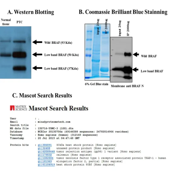
reverse 5′-ATT TTT GTG AAT ACT GGG GAA-3′. The amplified products were purified with the MinElute PCR Purification Kit (Qiagen) and were then sequenced on an ABI PRISM 3730XL automated capillary DNA Sequencer using the BigDye Terminator Cycle Sequencing Ready Reaction Kit (Applied Biosystems, Foster City, CA, USA).Western blotting analysis was performed according to standard methods with commercially available antibodies: B-RAF rabbit polyclonal antibody (sc-9002, Santa Cruz Biotechnology, Inc., Dallas, Texas, USA).
For the Mass spectrometry, Thawed cell samples were first suspended in HEPES buffer (10 mM HEPES, 15 mM MgCl2, 10 mM KCl, and 0.2% DTT) containing protease inhibitors and sonicated. Dissolved proteins were subjected to free-flow electrophoresis (FFE) using a BD™ FFE System (BD Diagnostics, Munich, Germany) with a stable pH gradient between pH 3 (anode) and pH 10 (cathode). After this first separation according to the isoelectric point, fractionated proteins were subjected to SDS-PAGE. Following electrophoresis, the gels were stained with Coomassie Brilliant Blue G-250 (BioRad, Munich, Germany), the lower BRAF bands cut out of the gel and the proteins digested. The proteins of each gel band were analyzed using a nano-LC-MS/MS. The mass spectra obtained were used to identify the corresponding peptides/proteins by the MASCOT™ algorithm. A protein was considered identified when the cumulative score was at least 100.
10
4. Analysis of CRAF Overexpression and Aberrant Expression of COT Total RNA was extracted using Trizol reagent (Invitrogen, Carlsbad, CA, USA), and complementary DNA (cDNA) was prepared from total RNA using M-MLV reverse transcriptase (Invitrogen) and oligo-dT primers (Promega, Madison, WI, USA). Quantitative RT-PCR (qRT-PCR) was performed on cDNA using the QuantiTect SYBR Green RT-PCR Kit (Qiagen, Valencia, CA, USA) with the following primers: C RAF, 5′-GGG AGC TTG GAA GAC GAT CAG-3′ and 5′-ACA CGG ATA GTG TTG CTT GTC-3′; COT, 5′-ATG GAG TAC ATG AGC ACT GGA-3′ and 5′-GCT GGC TCT TCA CTT GCA TAA AG-3′. The qRT-PCR sample measurements were carried out in triplicate, and three independent trials of each experiment were performed.
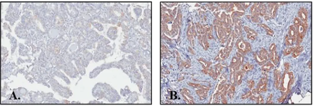
Immunohistochemical staining for COT was performed in 167 cases of PTC and matched normal tissues. Briefly, 4-mm tissue sections were heated at 60°C, deparaffinized in xylene, and hydrated in a graded series of alcohol. Antigen retrieval was performed by microwaving in citrate buffer for 10 min. Endogenous peroxidase activity was inactivated by incubating in 3% hydrogen peroxide for 10 min. Nonspecific binding sites were blocked by incubating in 10% normal goat serum diluted with phosphate-buffered saline (PBS). Tissue sections were then incubated with primary antibodies: COT rabbit polyclonal antibody (sc-720) for 60 min at room temperature. All sections were sequentially treated with biotinylated anti-rabbit immunoglobulin for 30 min, peroxidase-labeled streptavidin for 30 min, and diaminobenzidine in the
11
presence of hydrogen peroxide. Staining was scored as follows: 1, no staining; 2, weak or focal staining; 3, moderate staining in most cells; and 4, strong staining in most cells (Fig.2). To support the data obtained from Immunohistochemistry- Paraffin Embedded Tissue (IHC-P), we reviewed the representative images of IHC-P for COT from the Human Protein Atlas program (http://www.proteinatlas.org/).
Figure 2. Representative image of immunohistochemical staining of COT in PTC; (A) No staining, (B) Strong staining
5. Statistical Analysis
Statistical analysis was carried out using either SPSS version 18.0 for Windows (IBM Corporation, Armonk, NY, USA) or GraphPad Prism (GraphPad Software, Inc., San Diego, CA, USA). Relative mRNA expression was calculated using the StepOneTM Real-time PCR System (Applied Biosystems, Foster City, CA, USA). Average ratios were compared with the paired t test. The relationship between 2 groups was analyzed by Pearson correlation analysis. Group comparisons were performed by linear-by-linear association. All p values are 2-sided.
12
III. RESULTS
1. Mutations in NRAS and MEK
Exome sequencing was performed on tumor samples obtained from 30 patients with PTC with an average target depth of 120 per sample. However, oncogenic NRAS (Q61K and Q61R) and the MEK1C121S mutation were not detected in 30 of the PTC patients in this study. (Table. 1)
Gene Samples, n SNVs in splice site, n Indels, n
NRAS 30 0 0
MEK 30 0 0
SNVs: single nucleotide variations, Indels: small insertions or deletions
Table 1. Exome sequencing for detection of mutation in NRAS and MEK.
2. p61BRAFV600E splice variant
Analysis of DNA and complementary DNA derived from 40 patients with PTC showed that all retained expression of BRAFV600E. Analysis of BRAF protein expression revealed a 93 kDa band predicted by wild-type braf, and a new band at approximate molecular weights of 50 kDa and 37 kDa was identified in tumor samples from 4 patients. The lower BRAF bands were expected to be splicing variant products of the detected by immunoblotting protein samples, but p61BRAFV600E could not be identify the corresponding proteins by
the MASCOT™ algorithm
in mass spectrometry (Fig. 3).13
Figure 3. Detection of splicing variants of B-RAF; Western Blotting (A), Coomassie Brilliant Blue Stanining (B) and Mascot Search Results (C)
The lower BRAF bands indicate splicing variant products of the detected by
immunoblotting protein but, p61BRAFV600E could not be
identify the
corresponding proteins by the MASCOT™ algorithm
in mass spectrometry.14
3. CRAF Overexpression and Aberrant Expression of COT
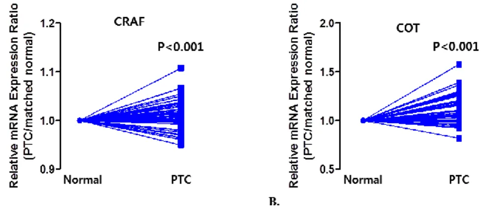
To investigate the expression of CRAF and COT in PTC, we first performed qPCR using mRNA derived from primary PTC tissues. Excluding 32 PTCs, from which we were only able to isolate mRNA of a quality too poor for further analysis, we conducted mean comparisons to compare the expression of CRAF and COT in PTCs using a paired t-test (n = 135). As shown in Figure 4, the relative mRNA expression of CRAF and COT were higher in PTCs than in normal tissues.
Figure 4. Relative mRNA expression in PTC and matched normal thyroid tissues for CRAF (A), and COT (B) in 135 patients with PTC. The relative
mRNA expression of C-RAF and COT were higher than normal tissues. Average ratios were compared with the paired t test. All p values are two sided. All sample measurements were carried out in triplicate, and three independent trials of each experiment were performed.
15
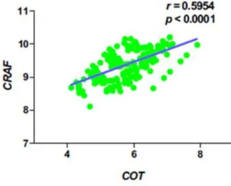
The expression of COT mRNA in PTC showed positive correlation with CRAF (r = 0.5954, p <0.001; Fig. 5)
Figure 5. Correlation of COT expression with C-RAF expression in PTC.
The expression of COT mRNA in PTC showed positive correlation with C-RAF (r = 0.5954, p <0.001). The relationship between 2 groups was analyzed by Pearson correlation analysis. r=Pearson correlation coefficient. Statistical analysis was performed using GraphPad Prism.
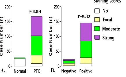
PTC tissues showed various staining intensities of COT that ranged from no staining to strong staining and COT expression group comparisons indicated that PTC had significantly higher staining of COT compared with normal thyroid tissues (p<0.001, Fig. 6A). Aberrant expression of COT was more frequently detected in BRAFV600E-positive PTC. (p=0.013, Fig. 6B).
16
Figure 6. Correlation of COT expression with B-RAFV600E mutation
(A) Comparison of COT expression in normal thyroid tissues and PTC; PTC had significantly higher staining of COT compared with normal thyroid tissues (p<0.001). (B) COT expression status according to the absence or presence of
B-RAFV600E mutation; Aberrant expression of COT was more frequently
17
IV. DISCUSSION
Following the discovery of the BRAFV600E mutation as an oncogenic kinase in various cancers including melanoma, thyroid, lung, and cholangiocarcinoma, targeted agents against the BRAFV600E kinase have taken a central role in cancer therapy. In this regard, sorafenib has activity against BRAFV600E and is licensed to treat radioactive iodine (RAI)-refractory PTC.23
In contrast to the high response rate of metastatic melanomas to BRAF inhibitors, RAF or MEK inhibitors show limited efficacy in RAI-refractory thyroid cancer and thyroid cancer cell lines harboring BRAFV600E.24-26 One of the possible explanations for the poor response to B-RAF inhibitors in thyroid cancer is related to feedback-induced ligand-dependent activation of human epidermal growth factor receptor (HER)2/HER3 signaling.25 In fact, recent biological and clinical studies have revealed multiple mechanisms of drug resistance: elevated expression of CRAF, COT1, or mutant BRAF kinases; activating mutations in NRAS, MEK1, or AKT1; aberrant splicing of BRAF (p61BRAF); activation of phosphatidylinositol-3-OH kinase by loss of phosphatase and tensin homolog; and activation of receptor tyrosine kinases, including platelet-derived growth factor receptor, beta polypeptide, insulin-like growth factor 1 receptor, and epidermal growth factor receptor. Interactions between tumors and their microenvironment also impact innate drug resistance to BRAF inhibitors.25
18
In the normal physiological setting, activated RAS signaling promotes the dimerization and activation of RAF proteins. In the presence of BRAF inhibitors and RAS signaling, the binding of drug to one molecule in a non-mutated RAF dimer can promote activation of the second RAF molecule. Thus, in the presence of RAS activation, the activity of homo - and hetero-dimeric RAF complexes can be paradoxically activated by RAF inhibitors.19,27,28 In melanomas with BRAFV600E, any alterations promoting RAF dimerization are predicted to confer resistance to RAF inhibitors. As expected, activating NRAS mutations mediate resistance to vemurafenib,15 a mutated BRAF inhibitor, and dabrafenib,29 a selective B-RAF kinase inhibitor. Although oncogenic N-RAS (Q61K and Q61R) was not detected in the PTC patients in this study, oncogenic N-RAS (Q61K and Q61R) was only detected in a melanoma patient with acquired resistance to vemurafenib. Also, the alteration frequency of the NRAS gene for thyroid carcinoma sets in the cBioPortal tool (http://www.cbioportal.org/) was only 8.4% (34 of 405 cases).
MEK1 have also been shown to confer resistance to MAPK inhibitors. The existence of MEK1 mutations in MAPK inhibitor-resistant cancers was independently verified in ex vivo cell lines established from tumor material, and its activity in conferring MEK- and BRAF inhibitor resistance validated in transfected melanoma cells. A MEK1C121S mutation was detected via mutational profiling in a melanoma sample from a patient with acquired resistance to vemurafenib.30 This mutation was not detected in the pre-treatment biopsy, and
19
was found to confer increased intrinsic kinase activity and resistance to BRAF and MEK inhibition in vitro.30 The alteration frequency of the MEK1C121S gene for thyroid carcinoma sets in cBioPortal was 0% (0 of 405 cases). Consistent with this, MEK1C121S mutation were not detected in the PTC patients in this study.
Several in-frame BRAFV600E splice variants lacking the RAS-binding domain have been detected in vemurafenib-resistant melanoma tumor specimens.18 These splice variants contain an in-frame deletion of exons 2–10. These variants lack the RAS-binding domain and can activate ERK in the presence of the inhibitor.18 In-frame deletion in the p61BRAFV600E variant leads to the constitutive dimerization of BRAF in the absence of activated RAS.31 Dimerization of p61BRAFV600E, lacking exons 4-8, was critical for mediating B-RAF inhibitor resistance.21
In this study, the lower BRAF bands were expected to be splicing variant products of the detected by immunoblotting protein samples, but p61BRAFV600E could not be identify the corresponding proteins by mass spectrometry. It was speculate that BRAF splice variants, as well as NRAF and MEK mutations, may be present, albeit at low levels in BRAF inhibitor treatment-naïve tumors, and, that tumors harboring these alterations likely underwent clonal selection during the course of treatment with a BRAF inhibitor.
20
RAF proteins such as A-, B-, and CRAF regulate the ERK signaling pathway, and individual RAF isoforms can be differentially regulated in a cell type-specific or context-dependent manner.32-34 In addition, the RAF isoforms have strikingly different phosphorylation sites.6 Recently, A-RAF has been reported to act as a scaffold to stabilize BRAF/CRAF heterodimers, whereas ARAF dimerization also promotes MAPK activation.35,36 Besides the 3 RAF isoforms, COT; (also called MAP3K8), a serine/threonine kinase, was shown to play a role in MAPK activation.22 To explain the regulatory mechanism of MAPK activation by COT, it has been suggested that COT can phosphorylate MEK1.37
In this study, qPCR was performed to estimate the expression of CRAF and COT mRNAs in PTC. Expression of CRAF and COT was increased in PTC. Consistent with the mRNA expression data, western blotting analysis indicated that the expression of CRAF and COT proteins was also increased in PTC. Immunohistochemical analysis clearly demonstrated overexpression or aberrant expression of BRAF and/or COT in PTC, compared with normal thyroid tissues. Furthermore, the expression of COT showed a strong positive correlation with C-RAF expression, suggesting that this de novo drug resistance mechanism is coordinately regulated in PTC. Taken together, we postulated that de novo drug resistance mechanisms to RAF inhibitors might be active in a significant proportion of PTC, coincident with the expression of COT.
21
lacking the RAS-binding domain was not detected in the PTC tissues in this study, further genotyping and molecular studies in a larger population of P T C p a t i e n t s , a t v a r i o u s s t a g e s o f t r e a t m e n t , a r e n e c e s s a r y . De novo drug resistance mechanisms also need to be investigated further so that unnecessary treatment can be avoided.
V. CONCLUSION
CRAF and COT expression levels are higher in PTCs than in normal thyroid tissues. Our data suggest that increased expression of COT might correlate with the innate drug resistance against RAF inhibitors in patients with PTC.
22
REFERENCES
1. Adeniran AJ, Zhu Z, Gandhi M, Steward DL, Fidler JP, Giordano TJ, et al. Correlation between genetic alterations and microscopic features, clinical manifestations, and prognostic characteristics of thyroid papillary carcinomas. Am J Surg Pathol 2006;30:216–22.
2. Kimura ET, Nikiforova MN, Zhu Z, Knauf JA, Nikiforov YE, Fagin JA. High prevalence of BRAF mutations in thyroid cancer: genetic evidence for constitutive activation of the RET/PTC-RAS-BRAF signaling pathway in papillary thyroid carcinoma. Cancer Res 2003;63:1454–7
3. Soares P, Trovisco V, Rocha AS, Lima J, Castro P, Preto A, et al. BRAF mutations and RET/PTC rearrangements are alternative events in the etiopathogenesis of PTC. Oncogene 2003;22:4578–80.
4. Frattini M, Ferrario C, Bressan P, Balestra D, De Cecco L, Mondellini P, et al. Alternative mutations of BRAF, RET and NTRK1 are associated with similar but distinct gene expression patterns in papillary thyroid cancer. Oncogene 2004;23:7436–40.
5. Leicht DT, Balan V, Kaplun A, Singh-Gupta V, Kaplun L, Dobson M, et al. Raf kinases: function, regulation and role in human cancer. Biochim Biophys Acta. 2007;1773:1196-212.
23
6. Wellbrock C, Karasarides M, Marais R. The RAF proteins take centre stage. Nat Rev Mol Cell Biol. 2004;5:875–85.
7. American Thyroid Association Guidelines Taskforce on Thyroid Nodules and Differentiated Thyroid Cancer. Cooper DS, Doherty GM, Haugen BR, Kloos RT, Lee SL, Mandel SJ, et al. Revised American Thyroid Association management guidelines for patients with thyroid nodules and differentiated thyroid cancer. Thyroid. 2009;19:1167–214.
8. Davies H, Bignell GR, Cox C, Stephens P, Edkins S, Clegg S, et al.Mutations of the BRAF gene in human cancer. Nature. 2002;417:949–54.
9. Tsai J, Lee JT, Wang W, Zhang J, Cho H, Mamo S, et al. Discovery of a selective inhibitor of oncogenic B-Raf kinase with potent antimelanoma activity Proc Natl Acad Sci U S A. 2008;105:3041–6.
10. Namba H, Nakashima M, Hayashi T, Hayashida N, Maeda S, Rogounovitch TI, et al. Clinical implication of hot spot BRAF mutation, V599E, in papillary thyroid cancers. J Clin Endocrinol Metab. 2003;88:4393–7.
11. Puxeddu E, Moretti S, Elisei R, Romei C, Pascucci R, Martinelli M, et al. BRAF(V599E) mutation is the leading genetic event in adult sporadic papillary thyroid carcinomas. J Clin Endocrinol Metab. 2004;89:2414–20.
24
12. Yang H, Higgins B, Kolinsky K, Packman K, Go Z, Iyer R, et al. RG7204 (PLX4032), a selective BRAFV600E inhibitor, displays potent antitumor activity in preclinical melanoma models. Cancer Res. 2010;70:5518–27.
13. Xing M. BRAF V600E mutation and papillary thyroid cancer. JAMA. 2013;310:535.
14. Chapman PB, Hauschild A, Robert C, Haanen JB, Ascierto P, Larkin J, et al. Improved survival with vemurafenib in melanoma with BRAF V600E mutation. N Engl J Med. 2011;364:2507–16.
15. Nazarian R, Shi H, Wang Q, Kong X, Koya RC, Lee H, et al. Melanomas acquire resistance to B-RAF(V600E) inhibition by RTK or N-RAS upregulation Nature. 2010;468:973–7.
16. Bollag G, Hirth P, Tsai J, Zhang J, Ibrahim PN, Cho H, et al. Clinical efficacy of a RAF inhibitor needs broad target blockade in BRAF-mutant melanoma. Nature. 2010;467:596–9.
17. Villanueva J, Vultur A, Herlyn M. Resistance to BRAF inhibitors: unraveling mechanisms and future treatment options. Cancer Res. 2011;71:7137–40.
18. Lito P, Rosen N, Solit DB. Tumor adaptation and resistance to RAF inhibitors. Nat Med. 2013;19:1401–9.
25
19. Hatzivassiliou G, Song K, Yen I, Brandhuber BJ, Anderson DJ, Alvarado R, et al. RAF inhibitors prime wildtype RAF to activate the MAPK pathway and enhance growth. Nature. 2010;464:431–5.
20. Montagut C, Sharma SV, Shioda T, McDermott U, Ulman M, Ulkus LE, et al. Elevated CRAF as a potential mechanism of acquired resistance to BRAF inhibition in melanoma. Cancer Res. 2008;68:4853–61.
21. Poulikakos PI, Persaud Y, Janakiraman M, Kong X, Ng C, Moriceau G, et al. RAF inhibitor resistance is mediated by dimerization of aberrantly spliced BRAF(V600E). Nature. 2011;480:387–90.
22. Johannessen CM, Boehm JS, Kim SY, Thomas SR, Wardwell L, Johnson LA, et al. COT drives resistance to RAF inhibition through MAP kinase pathway reactivation. Nature. 2010;468:968–72.
23. Brose MS, Nutting CM, Jarzab B, Elisei R, Siena S, Bastholt L, et al. Sorafenib in radioactive iodine-refractory, locally advanced or metastatic differentiated thyroid cancer: a randomised, double-blind, phase 3 trial. Lancet. 2014;384:319–28.
24. Hayes DN, Lucas AS, Tanvetyanon T, Krzyzanowska MK, Chung CH, Murphy BA, et al. Phase II efficacy and pharmacogenomic study of Selumetinib (AZD6244;ARRY142886) in iodine-131 refractory papillary thyroidcarcinoma with or without follicular elements. Clin Cancer Res. 2012;18:2056–65.
26
Sherman EJ, et al. Relief of feedback inhibition of HER3 transcription by RAF and MEK inhibitors attenuates their antitumor effects in BRAF-mutant thyroid carcinomas. Cancer Discov. 2013;3:520–33.
26. Shen CT, Qiu ZL, Luo QY. Sorafenib in the treatment of radioiodine refractory differentiated thyroid cancer: a meta-analysis. Endocr Relat Cancer. 2014;21:253–61.
27. Heidorn SJ, Milagre C, Whittaker S, Nourry A, Niculescu-Duvas I, Dhomen N, et al. Kinase-dead BRAF and oncogenic RAS cooperate to drive tumor progression through CRAF. Cell. 2010;140:209-21.
28. Poulikakos PI, Zhang C, Bollag G, Shokat KM, Rosen N. RAF inhibitors transactivate RAF dimers and ERK signalling in cells with wild-type BRAF. Nature. 2010;464:427 30.
29. Gowrishankar K, Snoyman S, Pupo GM, Becker TM, Kefford RF, Rizos H. Acquired Resistance to BRAF Inhibition Can Confer Cross-Resistance to Combined BRAF/MEK Inhibition. J Invest Dermatol. 2012;132(7):1850-9.
30. Wagle N, Emery C, Berger MF, Davis MJ, Sawyer A, Pochanard P, et al. Dissecting Therapeutic Resistance to RAF Inhibition in Melanoma by Tumor Genomic Profiling. J Clin Oncol. 2011;29:3085-96.
31. Weber CK, Slupsky JR, Kalmes HA, Rapp UR. Active Ras induces heterodimerization of cRaf and BRaf. Cancer Res. 2001;61:3595 -8.
27
32. Bogoyevitch MA, Marshall CJ, Sugden PH. Hypertrophic agonists stimulate the activities of the protein kinases c-Raf and A-Raf in cultured ventricular myocytes. J Biol Chem. 1995;270:26303–10.
33. Corbit KC, Soh JW, Yoshida K, Eves EM, Weinstein IB, Rosner MR. Different protein kinase C is forms determine growth factor specificity in neuronal cells. Mol Cell Biol. 2000;20:5392–403.
34. Kao S, Jaiswal RK, Kolch W, Landreth GE. Identification of the mechanisms regulating the differential activation of the mapk cascade by epidermal growth factor and nerve growth factor in PC12 cells. J Biol Chem. 2001;276:18169–77.
35. Rebocho AP, Marais R. ARAF acts as a scaffold to stabilize BRAF:CRAF heterodimers. Oncogene. 2013;32:3207–12.
36. Mooz J, Oberoi-Khanuja TK, Harms GS, Wang W, Jaiswal BS, Seshagiri S, et al. Dimerization of the kinase ARAF promotes MAPK pathway activation and cell migration. Sci Signal. 2014;7:ra73.
37. Salmeron A, Ahmad TB, Carlile GW, Pappin D, Narsimhan RP, Ley SC. Activation of MEK-1 and SEK-1 by Tpl-2 proto-oncoprotein, a novel MAP kinase kinase kinase. EMBO J. 1996;15:817–26.
28
ABSTRACT(IN KOREAN)
유두상 갑상선암 환자에서 RAF 억제제의
자발적 약제내성 확인
<지도교수 정웅윤>
연세대학교 대학원 의학과
박 재 현
최 근 BR AF 돌 연 변 이 양 성 암 치 료 를 위 한 여 러 표 적 약 물 이 개발되어 사용되고 있으며 전이성 악성 흑색종 환자에서 무진행 생존기간 연장효과를 확인할 수 있었다. 하지만 유두상 갑상선 암의 경우 RAF 억제제에 대한 효과는 악성 흑색종 환자와 비교해 볼 때 제한적인 것으로 보고되고 있다. 대표적인 RAF 억제제에 대한 약제내성 기전으로는 RAF 이합체화29
의존성 기전으로 NRAS 돌연변이, splice 변종 BRAF 돌연변이 (p61BRAFV600E splice variant), CRAF 과발현 등이 있고, RAF 이합체화 독립 기전으로 Cancer Osaka Thyroid Oncogene mitogen-activated protein kinase kinase kinase 8 (COT) 의 비정상적인 발현과 mitogen-activated
protein kinase kinase (MEK) 돌연변이 등이 있다. 본 연구에서는 유두상
갑상선 암의 RAF 억제제에 대한 자발적 약제내성 존재를 확인하기 위한 연구를 진행하였다.
본 연구는 유두상 갑상선 암으로 갑상선 전절제 수술을 시행 받은 167명을 대상으로 하였으며 86.8%의 환자에서 BRAF 돌연변이를
동반하였다. NRAS와 MEK에서 돌연변이 검출을 위해 Exome
Sequencing 을 시행하였으며, splice 변종 BRAF 돌연변이 분석을 위해
DNA 염기서열 분석 및 Western blot, 질량 분석법 (mass spectrometry)
을 시행하였다. CRAF 과발현과 COT의 비정상적인 발현은 역전사
중합효소 연쇄반응 (reverse transcription polymerase chain reaction,
RT-PCR) 과 면역 조직 화학 염색으로 분석 하였다.
본 연구결과 유두상 갑상선암 조직에서 NRAS 돌연변이, MEK
돌연변이, splice 변종 BRAF 돌연변이 등은 검출되지 않았다. 하지만
정상 갑상선 조직과 유두상 갑상선암 조직에서 역전사 중합효소 연쇄반응과 면역 조직 화학 검사를 이용하여 CRAF 와 COT 발현
30 정도를 비교한 결과 유두상 갑상선 암조직에서 CRAF 와 COT 발현이 증가되었다 (p <0.01). 또한 COT 발현의 발현은 CRAF의 발현과 양의 상관 관계가 있었으며 (p <0.001), 면역 조직 화학 검사결과 정상 갑상선 조직에 비해 유두상 갑상선암 조직에서 COT 염색 강도가 증가되었고 (p <0.01), BRAF 돌연변이 양성 유두상 갑상선암 조직에서 COT의 비정상적인 발현 빈도가 증가하였다 (p =0.013). 이상의 결과를 통하여 유두상 갑상선암 조직에서 COT 발현의 증가는 RAF 억제제에 대한 자발적 약제내성 기전과 관련이 있을 것으로 생각된다.
![Figure 1. Mechanisms of resistance to RAF inhibitors. (adapted from [18]) (A) RAF dimerization-dependent mechanism](https://thumb-ap.123doks.com/thumbv2/123dokinfo/5074984.72692/15.786.121.660.173.836/figure-mechanisms-resistance-inhibitors-adapted-dimerization-dependent-mechanism.webp)