IImmaaggiinngg
급성 충수돌기염 산모에서 초음파 검사와 MRI 검사의 비교 연구
연세대학교 의과대학 응급의학교실유복열∙정성필∙유제성∙구재은∙주영선
Ultrasonography Versus MRI for
Diagnosing Acute Appendicitis During
Pregnancy
Pok Yeol Ryu, M.D., Sung Phil Chung, M.D., Je Sung You, M.D., Jae Eun Ku, M.D., Young Seon Joo, M.D.
Purpose: The purpose of this study is to compare the
sen-sitivity, specificity, positive predictive value, and negative
predictive value of ultrasonography versus magnetic
reso-nance imaging (MRI) in evaluation of pregnant patients with
clinically suspicious acute appendicitis.
Methods: This study was a retrospective cohort study. A
total of 60 pregnant patients who presented to the
emer-gency department with suspected appendicitis and
under-went ultrasonography or MRI were included. The official
interpretation reports and pathologic reports were extracted
and analyzed. The sensitivity, specificity, positive predictive
value, and negative predictive value were calculated for
ultrasonography and MRI for diagnosis of acute
appendici-tis. We also calculated and compared area under the curve
(AUC) of both diagnostic tests with the receiver operating
characteristic (ROC) curve analysis.
Results: Among 60 patients, 43 (71%) underwent
ultra-sonography, 37 (61%) underwent MRI, and 20 (33%)
underwent both diagnostic tests. Twenty patients were
con-firmed as pathologically-proven acute appendicitis. The
sensitivity, specificity, positive predictive value, and
nega-tive predicnega-tive value of ultrasonography for diagnosing
acute appendicitis were 67%, 77%, 53%, and 86%, while
those of MRI were 100% for all parameters. In ROC
analy-sis, the AUC was 0.656, respectively, for ultrasonography,
and 1.000 for MRI (p value<0.0001).
Conclusion: This study suggests that MRI is more
accu-rate than ultrasonography for the diagnosis of acute
appen-dicitis in pregnant women, especially when the appendix is
invisible with ultrasonography.
Key Words: Appendicitis, Pregnancy, Magnetic resonance
imaging, Ultrasonography
Department of Emergency Medicine, Yonsei University College of Medicine, Seoul, Korea
Article Summary
What is already known in the previous study
Because of radiation exposure during computed tomogra-phy, ultrasonography is often the initial diagnostic imag-ing test for the pregnant patient suspicious of acute appen-dicitis. Magnetic resonance imaging (MRI) is more expen-sive but can observe the anatomical structure more accu-rately without radiation exposure.
What is new in the current study
This study suggests that MRI is more accurate than ultra-sonography for the diagnosis of acute appendicitis in preg-nant women, especially when the appendix is invisible with ultrasonography.
서
론
산모에서 복통의 원인을 정확히 진단하는 것은 어렵다. 산모에서 산과적 문제 이외에 수술적 치료를 필요로 하는 가장 흔한 복통의 원인은 충수돌기염이며, 약 1,700건의 임신마다 발생한다고 한다1). 산모는 임신 자체의 생리학 적, 해부학적 변화와 충수돌기염에 의한 발열, 오심, 구토, 우하복부 통증 등의 증상이나 백혈구 증가, 적혈구 침강속 책임저자: 정 성 필 서울특별시 강남구 언주로 211 연세대학교 의과대학 응급의학교실 Tel: 02) 2019-3030, Fax: 02) 2019-4820 E-mail: [email protected] 접수일: 2014년 10월 1일, 1차 교정일: 2014년 10월 2일 게재승인일: 2015년 1월 21일한국학술정보
도 증가 등의 검사 소견을 명확히 구분하기 어렵다2) . 따라 서 산모에게 발생된 충수돌기염은 일반 환자에 비해 천공 의 빈도가 더 높다고 하며 천공이 발생하면 태아사망률이 증가한다3,4) . 그렇다고 충수돌기염이 의심되는 모든 산모에 게 충수절제술을 권장하면 불필요한 수술의 시행 빈도를 증가시키며, 수술적 처치로 인한 조산과 태아사망이 증가 되므로 수술 전 진단의 정확도를 높이는 것은 매우 중요하 다5-7). 최근에 충수돌기염의 진단검사로는 전산화 단층촬영 (CT)이 많이 사용된다. 그렇지만 방사선 피폭의 위험 때문 에 산모에게 가장 흔히 시행되는 검사법은 초음파 검사이 다. 그렇지만 초음파 검사의 경우 검사자의 경험과, 태아의 크기 및 체위, 양수의 양, 임신 주수 등이 초음파 검사의 진 단능력에 영향을 미칠 수 있다8) . 임신 주수가 길수록 자궁 의 크기가 커져 충수돌기의 해부학적 위치가 변동되며 임 신 중에는 확장된 자궁 부속기 혈관이 충수돌기와의 감별 을 어렵게 한다9,10) . 자기 공명 영상(MRI)은 초음파 검사보다 더 고가의 검 사이나, 방사선에 의한 위험이 없고 해부학적 구조를 초음 파보다 정확하게 관찰할 수 있다는 장점이 있다. 하지만 충 수돌기염이 의심되어 응급실로 내원한 산모를 대상으로 초 음파와 MRI를 비교한 연구는 외국에서는 몇몇 연구가 있 으나 국내에서는 이루어지지 않았다11-13) . 저자들은 응급실 에 내원한 산모들을 대상으로 충수돌기염이 의심되어 시행 된 초음파 검사와 MRI 검사의 민감도, 특이도, 양성 예측 도, 음성 예측도를 비교해보고자 본 연구를 시행하였다.
대상과 방법
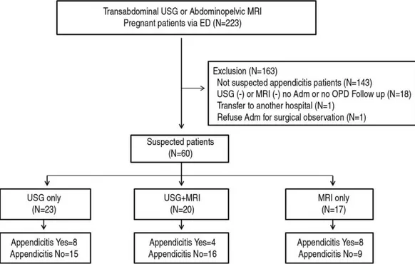
2007년 1월 1일 부터 2012년 12월 30일까지 서울 소 재 2곳의 지역응급의료센터에 방문한 산모 가운데 급성 충 수돌기염이 의심되어 복부 초음파 또는 MRI를 시행한 환 자를 대상으로 후향적 연구를 진행하였다. 복부 초음파, 복 부 및 골반 비조영 MRI 검사를 시행한 환자 223명의 의무 기록을 검토하였다. 이중 내원 시 추정진단이 요로결석, 급 성 담낭염, 복강 내 농양, 난소 염전 등과 같이 충수돌기염 이 아니었던 환자 143명을 제외하였으며, 18명은 초음파 또는 MRI 검사에서 급성 충수돌기염 음성이지만 입원 또 는 외래에서 경과 관찰을 하지 않아 최종진단을 알 수 없어 제외하였으며, 다른 병원으로 전원된 환자 1명과 자의 퇴 원 한 1명을 포함한 총 163명이 제외되었다(Fig. 1). 급성 충수돌기염이 의심되는 환자들은 복강경 충수절제 술 또는 진단적 개복술을 받았으며 대상 환자의 나이, 재태 연령, 복부 초음파 및 복부 MRI 검사 결과, 수술 여부, 병 리 진단검사 결과, 외래 경과 관찰 또는 입원 경과 관찰 결 과를 추출하였다. 충수돌기염의 확진은 수술 후 병리 검사 결과로 하였으며 충수돌기염 음성은 퇴원 후 외래 경과관 찰 후 이상이 없는 경우와 외과적 관찰을 위해서 입원 후 수술 받지 않고 퇴원한 경우로 정의하였다. 복부 초음파는 영상의학과 3년차 이상의 전공의가 12~15 MHz linear probe, 5~8 MHz curved probe를 이용하여 시행하였으며, 초음파 영상은 영상의학과 전문의Fig. 1. Study flow diagram showing patient enroll process.
의 재확인을 받았다. 사용한 초음파 기기는 Table 1과 같 다. 초음파 검사 시행 후 판독은 충수돌기 직경 및 압통 여 부, 충수돌기 주변 액체 집적 여부, 충수돌기 주변 농양 형 성 유무, 지방 침윤 여부, 충수돌기 통기 여부, 충수돌기 벽 비후 및 천공 여부를 고려하여 최종 판독에서 충수돌기염, 충수돌기염 의증라는 언급이 있는 경우 양성, 정상 충수돌 기, 관찰되지 않는 충수돌기(invisible appendix), 충수돌 기염의 근거가 없는 경우(no evidence of appendicitis) 음성으로 간주하였다.
MRI 촬영은 누운 자세로 시행하였으며, 조영제는 사용 하지 않았고 T2 coronal, sagittal, axial, single-shot fast spin-echo sequence, T1 thrive (3D) 촬영을 시행 하였다. 사용한 MRI 기기는 Table 1과 같다. 모든 MRI 판
독은 영상의학과 당직 전문의가 판독하였다. MRI 판독결 과 appendicitis, R/O appendicitis 의 경우 양성으로, normal appendix, no evidence of appedicitis의 경우 음 성으로 간주하였다.
Microsoft Excel� (Microsoft corp., Washington,
USA)을 이용하여 민감도, 특이도, 양성예측도, 음성예측도
를 계산하였다. SPSS�
version 22 (IBM Inc., Chicago, IL, USA)를 이용하여 receiver operating characteristic (ROC) curve와 area under curve (AUC)를 계산하였다.
Medcalc�
version 14.8.1 (Medcalc software, Ostend, Belgium)을 이용하여 두 검사의 AUC 값을 비교하였다.
Table 1. Equipments and software version of ultrasonography and MRI using this study.
Equipment Software version Philips 453561673701 5.2.2.44
GE R3.1.2
Philips 453561712581 6.3.5.70 Siemens 400.1.016
Magnetic resonance imaging
Philips 3.0 Tesla MR systems Achieva release 3.2.1.1 Philips 3.0 Tesla MR systems Achieva release 2.6.3.9 Philips 1.5 Tesla MR systems Achieva release 2.6.3.9 Philips 3.0 Tesla MR systems Achieva release 3.2.1.0 GE discovery 750 w DV 24
GE discovery 75 DV 24 MRI: magnetic resonance imaging
Table 2. Summary of patient characteristics.
Characteristics Value
Number 60
Mean age 31 y (19~40)
Mean gestational age 17 wk (4~35 wk) 1st trimester 21 (35%) 2nd trimester 32 (53%) 3rd trimester 07 (12%) Location of pain Right lower 44 (73%) Lower abdomen 14 (24%) Right upper 02 (04%) Diagnostic procedure Ultrasonography 43 (71%) MRI 37 (61%)
MRI: magnetic resonance imaging
Fig. 2. Evaluation diagram according to the results of imaging studies and pathology. USG: ultrasonography, MRI: magnetic resonance imaging
결
과
응급실에 복통으로 내원하여 급성 충수돌기염이 의심되 는 60명의 산모가 연구에 포함되었다. 평균 나이는 31세 (19~40세)였고, 재태 연령은 임신 1분기 21명(35%), 임 신 2분기 32명(53%), 임신 3분기 7명(12%)이었다. 재태 연령에 따른 분포는 Fig. 2와 같다. 주 호소는 우하복부 통 증을 호소하는 환자가 73%로 가장 많았고, 하복부 통증 24%, 우상복부 통증 4% 순으로 나타났다. 대상 환자 중 초음파 검사만 시행한 환자는 23명, MRI 검사만 시행한 환자는 17명, 둘 다 시행한 환자는 20명이었으며 충수돌기 염으로 진단된 환자는 20명이었다(Table 2).1. 초음파 시행 환자
초음파 검사를 시행한 43명의 환자 중에서 8명의 환자 는 정상 충수돌기로 판독되었다. 충수돌기염이 의심되어 수술한 환자는 14명이었으며, 14명중 10명은 초음파만 시 행 후 수술하였으며, 4명은 초음파 검사 결과가 invisible appendix로 MRI 촬영 후 수술하였다(Fig. 2). 초음파 검 사만 시행 후 수술한 환자 10명 중 8명이 충수돌기염으로 확진 되었으며, 1명은 난소 낭종, 1명은 충수돌기 자궁내 막증(appendix endometriosis)으로 확인되었다. 초음파 검사 후 13명의 환자는 음성으로, 외래에서 경과 관찰하거 나 입원 후 수술 받지 않고 퇴원하였다. 20명의 환자는 초 음파 검사 후 MRI 촬영을 추가로 시행하였다. 초음파 검사 의 민감도는 67%, 특이도는 77%, 양성예측도는 53%, 음 성예측도는 86%였다(Table 3).2. MRI 시행 환자
MRI 촬영한 환자는 37명이며 12명이 충수돌기염 진단 하에 복강경하 충수절제술을 시행 받았으며 12명 모두 병 리소견 상 충수돌기염으로 확진되었다. MRI에서 음성으로 판정된 25명의 환자는 산부인과 또는 외과로 입원하여 관 찰 후 수술하지 않고 퇴원하거나, 외래를 통해 경과 관찰하 였으나 증상이 호전되어 수술하지 않았다. MRI의 경우 민 감도, 특이도, 양성예측도, 음성예측도 모두 100%였다.3. 초음파와 MRI 모두 시행한 환자
초음파와 MRI 검사를 모두 시행한 환자는 20명인데 그 중에 4명이 수술을 받았으며 모두 충수돌기염으로 확진 되 었다. 초음파 검사 소견은 15명에서 정상 충수돌기 또는 관 찰되지 않는 충수돌기(invisible appendix)였는데, 이 중 4 명은 MRI로 충수돌기염을 진단하여 수술하였고, 5명은 초 음파 검사상 충수돌기염이 의심되었으나 다시 MRI를 촬영 하여 정상 충수돌기가 확인되어 수술하지 않았다. 20명을 대상으로 계산한 초음파 검사의 민감도는 0%, 특이도는 68%, 양성예측도는 0%, 음성예측도는 73%이었다(Table 3). 초음파와 MRI 검사를 모두 시행한 환자 대상으로 ROCTable 3. Accuracy of transabdominl USG and abdominopelvic MRI.
TP FP TN FN Total Sensitivity Sepcificity PPV NPV
No. No. No. No. No. % % % %
Single study USG 08 7 24 4 43 067 077 053 086 MRI 12 0 25 0 37 100 100 100 100 USG+MRI USG 00 5 11 4 20 000 069 000 073 MRI 04 0 16 0 20 100 100 100 100
USG: ultrasonography, TP: true positive, FP: false positive, TN: true negative, FN: false negative PPV: positive predictive value, NPV: negative predictive value
Fig. 3. Receiver operating characteristic curves for predic-tion of acute appendicitis with ultrasonography and MRI. The area under the curve was 0.656 and 1.0, respectively.
USG: ultrasonography, MRI: magnetic resonance imaging
곡선을 분석하였다(Fig. 4). AUC는 0.656 (CI=0.394-919)으로 측정되었다. MRI의 민감도, 특이도, 양성예측도, 음성예측도 모두 100%였고 AUC는 1.0으로 두 검사의 AUC 값은 의미 있는 차이가 있었다(p<0.0001).
고
찰
CT와 초음파는 일반적으로 급성 충수돌기염이 의심되는 환자에서 일반적으로 시행되는 검사 방법이다14) . 하지만 임 신한 환자에게 방사선 노출은 태아의 유년기 암 발생률을 증 가시킨다는 연구도 있어 초음파 검사가 가장 먼저 시행되는 검사이다15). 초음파 검사는 MRI에 비해 1/4 정도로 저렴하고 태아와 산모에게 모두 안전한 검사 방법이나 초음파 검사로 충수돌기염을 배제하기 위해서는 정상 충수돌기를 관찰해야 한다. Barloon 등16)의 연구에 임신 1, 2분기 산모에서는 비임 신 여성에서와 같이 초음파 검사의 진단 정확도는 91%, Yilmaz 등17)의 연구에서는 임신 2~3분기의 산모에서는 초음 파 검사의 진단 정확도는 50% 정도라는 보고가 있었다. 본 연구에서 초음파의 민감도는 67%, 특이도는 77%, 양 성예측도는 53%, 음성예측도는 86%였다. Lim 등10) 의 연 구(민감도: 100% 특이도: 96% 양성예측도: 94%, 음성예 측도: 100%)보다 민감도, 특이도, 양성예측도, 음성예측도 모두 낮았으며, Israel 등13) 의 연구(민감도: 50%, 특이도: 100%, 양성예측도: 100%, 음성예측도: 66%)보다는 민감 도는 높게 측정되었지만, 특이도는 낮게 측정되었다. Gary 등13) 의 연구에서도 초음파 검사에서는 정상 충수돌기였으 나 MRI에서 충수돌기염으로 진단되어 수술 후 충수돌기염 으로 확진 된 예가 있다. 본 연구에서도 정상 충수돌기로 초 음파 검사 상 확인한 환자 중에 1명은 이학적 검사 상 우하 복부 압통 및 반발 압통이 있으며, 백혈구 증가 17,000 (90%)로 MRI 촬영하지 않고 복강경적 충수돌기 절제술을 시행하여 급성 충수돌기염으로 진단된 경우와 초음파 시행 후 충수돌기염 의증으로 진단적 복강경 수술 시행 후 우측 난소 낭종을 진단 받은 사례가 있었다. 따라서 초음파 검사 도 중요하나 이학적 검사 및 임상 양상도 중요하다. 본 연구에서는 43명 중 35명(81%)의 환자에서 초음파 에서 정상 충수돌기를 확인하지 못하여 충수돌기염을 확실 하게 배제할 수 없었다. 기존 연구에서도 초음파로는 충수 돌기염을 정확히 진단하기 어려웠다고 보고하였는데, 89~96%에서 정확한 진단이 어려웠다고 보고하였다11-13) . 본 연구에서 초음파 검사는 주로 영상의학과 3년차 이상의 전공의가 시행하였지만 전문의가 시행한 기존 연구보다 정 상 충수돌기를 더 많이 확인하였다. 산모의 재태 연령에 따 라 차이를 보일 수 있으나 본 연구에서 임신 1분기는 35% (21/60) 이며, 이는 Israel 등13)의 연구(12/33)와 비슷한 수준이며, Lim 등10)의 연구(28/45)보다는 낮은 수치였다. 이는 초음파 검사의 경우 검사자의 경험과, 태아의 크기, 체위, 자궁 부속기의 비대, 양수의 양의 차이가 초음파 검 사의 특이도, 민감도에 영향을 줄 수가 있다8). 본 연구에서 는 초음파 검사에서 정상 충수 돌기가 관찰되지 않아 MRI 를 시행하여 충수돌기염으로 진단된 4례가 있다. MRI는 탁월한 해상력으로 태아에게 방사선의 영향이 없 이 횡단면 평가가 가능한 검사이다12). MRI는 음성예측도 가 매우 높은 검사 방법으로 수술이나 중재적 시술이 필요 한 난소염전, 담낭염, 복강 내 농양 등의 다른 원인을 배제 하기 위한 좋은 검사 방법이다9). MRI 검사는 초음파 비용 의 4배 정도의 보험 적용이 되지 않는 고가의 검사이며, 촬 영을 하려면 예약된 MRI 스케줄을 변경이 필요하며, 폐쇄 공포증 환자의 경우 촬영이 어려운 경우가 있으며, 현재까 지 밝혀진 MRI의 치명적인 부작용은 보고되지 않았으나, 동물 연구에서는 MRI 촬영 후 머리 엉덩 길이 감소, 안구 기형을 유발한다고 보고되었다18,19). 본 연구에서 MRI의 경우 94% (35/37)에서 충수돌기를 확인할 수 있었으며 나머지의 경우는 invisible appendix 소견을 보였다. 이는 Pedrosa 등11)의 연구 83% (39/47), Israel 등13)의 연구 87% (20/23), Cobben 등12)의 연구 52% (17/33) 보다 높은 비율이었다. 이는 이전 연구에서 사용된 것은 자석 밀도가 1.5 tesla의 MRI였으나 본 연구 에서는 3.0 tesla의 MRI를 주로 사용하여 해상도 차이가 있어 MRI 판독 결과에 영향을 주었을 것이라 예상한다. 본 연구에서는 37명의 산모가 MRI 검사를 받았으며 MRI의 민감도, 특이도, 양성 예측도, 음성 예측도 모두 100% 였다. 기존 연구들에서 급성 충수돌기염이 의심되는 산모에게 시행된 MRI의 진단성능을 비교해보았다(Table 4). Cobben 등12)의 연구, Israel 등13)의 연구에서는 본 연Table 4. Comparing current study with previous studies evaluating MRI for the diagnosis of appendicitis during pregnancy. Study reference Patients Sensitivity (%) Specifity (%) PPV (%) NPV (%)
Oto et al19) 118 090 98.1 81.8 99.1
Cobben et al14) 012 100 100 100 100
Israel et al15) 033 100 100 100 100
Lan et al18) 019 050 100 100 94.4
Current study 037 100 100 100 100
구와 마찬가지로 100% 민감도를 보였다. Oto 등20) 의 연구 에서는 118명의 산모를 대상으로 연구를 진행하였으며 민 감도 90%, 특이도 98.1% 양성예측도 81.8%, 음성예측도 99.1%로 이 연구에서 위양성은 MRI 상에서는 충수돌기염 이 의심되었으나 내과적 치료로 호전된 경우가 2례 있었으 며 위음성은 MRI 상에서는 Invisible appendix이나 수술
후 급성 충수돌기염으로 확진된 1례가 있었다. Lan 등18) 의 연구에서는 19명의 산모를 대상으로 연구하였으며 민감도 50%, 특이도 100%, 양성예측도 100%, 음성예측도는 94.4%로 본 연구보다 낮은 수치를 보였다. 본 연구에는 몇 가지 제한점이 있다. 첫째, 후향적인 연구 이므로 선택편견이 개입되었을 가능성이 높다. 둘째, 초음파 시행자가 대부분 영상의학과 전공의로 시행자의 경험에 따 라 진단 결과의 차이가 있을 수 있다. 셋째, MRI 판독의사가 초음파 결과를 알고 있는 상태에서 판독을 하여 눈가림이 적 용되지 못하였다. 넷째, 연구에 포함된 산모의 수가 적고, 두 가지 검사를 동시에 시행한 경우가 33% (20/60)에 불과하 여 초음파와 MRI 검사를 비교하는데 제한이 있었다.
결
론
이번 연구에서 충수돌기염이 의심되는 산모에게서 MRI 는 매우 정확한 검사 방법임을 확인하였다. 임상 양상 및 이학적 검사 및 산모 재태 연령을 감안하여 초음파 검사 없 이 MRI 촬영을 먼저 고려할 수 있으며, 충수돌기염이 의심 되는 산모의 경우 초음파 검사에서 진단이 명확하지 않다 면 복부 MRI 촬영이 고려되어야 한다.참고문헌
01. Mourad J, Elliott JP, Erickson L, Lisboa L. Appendicitis in pregnancy: new information that contradicts long-held clinical beliefs. Am J Obstet Gynecol. 2000;182:1027-9. 02. Unal A, Sayharman SE, Ozel L, Unal E, Aka N, Titiz I, et
al. Acute abdomen in pregnancy requiring surgical man-agement: a 20-case series. Eur J Obstet Gynecol Reprod Biol. 2011;159:87-90.
03. Tamir IL, Bongard FS, Klein SR. Acute appendicitis in the pregnant patient. Am J Surg. 1990;160:571-5.
04. Babaknia A, Parsa H, Woodruff JD. Appendicitis during pregnancy. Gastroenterol Clin North Am. 1998;27:73-88. 05. Andersen B, Nielsen TF. Appendicitis in pregnancy:
Diagnosis, management and complications. Acta Obstet Gynecol Scand. 1999;78:758-62.
06. Maslovitz S, Gutman G, Lessing JB. The significance of clinical signs and blood indices for the diagnosis of
appen-dicitis during pregnancy. Gynecol Obstet Incest. 2003;56: 188-91.
07. McGory ML, Zingmond DS, Tillous A, Hiatt JR, Ko CY, Cryer HM. Negative appendectomy in pregnant women is associated with a substantial risk of fetal loss. J Am Coll Surg. 2007;205:534-40.
08. Bulas D, Egloff A. Benefits and risks of MRI in pregnan-cy. Semin Perinatol. 2013;37:301-4.
09. Oto A, Srinivasan PN, Ernst RD. Revisiting MRI for appen-dix location during pregnancy. AJR Am J Roentgenol. 2006;186:883-7.
10. Lim HK, Bae SH, Seo GS. Diagnosis of acute appendicitis in pregnant women: value of sonography. AJR Am J Roentgenol. 1992;159:539-42.
11. Pedrosa I, Levine D, Eyvazzadeh AD, Siewert B, Ngo L, Rofsky NM. MR imaging evaluation of acute appendicitis in pregnancy. Radiology. 2006;238:891-9.
12. Cobben LP, Groot I, Haans L, Blickman JG, Puylaert J. MRI for clinically suspected appendicitis during pregnan-cy. AJR Am J Roentgenol. 2004;183:671-5.
13. Israel GM , Malguria N, McCarthy S, Copel J, Weinreb J. MRI vs. Ultrasound for Suspected Appendicitis during Pregnancy. J Magn Reson Imaging. 2008;28:428-33. 14. Ames Castro M, Shipp TD, Castro EE, Ouzounian J, Rao
P. The use of helical computed tomography in pregnancy for the diagnosis of acute appendicitis. Am J Obstet Gynecol. 2001;184:954-7.
15. Hurwitz LM, Yoshizumi T, Reiman RE. Radiation dose to the fetus from body MDCT during early gestation. AJR Am J Roentgenol. 2006;186:871-6.
16. Barloon TJ, Brown BP, Abu-Yousef MM, Warnock N, Berbaum KS. Sonography of acute appendicitis in preg-nancy. Abdom Imaging. 1995;20:149-51.
17. Yilmaz HG, Akgun Y, Bac B, Celik Y. Acute appendicitis in pregnancy--risk factors associated with principal out-comes: a case control study. Int J Surg. 2007;5:192-7. 18. Heinrichs WL, Fong P, Flannery M, Heinrichs SC, Crooks
LE, Spindle A, et al. Midgestational exposure of pregnant mice to magnetic resonance imaging. Magn Reson imag-ing. 1988;6:305-13.
19. Tyndall DA, Sulik KK. Effects of magnetic resonance imaging on eye development in the C57BL/6 J mouse. Teratology. 1991;43:263-75.
20. Oto A, Ernst RD, Ghulmiyyah LM, Nishino TK, Hughes D, Chaljub G, et al. MR imaging in the triage of pregnant patients with acute abdominal and pelvic pain. Abdom Imaging. 2009;34:243-50.
21. Vu L, Ambrose D, Vos P, Tiwari P, Rosengarten M, Wiseman S. Evaluation of MRI for the diagnosis of appen-dicitis during pregnancy when ultrasound is inconclusive. J Surg Res. 2009;156:145-9.