Acta Derm Venereol 90 299
Letters to the Editor
© 2010 The Authors. doi: 10.2340/00015555-0830
Journal Compilation © 2010 Acta Dermato-Venereologica. ISSN 0001-5555
We report here a case of a patient with anti-α-enolase
antibodies-positive Behçet’s disease (BD), which was
reactivated as a pathergy reaction after carbon dioxide
laser therapy.
CASE REPORT
A 60-year-old woman presented with multiple encrusted, ery thematous ulcerations on her face. Her medical history was significant for BD, including recurrent oral ulceration, eryt-hema nodosum- and Sweet’s syndrome-like skin lesions, and a positive pathergy test (1). Pathergy testing was performed by the intra cutaneous injection of 0.1 ml isotonic salt solution using a 20-gauge needle on the patient’s volar forearm, with pustule formation observed within 48 h at the injection site. Screening for genital ulceration and ocular involvement, such as iritis and uveitis, was performed; however, active disease involvement was not observed. The patient had been treated with colchicine and had been maintained in an inactive disease state for over a year.
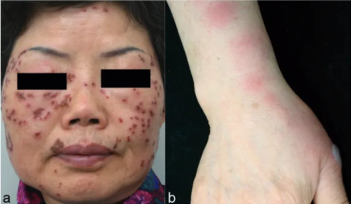
One week prior to presentation, the patient underwent carbon dioxide laser treatment for multiple seborrhoeic keratoses and lentigines at an outside clinic. However, 3 days after the procedu-re all of the tprocedu-reated sites became inflamed and ulcerated (Fig. 1a). In addition, multiple oral ulcerations, as well as several erythema nodosum-like erythematous tender nodules and a blister on the forearm and dorsum of hand were evident (Fig. 1b). The patient was afebrile, and laboratory blood tests revealed leukocytosis of 10,030/μl with neutrophilia (80.8%), elevated ery throcyte sedimentation rate of 40 mm/h, and positive C-reactive protein of 9.480 mg/dl (normal range 0–8 mg/l). Bacterial cultures of the ulcerated wounds were negative. Skin lesions were biopsied, and histology revealed septal infiltration of lymphohistiocytes and neutrophils with vasculitis, consistent with the findings of erythema nodosum (Fig. 2a, b).
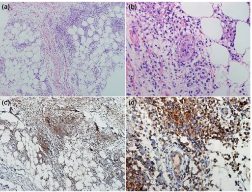
After obtaining informed consent, we collected serum samples from the patient, which were stored at –70˚C. The anti-α-enolase antibody test was performed according to the method described in a previous report (2). In an enzyme-linked immunosorbent assay (ELISA) for the anti-α-enolase antibody, the mean optical density (OD) (± 1 SD) for serum samples from 23 healthy controls was 0.139 ± 0.0682 (2). An OD exceeding this value by ≥ 3 SD was defined as positive reactivity. The patient’s sample was positive, with an OD of 0.6485. Immunohistochemical analysis of a biopsy specimen was also performed using a rabbit anti-human α-enolase antibody (Acris Antibodies, Acris Antibodies GmbH, Herford, Ger-many) in accordance with a previous report (3). The cytoplasm from lymphohistiocytes, neutrophils and endothelial cells were stained brown, whereas cell nuclei were counterstained blue (Fig. 2c, d).
The patient was treated with systemic low-dose prednisolone and colchicine. One month following this treatment, skin le-sions on both the face and forearm demonstrated improvement, leaving residual post-inflammatory hyperpigmentation and prominent depressed scars, without any evidence of recurrence over a 3-month follow-up period.
DISCUSSION
BD is a chronic, relapsing, multi-system vasculitis.
The diagnosis relies on the presence of recurrent oral
ulceration plus any two of the following: recurrent
genital ulceration, characteristic ocular lesions, typical
cutaneous lesions, or a positive pathergy test.
While the pathergy reaction, defined as an induction
of an inflammatory process following skin trauma, is not
a pathognomonic sign, it is a typical feature of BD (4).
Aside from its association with BD, the pathergy
reac-tion can also occur in patients with several condireac-tions,
including pyoderma gangrenosum, rheumatoid arthritis,
Crohn’s disease, genital herpes and Sweet’s syndrome
(1, 5, 6). A hypersensitivity reaction following trauma
has also been reported in patients with BD, including
long-lasting severe erythema and induration at the
surgically-incised site, as well as acute exacerbations
of arthritis following synovial biopsies (7, 8). In the
present case, we conclude that the extensive carbon
dioxide laser treatment induced a pathergy reaction,
which consequently resulted in the activation of BD
symptoms. According to a recent report, argon laser
photocoagulation can induce skin hyper-reactivity in
patients with BD (9).
Melikoglu et al. (10) reported that following a needle
prick, patients with BD displayed increased influxes
of cellular components, such as mature dendritic cells,
monocytes, and lymphocytes, and increased
expres-sion of cytokines, including interferon (INF)-γ,
inter-leukin (IL)-12 p40, and IL-15, chemokines, such as
macrophage inflammatory protein-3α, IFN-inducible
Reactivation of Anti-Human Alpha-Enolase Antibody-Positive Behçet’s Disease by Carbon
Dioxide Laser Treatment
Sung Bin Cho*, Jung U Shin*, Keun-Jae Ahn, Mi Ryung Roh, Dongsik Bang, Kwang Hoon Lee and Ju Hee Lee
Department of Dermatology and Cutaneous Biology Research Institute, Yonsei University College of Medicine, 250 Seongsanno, Seodaemoon-gu, Seoul, 120-752, Korea. E-mail: juhee@yuhs.ac
*The first two authors contributed equally to this work and should be considered as first authors.
Accepted November 30, 2009.
Fig. 1. (a) Multiple erythematous and encrusted ulcerations on the face
following carbon dioxide laser therapy, and (b) several tender erythematous subcutaneous nodules on the forearm and a bulla on the base of the thumb.
300 Letters to the Editor
protein-10, monokine induced by INF-γ, and interferon
inducible T-cell chemoattractant (ITAC), and adhesion
molecules, including intercellular adhesion molecule-1
and vascular cellular adhesion molecule-1 compared
with normal controls. This immune response ultimately
leads to an exaggerated lymphoit-helper (Th)1-type
response and clinically induces papules and pustules
on an erythematous base.
The glycolytic enzyme α-enolase is typically located
in the cytoplasm, but can be expressed in the cell
mem-branes of eukaryotic cells, including monocytes, T cells,
B cells, neuronal cells, and endothelial cells, following
an inflammatory stimulus through unknown mechanisms
(2, 11, 12). Testing for antibodies to α-enolase has been
proposed as a diagnostic tool or a biological marker for
various conditions, including BD, Kawasaki’s disease,
rheumatoid arthritis, and severe asthma (2, 11, 12). In
the present case, we identified serum reactivity against
human α-enolase as well as expressions of α-enolase
in the cytoplasm of lymphohistiocytes, neutrophils, and
endothelial cells from the biopsy specimen,
demonstra-ting the histological findings of erythema nodosum.
According to a previous report (2), serum anti-α-enolase
antibodies in BD can be associated with vascular system
involvement. However, the association between serum
anti-α-enolase antibodies and the risk of developing
pathergy reaction has not been proven.
The authors declare no conflict of interest.
REFERENCES
Criteria for diagnosis of Behçet’s disease. International 1.
Study Group for Behçet’s Disease. Lancet 1990; 335:
1078–1080.
Lee JH, Cho SB, Bang D, Oh SH, Ahn KJ, Kim J, et al. 2.
Human anti-α-enolase antibody in sera from patients with Behçet’s disease and rheumatologic disorders. Clin Exp Rheumatol 2009; 27: S63–66.
Kinloch A, Tatzer V, Wait R, Peston D, Lundberg K, 3.
Donatien P, et al. Identification of citrullinated alpha-enolase as a candidate autoantigen in rheumatoid arthritis. Arthritis Res Ther 2005; 7: 1421–1429.
Ozarmagan G, Saylan T, Azizlerli G, Ovul C, Aksungur VL. 4.
Re-evaluation of the pathergy test in Behçet’s disease. Acta Derm Venereol 1991; 71: 75–76.
Bi XL, Gu J, Yan M, Gao CF. A case of Sweet’s syndrome 5.
with slack skin and pathergy phenomenon. Int J Dermatol 2008; 47: 842–844.
Awan F, Hamadani M, Devine S. Paraneoplastic Sweet’s 6.
syndrome and the pathergy phenomenon. Ann Hematol 2007; 86: 613–614.
Bozkurt M, Torin G, Aksakal B, Ataoglu O. Behçet’s di-7.
sease and surgical intervention. Int J Dermatol 1992; 31: 571–573.
Giacomello A, Taccari E, Zoppini A. Marked synovial sen-8.
sitivity to pricking in Behçet’s syndrome. Arthritis Rheum 1980; 23: 259–260.
Sayarlioglu M, Calka O, Cinal A, Sayarlioglu H, Ak-9.
deniz N, Topcu N, et al. Comparison of argon laser photocoagulation-induced cutaneous inflammation and skin pathergy test in Behçet’s disease. Clin Rheumatol 2010; 29: 59–63.
Melikoglu M, Uysal S, Krueger JG, Kaplan G, Gogus 10.
F, Yazici H, Oliver S. Characterization of the divergent wound-healing responses occurring in the pathergy reac-tion and normal healthy volunteers. J Immunol 2006; 177: 6415–6421.
Lee KH, Chung HS, Kim HS, Oh SH, Ha MK, Baik JH, et 11.
al. Human alpha-enolase from endothelial cells as a target antigen of anti-endothelial cell antibody in Behçet’s disease. Arthritis Rheum 2003; 48: 2025–2035.
Pancholi V. Multifactorial α-enolase: its role in diseases. 12.
Cell Mol Life Sci 2001; 58: 902–920.
Fig. 2. (a–b) Biopsy specimen obtained from
the skin lesions on the forearm demonstrating septal infiltration of lymphohistiocytes and neutrophils with vasculitis consistent with the findings of erythema nodosum (haematoxylin and eosin stain, original magnification, a; ×100, b; ×400). (c–d) Immunohistochemistry of a biopsy section with rabbit anti-human α-enolase antibody showing expression of α-enolase in the cytoplasm of lymphohistiocytes, neutrophils, and endothelial cells (original magnification, c; ×100, d; ×400).