i
Effects of Biphasic Calcium Phosphate
Bone Substitute on Circumferential Bone Defects
around Dental Implants in Dogs
Sungtae Kim
The Graduate School
Yonsei University
Department of Dental Science
ii
Effects of Biphasic Calcium Phosphate
Bone Substitute on Circumferential Bone Defects
around Dental Implants in Dogs
A Dissertation
Submitted to the Department of Dental Science,
the Graduate School of Yonsei University
in partial fulfillment of the
requirements for the degree of
Doctor of Philosophy of Dental Science
Sungtae Kim
Feb 2011
iii
감사의 글
이 논문을 위해 처음부터 끝까지 열정적으로 지도해 주시고 격려해 주신 최성호 교수님께 진심으로 깊은 감사를 드립니다. 그리고 귀중한 시간을 내어주시어 작은 부분까지 세심하게 논문을 다듬어 주시고 심사해 주신 조규성 교수님, 김창성 교수님, 문홍석 교수님, 심준성 교수님께도 감사 드립니다. 또한 이 연구를 진행하는 과정에서 많은 도움을 주신 정의원 교수, 차재국 선생님, 손주연 선생님께도 감사를 드립니다. 또한 언제나 힘이 되어주는 가족들, 특히 여러모로 헌신적인 도움을 준 최연재, 힘들 때 마다 큰 기쁨이 되어 준 두 딸 김지원, 김예원과 이 기쁨을 함께 나누고자 합니다. 2011 년 2월 저자 씀iv
This Certifies that the dissertation theis of Sungtae Kim is
approved.
Thesis supervisor : Seong-Ho Choi
Kyoo-Sung Cho
Chang-Sung Kim
Hong-Seok Moon
June-Sung Shim
The Graduate School Yonsei University
v
Table of Contents
Abstract ... Vi
I. Introduction ... 1
II. Materials and Methods... 5
1. Animals... 5
2. Biomaterials... 5
3. Experimental design... 5
4. Surgical protocol ... 6
5. Clinical and Histological analysis...7
6. Histometric analysis... 7 7. Statistical methods... 8 III. Results... 9 1. Clinical findings... 9 2. Histological findings... 9 3. HIstometric analysis... 11 IV. Discussion... 12 V. Conclusion... 17 VI. References... 18
VII. Figure Legends... 22
Figures... 24
Tables... 29
vi Abstract
Effects of Biphasic Calcium Phosphate (BCP) Bone Substitute on
Circumferential Bone Defects around Dental Implants in Dogs
Sungtae Kim, DDS, MS, MS
Department of Dentistry
The Graduate School, Yonsei University
(Directed by Professor Seong Ho Choi, DDS., PhD)
The biphasic calcium phosphate (BCP) used in this study is a hydroxyapatite (HA) bone substitute coated with β-tricalcium phosphate (β-TCP) that has been used as an alternative to other types of bone substitutes or autogenous bone with favorable clinical outcomes. The aim of this study was to evaluate the effect of biphasic calcium phosphates(BCP) on the healing of circumferential bone defects surrounding dental implants. Defects were created in four mongrel dogs. Groups were divided into three; experimental, control groups A and B. In the experimental group, implants were placed in the edentulous area. Gap defects with 2-mm were surgically created around each implant and the defects were filled with BCP bone substitute. In control group A, gap defects were not made around the implants. In control group B, gap defects were prepared but were not filled with BCP bone substitute. Defects were histologically and histometrically evaluated after 8 and 16 weeks after implant placement. Histometric measurements in the groups after 8 weeks showed the smallest
vii
remaining defect depths (RDD) in control group A. The RDD in the experimental group was greater than that in control group A, and control groupB had the greatest RDD. There was no difference in bone to implant contact (BIC) among groups. Histometric measurements at 16 weeks showed that the RDD in control group B was greater than in both control group A and the experimental group. Within the limitations of this study, BCP bone substitute contributed to defect resolution and maintained space for new bone ingrowth without BIC improvement.
---Keywords: biphasic calcium phosphates, circumferential bone defect, implant, remaining defect depth, bone to implant contact
א
Effects of Biphasic Calcium Phosphate Bone Substitute on
Circumferential Bone Defects around Dental Implants in Dogs
Sungtae Kim, DDS, MS, MS
Department of Dentistry
The Graduate School, Yonsei University
(Directed by Professor Seong Ho Choi, D.D.S., M.S.D., Ph.D.)
I. INTRODUCTION
Roughened or coated dental implant surfaces result in faster osseointegration, better long-term predictability (Oates et al., 2007; Roccuzzo et al., 2009), and greater long-term success rates for dental implants(Albrektsson et al., 2004). With these advancements, the conventional surgical and loading protocol, which requires long periods of time, could be switched to an immediate placement and loading protocol. In the immediate placement protocol, the dental implant is placed in the extraction socket as soon the tooth is extracted. This protocol is known as a predictable treatment modality (de Sanctis et al., 2009; Gatti et al., 2000). Since the immediate implant placement protocol has became popular, the gap defects around the coronal area of the dental implant have been encountered (Akimoto et al., 1999). This results when the diameter of the extracted tooth is greater than that of dental implant, or
ב
when the shape of the extraction socket is not as round as that of the dental implant. Resolution of the gap defect depends on the surface of the implant and the size of the bone defect (Botticelli et al., 2005; Chae et al., 2008). Osseointegration of implants placed in sites with gap defects is affected by the surface characteristics of the implant (Botticelli et al., 2005). For example, implant surfaces coated with calcium phosphate showed more favorable bone responses (Chae et al., 2008). In studies investigating bone defect healing according to the dimension of the bone defect (Akimoto et al., 1999; Yoon et al., 2008), healing was more greatly compromised in deeper defects. Healing in defects 1.5 mm wide and 5 mm deep was poorer than in defects 1.5 mm wide and 2.5 mm deep (Yoon et al., 2008). There was less bone to implant contact (BIC) in wider defects in an animal study investigating gap defects that were 0.5 mm, 1.0 mm, and 1.4 mm. The poorest BIC was found in 1.4-mm gap defects (Akimoto et al., 1999).
Circumferential defects can be resolved without regenerative therapy as long as the buccal bone is intact and the defect is small (Botticelli et al., 2003; Jung et al., 2007). Gaps 1–1.25 mm wide and 5 mm deep around roughened-surface implants were favorably resolved with minor marginal bone resorption compared with turned-surface implants (Botticelli et al., 2003). However, when buccal bone was removed or the defect size was greater than 1.25 mm, additional regenerative therapy with barrier membranes or bone substitutes is required. The various results of each therapy have been reported previously. Using a membrane with a bone substitute leads to the most favorable results (Jung et al., 2007). However, using a bone substitute alone could be an alternative treatment modality as long as the space for regeneration is properly maintained (Carmagnola et al., 2000).
ג
Bone substitutes can be classified as allograft-based, factor-based, cell-based, ceramic-based, and polymer-based bone substitutes (Laurencin et al., 2006). Although autogenous bone is considered to be the gold standard in bone grafting (Marx et al., 1984), the use of autogenous bone also has potential disadvantages such as restricted donor sites (Schallhorn RG et al., 1972). Alloplastic calcium phosphates (CP) in the form of hydroxy apatite (HA) has a equal Ca/P ratio (1.67) to the inorganic phase of bone. Generally, HA-based bone substitute are known to be non-resorbable. On the other hand, Tricalcium phosphate (TCP) has a closer Ca/P ratio(1.5) to that of the amorphous biological precurssor to bone. TCP is considered to be substituted by newly formed bone at a high rate in the defect condition with high osteogenic potential. However, TCP is considered to have too high rate of degradation to preserve proper space for regeneration at the augmented site. Biphasic calcium phosphate (BCP) was developed by combining HA and TCP to modulate the degradation rate and the bioactivity. According to the ratio of HA/TCP, the degradation rate and the bioactivity change. In the study by LeGeros (2003) and Jensen (2007), pure TCP formed more new bone than HA-containing materials, whereas the rate of resorption has been known to exceed that of particulated autograft. In contrast, pure HA and BCP exhibited similar resorption patterns at a very limited rate. The BCP formed more new bone than the pure HA at the early healing phases, which was attributed to the TCP content. The biphasic calcium phosphate (BCP) used in this study is a hydroxyapatite (HA) bone substitute coated with β-tricalcium phosphate (β-TCP) that has been used as an alternative to other types of bone substitutes or autogenous bone with favorable clinical outcomes. The ratio of HA/TCP was 7:3. This ratio is considered to be ideal to be applied for perio defect or
ד
the defect around dental implant or ridge augmentation or sinus elevation when considering bone regeneration and degradation rate(Kim et al., 2008).
The purpose of this study was to evaluate the effect of the BCP bone substitute on the healing of circumferential bone defects around dental implants histologically.
ה
Ⅱ. MATERIALS AND METHODS
1. Animals
Four male mongrel dogs, 18–24 months old and weighing about 30 kg each were chosen. The dogs had intact dentition without any inflammation in periodontium. Management, preparation, and surgical protocols for animals followed the routine procedures approved by the Animal Care and Use Committee, Yonsei Medical Center, Seoul, Korea.
2. Biomaterials
Implants
Grade 2 titanium implants with surfaces modified by thermal etching and corundum particle blasting (Xive, Friadent GmbH, Mannheim, Germany) were used in this study. Implants were 3.4 mm in diameter and 9.4 mm in length.
Bone substitute
The BCP bone substitute (OSTEON ® , Dentium, Seoul, Korea) used in this study was HA scaffold (70%) coated with ß-TCP (30%). The pore size corresponded to 300–500 μm.
3. Experimental design
ו
substitute. Healing was observed after 8 weeks and 16 weeks. Control group A was without circumferential bone defects; control group B had a circumferential bone defect only; and the experimental group had both a circumferential bone defect and bone substitute.
4. Surgical protocol
Premolars and molars were extracted under general anesthesia and sterile conditions. Atropine 0.05 mg/kg was injected subcutaneously. Xylazine (Rompun, Bayer Korea, Seoul, Korea) 2 mg/kg and ketamine hydrochloride (Ketalar, Yuhan Co., Seoul, Korea) 10 mg/kg were administered intravenously. The dogs were laid on a heating pad during surgery. An endotracheal tube was used for intubation and enflurane 2% was administered. The dogs were monitored with an electrocardiogram. In total, 24 titanium implants with surfaces treated by grit-blasting and thermal etching (Xive, Friadent) were placed on edentulous mandibles after a 2-month healing period. The animals were anesthetized as described above. A crestal incision was made and a full mucoperiosteal flap was raised. Three implant sites on each side of the mandible were identified, and surgical preparation was performed according to the manufacturer’s instructions using pilot and twist drills. Circumferential defects with 2.0-mm gaps and 5.0-mm depths (Fig 1, Fig 2) were surgically created with a customized step-drill (Fig 3) around each implant in control group B and the experimental group (Fig 1). No circumferential defect was created around implants in control group A. At test sites in the experimental group, bone substitute was placed in the defect.
ז
were placed without any bone grafting material. Postoperative care was similar to that for tooth extraction. The sutures were removed after 7 to 10 days, and the dogs were fed a soft diet for 2 weeks. The dogs were sacrificed with an anesthesia overdose 8 or 16 weeks after surgery.
5. Clinical and histologic analysis
The block sections, including the segments with implants, were preserved. They were fixed in 10% neutral buffered formalin, dehydrated in ethanol, embedded in methacrylate, and sectioned in the mesio-distal plane. The central section from each specimen was reduced to 35μm thickness by microgrinding (Exakt, Apparatebau, Norderstedt, Germany). The sections were stained with hematoxylin and Eosin. Histologic evaluation was performed under an optical microscope (Leica DM-LB, Leica, Wetzlar, Germany).
6. Histometric analysis
Computer-assisted histometric measurements were performed with an automated image analysis system (Image-Pro Plus, Media Cybernetics, Silver Spring, MD). Bone-to-implant contact (BIC) within the most coronal 5-mm area of the implants was measured. The remaining defect depth (RDD) was measured. RDD is the distance from the shoulder of the implant to the most coronal point of osseointegration (Fig 4).
ח
7. Statistical methods
One way analysis of variance (ANOVA) was used to determine whether mean values of BIC and RDD differed significantly (α=.01) among groups at each time point. The independent t test was used to determine whether the mean values of BIC and RDD differed significantly (α=.01) between the 8-week and 16-week time points in each group.
ט
Ⅲ. RESULTS
1. Clinical findings
During the postoperative period, healing was uneventful and implants were well maintained. No signs of inflammation were observed in the mucosa adjacent to the implants.
2. Histological findings
8-week time point
Control group A: The bone tissue surrounding implants at 8 weeks in control group A
is shown in Fig. 5. Wide, dense layers of cortical bone were found around the shoulder of the implant. A high percentage of direct contact between bone and the implant surface was found around the implant.
Control group B: Less than half of the defect depth was filled with newly formed
bone, which was mature bone with nearby marrow spaces originating from the defect base (Fig.6). Newly formed trabeculae and direct apposition of bone to the implant surface were also observed adjacent to the lateral bone walls. Apical migration of epithelium and loose connective tissue in the remaining defects were found in the coronal half of the original defect.
Experimental group: Direct bone contact with the implant with a thin rim of
י
Apical migration of epithelium and loose connective tissue in the remaining defects were less than that found in control group B at 8-weeks (Fig. 7A). Appositional bone formation was found around particles of bone substitute beside the implant surface (Fig. 7B). Direct bone contact with the implant surface was observed around particles of bone substitute.
16-week time point
Control group A: The histological findings were similar to those observed at 8-weeks
except that there was slightly more marginal bone loss (Fig 8).
Control group B: Newly formed bone failed to reach more than half of the defect.
The remaining defect depth seemed similar to that observed at 8-weeks. Connective tissue separating the new bone from the implant was observed (Fig. 9).
Experimental group: Bone was in direct contact with the implant except in the
coronal quarter of the defect. Bone substitute still remained and maintained space to prevent the apical migration of soft tissue. The degradation of the bone substitute could be estimated, progressing from the rounded margin of the particles encapsulated by newly-formed bone (Fig. 10A). However, it was not completely degraded after the 16-week healing time. Particles of bone substitute around potential new bone were found (Fig. 10B). Connective tissue separated the new bone from the
אי
implant in the coronal part of the implant. More direct contact between new bone and the implant surface was observed near the bone substitute than was observed after 8-weeks in the control groups (Fig 10B).
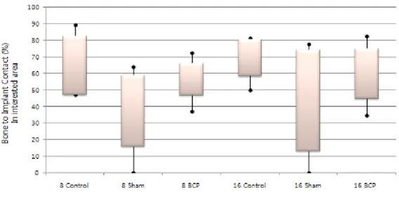
3. Histometric analysis (Table 1).
There were no statistically significant differences between healing after 8 weeks and 16 weeks. At 8 weeks, the difference in RDD among groups was statistically significant. The RDD was 0.89 ± 0.72 mm in control group A, 3.11 ± 1.02 mm in control group B, and 1.99 ± 0.66 mm in the experimental group. The difference in the BIC between control group A and control group B was statistically significant. The BIC was 64.81 ± 17.74 % in control A group and 37.11 ± 21.42% in control B group. At 16-weeks, the RDD in control group B (2.86 ± 1.53 mm) was greater than that in control group A (0.64 ± 0.45 mm) and the experimental group (1.59 ± 0.87 mm) and the difference was statistically significant. No difference in BIC was found among groups.
בי
Ⅳ. DISCUSSION
Various dimensions of the gap defect model in dogs were used in previous studies. Defects 1.25 mm or 1.0 mm in width and 5.0 mm in depth were used by Botticelli (2005, 2003); defects 3 mm wide and 5.0 mm deep were used by Stentz (1997); defects 0.5 mm, 1.0 mm, or 1.4 mm wide and 6.0 mm deep were used by Akimoto (1999); defects 2 mm wide and 5.0 mm deep were used by Veis (2004); defects 1 mm, 1.5 mm, and 2.0 mm wide were used by Jung (2007); and defects 2.5 mm or 5.0 mm deep and 1.5 mm wide were used by Yoon (2008). According to Botticelli’s studies (2005, 2003), defects more than 1.25 mm wide and 5 mm deep are poorly resolved without additional regenerative therapy. Therefore, the gap defect needed to evaluate the effect of a bone substitute should be more than 1.25 mm wide and 5 mm deep. In addition, considering the width of the alveolar ridge in the mandible of mongrel dogs, and the minimum bone height for the initial stability of a dental implant, defects 2.0 mm wide and 5.0 mm deep were used in the present study as an effective dimension to evaluate the effect of a bone substitute on the resolution of circumferential bone defects around dental implants.
The BCP bone substitute used in the present study is a degradable polymer-based bone substitute, which allows ingrowth and remodeling of new bone. The HA in this bone substitute is a synthetic apatite and a synthetic calcium phosphate (CaP) ceramic. The β-tricalcium phosphate is also a synthetic CaP ceramic. The present study demonstrated that BCP bone substitute placed in circumferential bone defects around dental implants was incorporated in the newly-formed bone tissue. Relatively high degrees of contact were established between the particles of bone substitute and the
גי
lamella and woven bone.
BCP bone substitute was selected because it has features similar to those of natural bone. The interconnected pore structure and porosity are 300–500μm, 77% of which are similar to those in human cancellous bone (Kim et al., 2008). The morphological characteristics and structure of the bone substitute play an essential role in tissue ingrowth (Habibovic et al., 2005). Cell migration and tissue ingrowth occur in the pores of the bone substitute. The pores help improve the integration of the bone substitute into the surrounding tissue, and are also related with the subsequent degradation of the bone substitute (Fabbri et al., 1995; Hulbert et al., 1972). In the current literature, a minimum pore size of 200 to 400 μm was determined to be sufficient for vascularization and osteoconduction (Dong et al., 2002; Karageorgiou et al., 2005). Therefore, the bone substitute used in the present study seems to match the pore size-related requirements for bone regeneration.
Various healing times for the gap defect model in dogs were used in previous studies. The healing time in Botticelli (2005) and Stentz (1997)’s studies was 16 weeks, and was 12 weeks in Akimoto’s study (1999). A healing time of 20 weeks was used by Veis (2004). In the present study, the healing time was 16 weeks. There was no significant difference between the 8-week and 16-week healing times in each control group. The majority of healing in the control group occurred within the first 8 weeks. This healing is faster than that required for defect resolution around the machine-surface implant. Therefore, the implant surface modified by thermal etching and corundum particle blasting seems to enhance healing and shorten the healing time required to fill the defect. This surface-modified implant, as demonstrated in this study, can be used for immediate placement and in an early loading protocol. It
די
shortens the total treatment time and minimizes patients’ discomfort. However, in the experimental group, the 16-week healing time was not long enough to observe particle degradation, irrespective of the defect fill. Most of the remaining particles of bone substitute were still present after 16 weeks. The degradation rate is known to vary according to the solubility of BCP. The solubility of BCP depends on the ratio of HA/β-TCP. In previous studies, greater proportions of HA were observed to lower the resorption rate (Yamada et al., 1997; Jensen et al., 2009). In an osteoclast culture model used by Yamada(1997), less soluble ceramics (HA/ β-TCP: 75/25) were not resorbed well by osteoclasts, whereas faster and more extensive resorption by osteoclasts was found with more soluble ceramics (HA/ β-TCP: 25/75). Lacunae similar to what are ordinarily formed by osteoclasts on natural mineralized organic tissues were observed. In an animal model, Jensen(2009) showed that BCP (HA/ β-TCP: 20/80) had a higher resorption rate, higher percentage of graft surface coverage throughout the study period, and more bone formation that was similar to autografts during the initial healing phase than BCP (HA/ β-TCP: 80/20 or 60/40). During the total 52-week healing time, the total resorption of BCP(HA/ β-TCP: 80/20 or 60/40), which has a slow resorption rate, was not complete. However, qualitative differences were observed among BCPs with different HA/ β-TCP ratios after 4 weeks.
The degradation of bone substitute was not complete in the present study. The rounded margins of particles incorporated into newly-formed bone showed that resorption was taking place. A healing time of more than 52 weeks would be required for total degradation of the bone substitute. However, the 8- and 16-week healing times were sufficient to observe qualitative and quantitative differences between the
וט
groups used in the present study. At 8 and 16 weeks in the experimental group, the RDD was less than that of control group B and the difference was statistically significant for the corresponding time. Newly-formed bone incorporated in the BCP particles were found in areas relatively close to the base and lateral walls of the defect in the 8-week experimental group. Newly-formed bone incorporated with BCP particles was found not only in relatively close areas, but also in areas relatively distant from the base and lateral wall of the defect at 16-weeks in the experimental group. This finding demonstrates that defect resolution was in progress between weeks 8 and 16. Apical migration of epithelium and loose connective tissue was less advanced in the experimental group than in control group B. This finding suggests that the space required for bone ingrowth was properly maintained by the bone substitute. New bone incorporated with bone substitute particles was found on the implant surface (Fig. 5B). Davies (1998) proposed that there are two different osteogenesis phenomena around dental implants: distance and contact osteogenesis. Osteogenesis is the process of laying down new bone material by osteoblasts. Distance osteogenesis occurs on the surface of bone around the implant through appositional growth. Contact osteogenesis is new bone formation that occurs directly on the surface of the implant. A rough implant surface advanced contact osteogenesis compared with a smooth implant surface by increasing the surface area for fibrin attachment and by providing surface features with which fibrin could be tangled. In the experimental group at 16-weeks, both distance osteogenesis due to the BCP and contact osteogenesis assisted by the roughened implant surface was distinct when compared with control group B. Most of the healing of the defect occurred within 8 weeks in all three groups, explaining the lack of a statistically significant difference
זט
between the 8-week and 16-week healing times in each group.
The majority of healing in the control group occurred within 8 weeks. Further outstanding improvement was not observed in the 16-week. The
healing in the present study was faster than the healing observed in the smooth-surfaced implant by Botticelli(2005). The roughened implant surface may have enhanced the healing time.
Healing retardation because of bone substitute was reported by Araujo (2009) in a study investigating extraction sockets filled with bone substitutes (Araújo et al., 2009). Future studies will be needed to determine how bone substitutes may retard tissue healing around dental implants, since this was not ascertained in the present study. Healing times of more than 52 weeks may be needed, and greater sample sizes will be required because of the relatively high standard deviation caused by biological variation among animals.
זי
Ⅴ. Conclusion
Within the limitations of this study, BCP bone substitute contributed to defect resolution and maintained space for new bone ingrowth before 8 weeks. No more contribution to defect resolution by bone substitute was found after 8weeks.
חי
ⅤI. References
Akimoto K, Becker W, Persson R, Baker DA, Rohrer MD, O'Neal RB: Evaluation of titanium implants placed into simulated extraction sockets: a study in dogs. Int J Oral
Maxillofac Implants 14: 351-360, 1999.
Albrektsson T, Wennerberg A: Oral implant surfaces: Part 2--review focusing on clinical knowledge of different surfaces. Int J Prosthodont 17: 544-564, 2004.
Araújo M, Linder E, Lindhe: Effect of a xenograft on early bone formation in extraction sockets: an experimental study in dog. J Clin Oral Implants Res 20: 1-6, 2009.
Botticelli D, Berglundh T, Persson LG, Lindhe J: Bone regeneration at implants with turned or rough surfaces in self-contained defects. An experimental study in the dog. J
Clin Periodontol 32: 448-455, 2005.
Botticelli D, Berglundh T, Buser D, Lindhe J: The jumping distance revisited: An experimental study in the dog. Clin Oral Implants Res 14: 35-42, 2003.
Carmagnola D, Berglundh T, Araújo M, Albrektsson T, Lindhe J: Bone healing around implants placed in a jaw defect augmented with Bio-Oss. An experimental study in dogs. J
Clin Periodontol 27: 799-805, 2000.
Chae GJ, Jung UW, Jung SM, Lee IS, Cho KS, Kim CK, Choi SH: Healing of surgically created circumferential gap around nano-coating surface dental implants in dogs. Surf
Interface Anal 40: 184-187, 2008.
טי
extraction sockets: bone healing in four different implant systems. J Clin Periodontol 36: 705-711, 2009.
Davies JE: Mechanisms of endosseous integration. Int J Prosthodont 11: 391-401, 1998. Dong J, Uemura T, Shirasaki Y, Tateishi T: Promotion of bone formation using highly pure porous beta-TCP combined with bone marrow-derived osteoprogenitor cells.
Biomaterials 23: 4493-4502, 2002.
Fabbri M, Celotti GC, Ravaglioli A: Hydroxyapatite-based porous aggregates: physico-chemical nature, structure, texture and architecture. Biomaterials 16: 225-228, 1995.
Gatti C, Haefliger W, Chiapasco M: Implant-retained mandibular overdentures with immediate loading: a prospective study of ITI implants. Int J Oral Maxillofac Implants 15: 383-388, 2000.
Habibovic P, Yuan H, van der Valk CM, Meijer G, van Blitterswijk CA, de Groot K: 3D microenvironment as essential element for osteoinduction by biomaterials. Biomaterials 26: 3565-3575, 2005.
Hulbert SF, Morrison SJ, Klawitter JJ: Tissue reaction to three ceramics of porous and non-porous structures. J Biomed Mater Res 6: 347-374, 1972.
Jensen SS, Yeo A, Dard M, Hunziker E, Schenk R, Buser D. Evaluation of a novel biphasic calcium phosphate in standardized bone defects. A histologic and histomorphometric study in the mandibles of minipigs. Clin Oral Implants Res 2007;18:752-760.
Jensen SS, Bornstein MM, Dard M, Bosshardt DD, Buser D: Comparative study of biphasic calcium phosphates with different HA/TCP ratios in mandibular bone defects. A long-term histomorphometric study in minipigs. J Biomed Mater Res B Appl Biomater 90:
כ
171-181, 2009.
Jung UW, Kim CS, Choi SH, Cho KS, Inoue T, Kim CK: Healing of surgically created circumferential gap around non-submerged-type implants in dogs: a histomorphometric study. Clin Oral Implants Res 18: 171-178, 2007.
Karageorgiou V, Kaplan D: Porosity of 3D biomaterial scaffolds and osteogenesis.
Biomaterials 26: 5474-5491, 2005.
Kim YK, Yun PY, Lim SC, Kim SG, Lee HJ, Ong JL: Clinical evaluations of OSTEON as a new alloplastic material in sinus bone grafting and its effect on bone healing. J Biomed
Mater Res B Appl Biomater 86: 270-277, 2008.
Laurencin C, Khan Y, El-Amin SF: Bone graft substitutes. Review. Expert Rev Med
Devices 3: 49-57, 2006.
LeGeros RZ, Lin S, Rohanizadeh R, Mijares D, LeGeros JP. Biphasic calcium phosphate bioceramic: Preparation, properties and applications. J Mater Sci Mater Med 2003;14:201-209.
Marx RE, Miller RI, Ehler WJ, Hubbard G, Malinin TI: A comparison of particulate allogeneic and particulate autogenous bone grafts into maxillary alveolar clefts in dogs. J
Oral Maxillofac Surg 42: 3-9, 1984.
Oates TW, Valderrama P, Bischof M, Nedir R, Jones A, Simpson J: Enhanced implant stability with a chemically modified SLA surface: a randomized pilot study. Int J Oral
Maxillofac Implants 22: 755-760, 2007.
Roccuzzo M, Wilson TG Jr: A prospective study of 3 week’s loading of chemically modified titanium implants in the maxillary molar region: 1-year results. Int J Oral
Maxillofac Implants 24: 65-72, 2009.
אכ
periodontal osseous defects. II. Clinical observations. J Periodontol 43: 67-81, 1972. Stentz WC, Mealey BL, Gunsolley JC, Waldrop TC: Effects of guided bone regeneration around commercially pure titanium and hydroxyapatite-coated dental implants. II. Histologic analysis. J Periodontol 68: 933-949, 1997.
Veis AA, Trisi P, Papadimitriou S, Tsirlis AT, Parissis NA, Desiris AK: osseointegration of Osseotite and machined titanium implants in autogenous bone graft. A histologic and histomorphometric study in dogs. Clin Oral Implants Res 15: 54-61, 2004.
Yamada S, Heymann D, Bouler JM, Daculsi G: Osteoclastic resorption of calcium phosphate ceramics with different hydroxyapatite/beta-tricalcium phosphate ratios.
Biomaterials 18: 1037-1041, 1997.
Yoon HC, Choi JY, Jung UW, Bae EK, Choi SH, Cho KS: Effects of different depths of gap on healing of surgically created coronal defects around implants in dogs: a pilot study.
בכ
ⅤII. Figure Legends
Figure 1. Schematic drawing of the gap defect around the dental implant. Figure 2. Schematic drawing of the measured area.
Figure 3. Schematic drawing illustrating gap defects around dental implants. Figure 4. Customized step drill for preparing gap defect
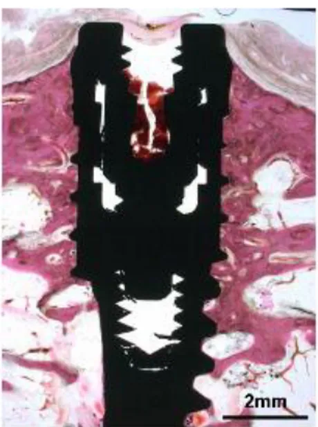
Figure 5. Histologic view of control group A at 8 weeks (8 Control A). Bone was in direct contact with the implant up to the coronal shoulder of the implant (12.5x, H-E staining).
Figure 6. Histologic view of control group B at 8 weeks (8 Control B). Bone was in direct contact with the implant in one third of the defect originating at the defect base. Connective tissue separated the new bone from the implant (12.5x, H-E staining). Figure 7. Histologic view of the Experimental group at 8 weeks (8 Experimental): (A) Bone was in direct contact with the implant in the apical half of the defect. Space was maintained by the bone substitute for bone formation (12.5x, H-E staining). Bone substitute was not degraded during this period of time. (B) Particles of bone substitute (small arrow) surrounded by connective tissue were found. A thin, continuous layer of newly-formed bone in direct contact with the implant surface (large arrow) was observed beside the bone substitute (40x, H-E staining).
Figure 8. Histologic view of the control group A at 16 weeks (16 Control A). Slight marginal bone loss was found (12.5x, H-E staining).
גכ
Figure 9. Histologic view of control group B at 16 weeks (16 Control B). Newly-formed bone failed to reach more than half of the defect. The remaining defect depth was similar to that observed at 8-weeks. Connective tissue separated the new bone from the implant (12.5x, H-E staining).
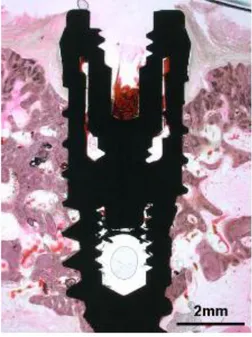
Figure 10. Histologic view of the experimental group at 16 weeks (16 Experimental). (A) Bone was in direct contact with the implant except at the coronal quarter of the defect. Space was successfully maintained for bone formation by the bone substitute (12.5x, H-E staining). (B) The margin of bone substitute became more rounded than it was at 8 weeks (small arrow), but was not completely degraded at this time. Bone substitute particles incorporated with newly formed bone was observed (small arrow). Connective tissue separated the new bone from the implant in the coronal part of the implant (Large arrows) (40x, H-E staining). More direct contact between new bone and the implant surface was observed beside bone substitute than at 8-weeks in the control group.
דכ
Figures
Figure1
הכ
Figure 3
וכ
Figure 5 Figure 6
זכ
חכ
טכ
Table
ל
Table 1. Remaining defect depth (RDD) and bone-to-implant contact (BIC) in each group at two time points (mean ± standard deviation)
*Significant difference in RDD among groups at 8-weeks (P<0.01; one way ANOVA). †Significant difference in BIC between control group A and control group B (P<0.01; one way ANOVA).
‡Significant difference in RDD compared with control group B at 16 weeks (P<0.01; one way ANOVA).
אל 국 문 요 약
성견 임플란트 주위 환상형 골 결손부에서 이중 인산칼슘
합성골 이식재의 효과
연세대학교 대학원 치의학과 (지도 최성호 교수) 김성태이중인산 칼슘 골이식재는 β-tricalcium phosphate (β-TCP)로 코팅된 hydroxyapatite (HA) 골이식재 이고, 현재 자가골이나 다른 골이식재를 대신하여 널리 사용되고 있는 합성골이식재 이다. 이 연구의 목적은 성견 임플란트 주위 환상형 골 결손부에서 이중 인산칼슘 합성골 이식재의 효과를 평가하는 것이다. 4마리의 잡견의 무치악 부위에 임플란트가 식립 되었고 그 주위로 2mm gap distance의 환상형 골 결손부가 형성되었다. 군은 세가지로 나뉘어 졌다; 실험군, 대조군 A, 대조군 B. 실험군의 골 결손부에는 이중인산 칼슘 골이식재가 채워졌다. 대조군 A에는 임플란트 주위에 골 결손부가 만들어지지 않았다. 대조군 B에는 2mm gap distance의 환상형 골 결손부 형성 후 아무 것도 채워 넣지 않았다. 결손부는 8주 16주 후에 조직학, 조직계측학적으로 관찰 되었다. 8주 후에 조직계측학적 결과에서 남아 있는 결손부 양 (RDD)은 대조군A 에서 가장 적은 값을 보였다. 실험군 에서의 RDD는 대조군 A 보다 큰 값을 보였고 대조군 B에서 가장 큰
בל 값을 보였다. 8주째 임플란트와 골의 접촉은 군간 차이를 보이지 않았다. 16주째에 대조군B의 RDD는 다른 군들과 비교하였을 때 가장 큰 값을 보였다. 이 실험의 한계에도 불구하고 이중인산 칼슘 골이식재는 결손부 해소와 신생골 형성을 위한 공간 유지에 기여하는 바가 있는 것으로 사료된다. ---핵심되는 말: 이중인산칼슘 골이식재, 환상형 골결손부, 임플란트, 남아 있는 결손부 양, 골 임플란트 접촉