INTRODUCTION
Breast cancer is one of the most common cancers in women and a major cancer death in women. The incidence of breast cancer in Korea continues to rise year by year, and its clinical characteristics are becoming closer to those observed in West-ern countries [1].
Among the many genes that have been known to play im-portant roles in breast carcinogenesis, 14-3-3 sigma (σ) has been directly implicated in the tumorigenesis of breast cancer
[2]. 14-3-3 σ is a p53-dependent, negative regulator of the cell cycle and contributes to G2/M arrest [2] by inhibiting the for-mation of the Cdc2-cyclin B1 complex [2,3]. It can also stabil-ize p53 expression by blocking MDM2-mediated p53 ubiquiti-nation [4]. 14-3-3 σ inactivation has been reported in various types of cancers, such as lung, prostate, and vulvar and oral squamous cell carcinoma [5-7]. This suggests that 14-3-3 σ acts as a tumor suppressor and its inactivation contributes to tumorigenesis by promoting cell cycle progression. Studies have shown that 14-3-3 σ is selectively inactivated by epigene-tic silencing [5,6,8]. In fact, loss of 14-3-3 σ by hypermethyl-ation of its promoter is reported to be one of the most consis-tent molecular alterations discovered in breast cancer so far [9,10]. Recently, a few studies showed that 14-3-3 σ was also downregulated through ubiquitin-mediated proteolysis by es-trogen-responsive finger protein (Efp), an estrogen-dependent E3 ubiquitin ligase [11]. Efp is a downstream target of estro-gen receptor and mediates estroestro-gen-induced cell growth, which implies possible involvement in the development of
The Role and Regulatory Mechanism of 14-3-3 Sigma in Human Breast Cancer
SeungSang Ko*, Ji Young Kim1,*, Joon Jeong2, Jong Eun Lee3, Woo Ick Yang4, Woo Hee Jung4Department of Surgery, Cheil General Hospital & Women’s Health Care Center, Catholic Kwandong University College of Medicine, Seoul; 1Department of
Pathology, CHA Gangnam Medical Center, CHA University, Seoul; Departments of 2Surgery, 3Anatomy, and 4Pathology, Yonsei University College of
Medicine, Seoul, Korea ORIGINAL ARTICLE
Purpose: 14-3-3 sigma (σ) is considered to be an important tu-mor suppressor and decreased expression of the same has been reported in many malignant tumors by hypermethylation at its promoter or ubiquitin-mediated proteolysis by estrogen- responsive ring finger protein (Efp). In this study, we investigated the significance of 14-3-3 σ expression in human breast cancer and its regulatory mechanism. Methods: Efp was silenced using small interfering RNA (siRNA) in the MCF-7 breast cancer cell line in order to examine its influence on the level of 14-3-3 σ protein. The methylation status of the 14-3-3 σ promoter was also evalu-ated by methylation-specific polymerase chain reaction (PCR). The expression of Efp and 14-3-3 σ in 220 human breast carci-noma tissues was assessed by immunohistochemistry. Other clinicopathological parameters were also evaluated. Results: Si-lencing Efp in the MCF-7 breast cancer cell line resulted in in-creased expression of 14-3-3 σ. The Efp-positive human breast cancers were more frequently 14-3-3 σ-negative (60.5% vs.
39.5%). Hypermethylation of 14-3-3 σ was common (64.9%) and had an inverse association with 14-3-3 σ positivity (p= 0.072). Positive 14-3-3 σ expression was significantly correlated with poor prognosis: disease-free survival (p=0.008) and dis-ease-specific survival (p=0.009). Conclusion: Our data suggests that in human breast cancer, the regulation of 14-3-3 σ may in-volve two mechanisms: ubiquitin-mediated proteolysis by Efp and downregulation by hypermethylation. However, the inactiva-tion of 14-3-3 σ is probably achieved mainly by hypermethyl-ation. Interestingly, 14-3-3 σ turned out to be a very significant poor prognostic indicator, which is in contrast to its previously known function as a tumor suppressor, suggesting a different role of 14-3-3 σ in breast cancer.
Key Words: Breast neoplasms, Estrogen-responsive finger protein, Methylation, SFN protein
Correspondence to: Woo Hee Jung
Department of Pathology, Yonsei University College of Medicine, 211 Eonju-ro, Gangnam-gu, Seoul 135-720, Korea
Tel: +82-2-2019-3541, Fax: +82-2-3463-2103 E-mail: [email protected]
*These authors contributed equally to this work.
The present research has been supported by Korea Breast Cancer Foundation.
Received: March 31, 2014 Accepted: September 2, 2014
Cancer
human breast cancers. However, the exact mechanism of how 14-3-3 σ is inactivated and its association with Efp in breast cancer has not been clearly elucidated. Studies on Efp and 14-3-3 σ in human breast cancer are still very limited, and the bi-ological significance remains unclear.
Therefore, in this study we investigated the regulatory mech-anism of 14-3-3 σ in breast cancer. We also evaluated the ex-pression pattern of 14-3-3 σ using clinicopathological features of human breast cancer to deepen our understanding of the role of 14-3-3 σ in breast cancer tumorigenesis and biology. In order to do this, we investigated whether silencing Efp could affect 14-3-3 σ expression in vitro. We also examined the ex-pression of Efp and 14-3-3 σ in 220 cases of human breast can-cer tissues using immunohistochemistry. To investigate the epigenetic silencing of 14-3-3 σ by methylation, we examined the methylation status of 14-3-3 σ promoter sites in breast can-cer tissues by methylation-specific polymerase chain reaction (PCR). We subsequently correlated these findings with various clinicopathological variables, including the clinical outcome of the patients.
METHODS
Patients and tissue specimens
We analyzed the breast cancer registry program of the De-partment of Surgery, Gangnam Severance Hospital, Yonsei University College of Medicine. Two hundred twenty cases of pure invasive ductal carcinoma (‘NOS ductal’ according to World Health Organization classification) were obtained from female patients who underwent surgery between 1996 and 2001 in Gangnam Severance Hospital, Yonsei University Col-lege of Medicine. Inclusion criteria were cases of pure invasive ductal carcinoma without evidence of remote metastasis. Ex-clusion criteria were cases of specific histological types (medul-lary, mucinous, papil(medul-lary, tubular, lobular, etc.), inflammatory breast cancer, bilateral cases, and those in which tissue samples and clinical data, including the follow-up results, were unavail-able. The median follow-up period was 72 months.
Paraffin-embedded tissue sections were selected from the pathological files of the Department of Pathology. As men-tioned above, we obtained baseline data and follow-up results, including the clinicopathological characteristics. The histologic grade was determined using a Nottingham combined histo-logic grade (Elston-Ellis modification of Scarff-Bloom-Rich-ardson grading system) [12]: G1 (well-differentiated), G2 (moderately-differentiated), and G3 (poorly-differentiated). Using a reverse Black method, the nuclear grade was deter-mined to be grade 1 (well-differentiated), grade 2 (moderately-differentiated), or grade 3 (poorly-differentiated).
Disease-spe-cific survival was defined as the time that elapsed from the date of surgery to the date of death due to breast cancer. The follow-up period was defined as the time that elapsed between sur-gery and death or the last available date of follow-up. In cases in which there was no outpatient record during the most re-cent 1-year period, we determined the death of the corre-sponding cases by making an inquiry, based on the Korean so-cial security number, to the Korea National Statistical Office, the district office, and/or the police station.
This study was approved by Institutional Review Board of the Gangnam Severance Hospital, Yonsei University College of Medicine (approval number: 3-2014-0192).
Tissue microarray and immunohistochemical evaluation A tissue microarray was constructed by ISU Abxis Co. (Seoul, Korea) from the paraffin-embedded blocks of 220 breast cancer cases. Briefly, all cases were histologically re-viewed and representative tumor areas were marked in the corresponding paraffin blocks. Two selected cylinders (2 mm in diameter) from two different tumor areas were included for each case. Normal control tissues were obtained from sur-rounding normal breast tissue.
Immunohistochemical staining was done for estrogen re-ceptor (ER), progesterone rere-ceptor (PR), c-erbB-2, p53, Efp, and 14-3-3 σ. The tissue microarray slides were deparaffinized by xylene two times and treated with 100%, 95%, and 80% ethanol, and then hydrated with distilled water. These slides were then treated with citric acid buffer (pH 6.0) and boiled in a microwave for 20 minutes for antigen retrieval. Intrinsic peroxidase activity was blocked using a solution containing 3% hydrogen peroxide. Next, the slides were rinsed with tris-buffer solution (TBS) and incubated with the primary anti-bodies for ER, PR, c-erbB-2, p53, Efp, and 14-3-3 σ overnight at 4°C (Table 1). The slides were rinsed with TBS, and then the Vectastain universal elite ABC kit (Vector Laboratories, Burlingame, USA) was applied. The color development was
Table 1. Primary antibodies for the immunohistochemical staining Antibody Manufacturer Isotype Dilution Efp BD Bioscience
(Franklin Lakes, USA) Mouse monoclonal 1:300 14-3-3 σ IBL (Takasaki, Japan) Rabbit polyclonal 1:100 ER Thermo Fisher Scientific
(Fremont, USA) Mouse monoclonal 1:100 PR Invitrogen (Carlsbad, USA) Mouse monoclonal 1:100 c-erbB-2 Thermo Fisher Scientific
(Fremont, USA) Mouse monoclonal 1:100 p53 Invitrogen (Carlsbad, USA) Mouse monoclonal 1:100 Efp=estrogen-responsive ring finger protein; ER=estrogen receptor; PR= progesterone receptor.
done with NovaRed (Vector Laboratories), and then the slides were counterstained with hematoxylin. The slides were finally mounted and observed under a microscope.
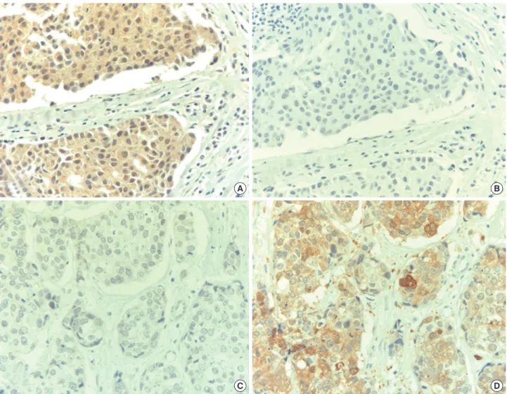
The immunohistochemical results were interpreted by two experienced pathologists. A positive reaction was determined by the ratio of cancer cells stained by the primary antibody to the total cancer cells in each slide (Figure 1). For ER and PR, a positive diagnosis was determined when intensely stained nu-clei were seen in more than 10% of the total cancer cells [13]. For c-erbB-2, a positive reaction was determined when the cell membranes were evenly stained (score 2 or 3 by the four-grade system) [14,15]. For p53 protein, a positive reaction was determined when intensely stained nuclei were seen in more than 5% of the total cancer cells. Efp and 14-3-3 σ expression was graded as 0 (if none of the tumor cells were positive), 1 (if up to 10% of the tumor cells were positive), 2 (if 10% to 50% of the tumor cells were positive), and 3 (if more than 50% of the tumor cells were positive) by the two pathologists inde-pendently. The final diagnosis was determined as positive when the sum of the two independent scores was equal or more than five [16,17].
Methylation-specific PCR
We analyzed the methylation status of 14-3-3 σ in randomly selected samples from 111 cases. The methylation status of the samples was investigated by methylation-specific PCR, as de-scribed previously [9,18]. Briefly, genomic DNA was extracted using Qiagen DNeasy Blood and Tissue Kit (Qiagen, Valencia, USA) from the formalin-fixed, paraffin-embedded tumor tis-sue after manual microdissection under a microscope to ob-tain a sample that consisted of more than 70% tumor cells. One microgram of genomic DNA was then treated with sodium bi-sulfite using an EZ DNA Methylation-Gold Kit (Zymo Re-search, Orange, USA) and was analyzed by methylation-specif-ic PCR (MSP) using a primer set that covered CG dinucleotide numbers 3, 4, 8, and 9. Primers specific for methylated DNA (59-TGGTAGTTTTTATGAAAGGCGTC-39 [sense] and 59-CCTCTAACCGCCCACCACG-39 [antisense]), and prim-ers specific for unmethylated DNA (59-ATGGTAGTTTT-TATGAAAGGTGTT-39 [sense] and 59-CCCTCTAAC-CACCCACCACA-39 [antisense]) yielded a 105 to 107-bp PCR product. The PCR conditions were as follows: 1 cycle of 95°C for 5 minutes; 31 cycles of 95°C for 45 seconds, 56°C for 30 seconds for the unmethylated primer, 60°C for 30 seconds
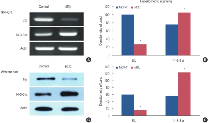
120 100 80 60 40 20 0 Efp MCF-7 siEfp 14-3-3 σ Densitometric scanning * * Densitometry of band 140 120 100 80 60 40 20 0 Efp MCF-7 siEfp 14-3-3 σ * * Densitometry of band Control siEfp Efp RT-PCR 14-3-3 σ Actin Control siEfp Efp Western blot 14-3-3 σ Actin A B C D
Figure 1. The increased expression of 14-3-3 σ by knocks down of estrogen-responsive ring finger protein (Efp) using small interfering RNA (siRNA). siEfp was transfected in human MCF-7 breast cancer cells. After transfection, total RNA and whole cell lysates were prepared and subjected to re-verse transcription polymerase chain reaction (A) and Western blot (C). Relative levels of mRNA and protein expression were determined by densito-metric scanning of the bands (B, D). The siEfp transfection resulted in increased levels of 14-3-3 σ mRNA and protein.
for the methylated primer, and 72°C for 30 seconds; and 1 cycle of 72°C for 4 minutes.
The MCF-7 cell line, previously reported to be ated at the 14-3-3 σ promoter [9], was used as the unmethyl-ated control. Commercially available methylunmethyl-ated DNA (Uni-versal Methylated Human DNA Standard; Zymo Research) was used as the methylated control. Methylated bands on MSP which were more dense than the corresponding unmethylated bands or the ones as dense as the universally methylated DNA control were determined as hypermethylated.
Small interfering RNA silencing of Efp
For the transfection procedure, MCF-7 cells were grown to 60% confluence, and Efp small interfering RNAs (siRNAs) (5'-GGTGGAGCAGCTACAACAATT-3') were transfected us-ing the RNAiMax reagent (Invitrogen, Carlsbad, USA) accord-ing to the manufacturer’s instructions. Briefly, the RNAiMax reagent was incubated with serum-free medium for 10 minutes. Subsequently, a mixture of the siRNA was added. After incuba-tion for 15 minutes at room temperature, the mixture was di-luted with medium and added to each well. The final concen-tration of siRNAs in each well was 100 nM. After culturing for 40 hours, cells were washed, resuspended in new culture media for reverse transcription (RT)-PCR and Western blot. Total RNA and whole cell lysates was prepared and subjected to RT-RCR and Western blot. Relative levels of mRNA and protein expression were determined by densitometry of the bands. RT-PCR analysis and Western blot
Confluent MCF-7 cell monolayers were washed with PBS. Total RNA (12 μg) was isolated using TRIzol reagent. One mil-liliter of TRIzol reagent (Invitrogen) per well was added to cells in 6-well plates, and total RNA was extracted using the manu-facturer’s protocol (Invitrogen). RT was performed using oligo (dT) primers and superscript reagent (Invitrogen). Five micro-grams of RNA were used for first strand cDNA synthesis using oligo (dT) in a final volume of 20 μL. Seven microliters of the cDNA mixture was used to amplify mRNA for Efp, 14-3-3 σ, and 18s ribosomal RNA (18s rRNA) as a loading control by PCR. Efp, 14-3-3 σ, and 18s rRNA were amplified using the fol-lowing primer sets: Efp (forward 5'-AACATCTCTCAAGGC-CAAGGT-3' and reverse 5'-AGATGCCTACCCCACAG AA-GT-3'), 14-3-3 σ (forward 5'-GTGTGTCCCCAGAGCATGG-3' and reverse 5'-ACCTTCTCCCGGTACTCA CG-3'), and 18s
rRNA (forward 5'-CGGCTACCACATCCAAGGAA-3' and
reverse 5'-GCTGGAATTACCGCGGCT-3'). The RT product (7 μL) was amplified in a 20-μL volume containing 10 pmol of primers and 2.5 units of Taq DNA polymerase. Reaction con-ditions were as follows: 95°C for 1 minute, 60°C for 1 minute,
72°C for 1 minute for 33 cycles, and then 72°C for 7 minutes. The PCR products were resolved on a 1.2% agarose gel and identified by ethidium bromide staining. Normalization of Efp and 14-3-3 σ expression was achieved by comparing the ex-pression of 18s rRNA for the corresponding sample.
For Western blot analysis, proteins were isolated from MCF-7 and siEfp-treated MCF-MCF-7 cells and lysed in solubilizing buffer (1×PBS, 1% nonidet P-40, 0.5% sodium deoxycholate, 0.1% SDS, protease inhibitors, PMSF, aprotinin, and sodium or-thovandate). Equal amounts of protein extracts were separated by 10% SDS-PAGE and transferred to PVDF membrane (Mil-lipore, Billerica, USA). The membrane was blocked with 5% nonfat milk in TBS containing 0.05% Tween, and then incu-bated with antibody mouse anti-Efp (1:5,000; BD Bioscience, Franklin Lakes, USA), rabbit anti-14-3-3 σ (1:1,000; IBL Co., Ltd., Takasaki, Japan), and mouse antiactin (1:5,000; Santa Cruz Biotechnology, Santa Cruz, USA). The membrane was then incubated with the secondary antibody and thoroughly washed. Immunoreactive bands were visualized with a Super-Signal (Thermo-Fisher Scientific, Waltham, USA).
Statistical analyses
All statistical analyses were done using SPSS version 13.0 for Windows (SPSS Inc., Chicago, USA). To evaluate correlations between Efp, and 14-3-3 σ expression and clinicopathologic parameters, data were cross-tabulation (chi-square test/2×2 table; Pearson or Fisher exact test) was done. Univariate analy-sis with patient survival was evaluated using life tables con-structed from survival data with Kaplan-Meier plots. Compar-isons of the different groups were done with the log-rank test. The end point in the present study was disease-specific surviv-al ranging from the date of surgery until the date of breast can-cer-related death or, if no information was documented, until the date of last follow up information (=censored). Multivari-ate survival analysis was carried out on samples where all clini-cal parameters were available using the Cox proportional haz-ard model to evaluate the independent power of each variable. Statistical significance was set at p-value <0.05 (95% level of confidence).
RESULTS
Demographic data of the subjects
Basic clinical and demographic data are summarized in Ta-ble 2. Mean patient age was 47.1±9.9 years old. 66.3% (146/ 220) are positive to at least one of the ER, PR, and HER2 (re-ceptor-positive group). ER-positive cases, 19.5% (43/220) were HER2-positive cases, and all receptor groups were negative in 26.4% (58/220), i.e., the triple-negative group. The
triple-nega-tive group frequently showed high nuclear grade, high histo-logic grade, and p53 positive cells.
Efp silencing upregulates 14-3-3 σ expression in vitro
Efp silencing was performed by transfection of siEfp in the human breast cancer cell line, MCF-7. Transfection of siRNA resulted in the downregulation of Efp mRNA levels by RT-PCR and increased the expression of 14-3-3 σ protein levels by Western blot (Figure 1). All three independent experiments gave similar results. This clearly shows that Efp is associated with decreased expression of 14-3-3 σ, and silencing of Efp can
increase the expression of 14-3-3 σ.
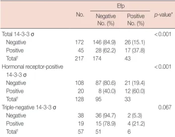
Expression of Efp and 14-3-3 σ in subtypes of breast cancer Both Efp and 14-3-3 σ showed cytoplasmic staining pat-terns. 14-3-3 σ was strongly expressed in the myoepithelial cells. In the normal breast epithelial cells, Efp was only weakly positive. About two-thirds of the total tumors (146/217 cases, 67.3%) were negative for both Efp and 14-3-3 σ. About one-third of the total cases (71/217 cases, 32.7%) were positive for at least one of the two genes. Fifty-four of 217 cases (24.9%) were positive for only one of the two genes, while only 17 of 217 cases (7.8%) showed positive staining for both. For the cases when at least one gene was positive, the majority (54/71 cases, 76%) demonstrated a mutually exclusive pattern of pos-itivity, i.e., positive to only one of the two proteins (Table 3, Figure 2). Only 17 cases (23.9%) of the positive 71 cases were positive for both Efp and 14-3-3 σ. The Efp-positive cases were more frequently negative (26/43 cases, 60.5%) for 14-3-3 σ than positive (17/43 cases, 39.5%).
The majority (36/43 cases, 85.7%) of the Efp-positive tumors were receptor-positive (p=0.019). Triple-negative tumors were more frequently positive for 14-3-3 σ (19/58 cases) than recep-tor-positive tumors (26/145 cases) (p=0.006) (Table 4). Correlation between Efp, 14-3-3 σ expression, and clinicopathological variables
Correlation between Efp, 14-3-3 σ expression status, and other clinicopathological parameters of patients are summa-rized in Table 5. Notably, Efp was negatively correlated with axillary lymph node metastasis (p=0.021) or positive p53
ex-Table 2. Baseline clinicopathologic characteristics of breast cancer pa-tients examined Characteristic Subgroup* p-value§ Receptor (+)† (n=129) No. (%) Triple (-)‡ (n=58) No. (%) HER2 only (+) (n=17) No. (%) Age (yr)II 46.9±10.1 47.0±10.1 49.1±8.1 0.939¶ Follow-up (mo)** 72 (1–144) 73 (5–135) 83 (7–135) 0.648¶ Tumor size (cm) ≤2 54 (74.0) 18 (24.7) 1 (1.4) 0.010 >2 75 (57.3) 40 (30.5) 16 (12.2) Axillary node Negative 59 (58.4) 33 (32.7) 9 (8.9) 0.353 Positive 70 (68.0) 25 (24.3) 8 (7.8) Stage I 33 (71.7) 13 (28.3) 0 0.178 II 70 (61.4) 33 (28.9) 11 (9.6) III 26 (59.1) 12 (27.3) 6 (13.6) Histologic grade I 36 (87.8) 3 (7.3) 2 (4.9) 0.001 II & III 93 (57.4) 54 (33.3) 15 (9.3) Nuclear grade 1 & 2 84 (79.2) 17 (16.0) 5 (4.7) <0.001 3 44 (45.8) 40 (41.7) 12 (12.5) ER Negative 43 (36.4) 58 (49.2) 17 (14.4) Positive 86 (100.0) 0 0 PR Negative 18 (19.4) 58 (62.4) 17 (18.3) Positive 111 (100.0) 0 0 HER2 Negative 103 (64.0) 58 (36.0) 0 Positive 26 (60.5) 0 17 (39.5) p53 Negative 98 (75.4) 26 (20.0) 6 (4.6) <0.001 Positive 27 (39.1) 31 (44.9) 11 (15.9)
ER=estrogen receptor; PR=progesterone receptor; HER2=human epidermal growth factor receptor 2.
*Excluding unknown cases; †Receptor-positive group: positive to at least one of the ER, PR, and HER2; ‡Triple-negative group: negative to all of the ER, PR, and HER2; §p-value from chi-square test (2×2 Pearson or Fisher exact test; except TNM stage); IIMean±SD; ¶p-value of age and follow-up duration: from independent sample t-test; **Median (range).
Table 3. Correlation between Efp and 14-3-3 σ expression No.
Efp
p-value*
Negative
No. (%) PositiveNo. (%)
Total 14-3-3 σ <0.001 Negative 172 146 (84.9) 26 (15.1) Positive 45 28 (62.2) 17 (37.8) Total† 217 174 43 Hormonal receptor-positive <0.001 14-3-3 σ Negative 108 87 (80.6) 21 (19.4) Positive 20 8 (40.0) 12 (60.0) Total† 128 95 33 Triple-negative 14-3-3 σ 0.067 Negative 38 36 (94.7) 2 (5.3) Positive 19 15 (78.9) 4 (21.2) Total† 57 51 6
Efp=estrogen-responsive ring finger protein.
*p-value from chi-square test (2×2 Pearson); †Total number of the group (ex-cluding unknown cases).
pression (p=0.009). There was an increase in the proportion of Efp-negative cases as the histologic grade and nuclear grade increased, but it was not statistically significant. No other
clin-icopathological variables, such as tumor size, stage, histologic grade, nuclear grade, ER, or c-erbB-2 appeared to be signifi-cantly related to Efp expression.
Positive 14-3-3 σ expression was significantly correlated with high histologic grade, high nuclear grade, and positive p53 (p=0.012, p=0.033, and p=0.001, respectively). There was a statistically significant inverse correlation between 14-3-3 σ expression and both ER-positivity (p=0.020) and PR-pos-itivity (p=0.032), which is in accordance with our previous finding that the triple-negative group was more frequently 14-3-3 σ-positive than the receptor-positive group.
MSP for the 14-3-3 σ promoter
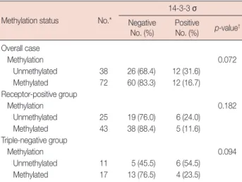
Methylation status of the 14-3-3 σ promoter was evaluated with MSP, as shown in Figure 3. Of the 111 cases examined, 72 cases (64.9%) were hypermethylated by MSP (Table 6). The
A B
C D
Figure 2. Expression of estrogen-responsive ring finger protein (Efp) and 14-3-3 σ in breast cancer. An estrogen receptor-positive breast cancer was positive to Efp (A) and negative to 14-3-3 σ (B) while a triple-negative breast cancer was negative to Efp (C) and positive to 14-3-3 σ (D) (NovaRed with Hematoxylin counterstain, ×400).
Table 4. Expression of Efp and 14-3-3 σ according to the subtype of breast cancer by receptor expression
Receptor-positive (n=128)* No. (%) Triple-negative (n=57)* No. (%) p-value † Efp 0.019 Negative 95 (74.2) 51 (89.5) Positive 33 (25.8) 6 (10.5) 14-3-3 σ 0.006 Negative 108 (84.4) 38 (66.7) Positive 20 (15.6) 19 (33.3) Efp=estrogen-responsive ring finger protein.
hormone receptor-positive group was more frequently hyper-methylated (45/71 cases, 63.4%) than the triple-negative group (17/28 cases, 60.7%), but the difference was not statistically sig-nificant (Table 6). The 14-3-3 σ positive results by immunohis-tochemistry were negatively associated with the hypermethyl-ation of the 14-3-3 σ promoter, but only showed marginal
sig-nificance (p=0.072) (Table 6).
Correlation between Efp, 14-3-3 σ expression, and clinical outcome
Univariate analysis of various clinicopathological factors that may be correlated with the expression of Efp and 14-3-3
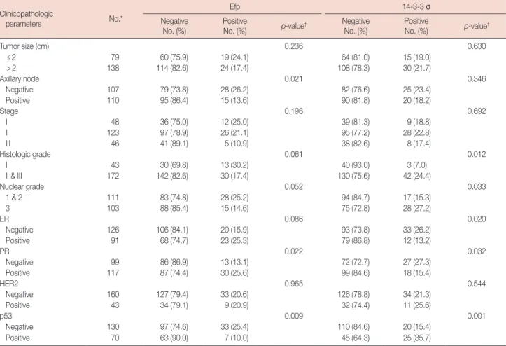
Table 5. The correlations between the Efp, 14-3-3 σ, and other clinicopathological parameters Clinicopathologic
parameters No.*
Efp 14-3-3 σ
Negative
No. (%) PositiveNo. (%) p-value† NegativeNo. (%) PositiveNo. (%) p-value†
Tumor size (cm) 0.236 0.630 ≤2 79 60 (75.9) 19 (24.1) 64 (81.0) 15 (19.0) >2 138 114 (82.6) 24 (17.4) 108 (78.3) 30 (21.7) Axillary node 0.021 0.346 Negative 107 79 (73.8) 28 (26.2) 82 (76.6) 25 (23.4) Positive 110 95 (86.4) 15 (13.6) 90 (81.8) 20 (18.2) Stage 0.196 0.692 I 48 36 (75.0) 12 (25.0) 39 (81.3) 9 (18.8) II 123 97 (78.9) 26 (21.1) 95 (77.2) 28 (22.8) III 46 41 (89.1) 5 (10.9) 38 (82.6) 8 (17.4) Histologic grade 0.061 0.012 I 43 30 (69.8) 13 (30.2) 40 (93.0) 3 (7.0) II & III 172 142 (82.6) 30 (17.4) 130 (75.6) 42 (24.4) Nuclear grade 0.052 0.033 1 & 2 111 83 (74.8) 28 (25.2) 94 (84.7) 17 (15.3) 3 103 88 (85.4) 15 (14.6) 75 (72.8) 28 (27.2) ER 0.086 0.020 Negative 126 106 (84.1) 20 (15.9) 93 (73.8) 33 (26.2) Positive 91 68 (74.7) 23 (25.3) 79 (86.8) 12 (13.2) PR 0.022 0.032 Negative 99 86 (86.9) 13 (13.1) 72 (72.7) 27 (27.3) Positive 117 87 (74.4) 30 (25.6) 99 (84.6) 18 (15.4) HER2 0.965 0.544 Negative 160 127 (79.4) 33 (20.6) 126 (78.8) 34 (21.3) Positive 43 34 (79.1) 9 (20.9) 32 (74.4) 11 (25.6) p53 0.009 0.001 Negative 130 97 (74.6) 33 (25.4) 110 (84.6) 20 (15.4) Positive 70 63 (90.0) 7 (10.0) 45 (64.3) 25 (35.7)
Efp=estrogen-responsive ring finger protein; ER=estrogen receptor; PR=progesterone receptor; HER2=human epidermal growth factor receptor 2. *Total number of the group (excluding unknown cases); †p-value from chi-square test (2×2 Pearson; except TNM stage).
Figure 3. Methylation-specific polymerase chain reaction (MSP) in breast cancer. Methylated and unmethylated bands on MSP. Methylated bands which are brighter than the corresponding unmethylated band or the ones as dense as the universally methylated DNA control were determined as hyper-methylated.
σ showed that 14-3-3 σ expression was significantly associated with an increased risk of recurrence and disease-related death (p=0.011 and p=0.022), while Efp was not. Other tradition-ally well-known prognostic factors, such as tumor size, axil-lary lymph node status, TNM stage, and histologic grade, were also significant prognostic variables for survival (data not shown).
Multivariate analysis revealed that the axillary lymph node status (p=0.028) and 14-3-3 σ expression (p=0.008) were the only two independent prognostic factors for recurrence with relative risks over 1.0, whereas tumor size, TNM stage, and histologic grade were not significant (Table 7). Only TNM stage (p=0.015) and 14-3-3 σ expression (p=0.009) were in-dependent prognostic factors in cancer-related deaths, but other factors including Efp were not significant prognostic in-dicators (Table 7).
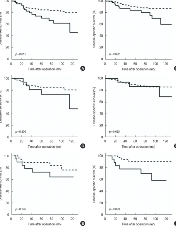
In both the univariate and multivariate analysis, 14-3-3 σ appeared to be the single most powerful prognostic indicator. Survival curves based on 14-3-3 σ expression also demon-strated a significant correlation between positive 14-3-3 σ ex-pression and adverse clinical outcome of the patients (Figure 4A and 4B). This correlation appeared to be more pronounced in the triple-negative group (Figure 4E and 4F) than in the re-ceptor-positive group (Figure 4C and 4D). In the receptor-positive group, the difference between the 14-3-3 σ receptor-positive group and negative group was not statistically significant.
DISCUSSION
The role of the 14-3-3 σ as a tumor suppressor has been shown by its known function as a negative cell cycle regulator, a G2/M checkpoint by inhibiting the formation of the
Cdc2-Table 6. 14-3-3 σ expression and its methylation status in breast cancer Methylation status No.*
14-3-3 σ Negative
No. (%) PositiveNo. (%) p-value† Overall case Methylation 0.072 Unmethylated 38 26 (68.4) 12 (31.6) Methylated 72 60 (83.3) 12 (16.7) Receptor-positive group Methylation 0.182 Unmethylated 25 19 (76.0) 6 (24.0) Methylated 43 38 (88.4) 5 (11.6) Triple-negative group Methylation 0.094 Unmethylated 11 5 (45.5) 6 (54.5) Methylated 17 13 (76.5) 4 (23.5)
*Total number of the group (excluding unknown cases); †p-value from chi-square test (2×2 Pearson).
Table 7. Multivariate analysis of recurrence and cancer-related death Coefficient SE RR (95% CI) p-value*
For recurrence Tumor size (cm) ≤2 - - 1.0 >2 0.854 0.493 2.350 (0.894–6.172) 0.083 Axillary node Negative - - 1.0 Positive 1.071 0.487 2.917 (1.123–7.576) 0.028 TNM stage 0.077 I - - 1.0 II -0.066 0.952 0.936 (0.145–6.051) 0.945 III 0.772 1.012 2.165 (0.298–15.743) 0.445 Histologic grade I - - 1.0 II & III 0.929 0.746 2.531 (0.586–10.926) 0.213 14-3-3 σ Negative - - 1.0 Positive 0.937 0.351 2.551 (1.283–5.075) 0.008 For cancer-related death Tumor size (cm) ≤2 - - 1.0 >2 -0.912 0.634 2.489 (0.719–8.620) 0.150 Axillary node Negative - - 1.0 Positive 0.449 0.586 1.567 (0.497–4.948) 0.443 TNM stage 0.015 I - - 1.0 II -0.279 1.046 0.756 (0.097–5.877) 0.789 III 1.144 1.133 3.139 (0.341–28.890) 0.313 Histologic grade I - - 1.0 II & III 1.052 1.042 2.864 (0.372–22.071) 0.312 14-3-3 σ Negative - - 1.0 Positive 1.116 0.428 3.502 (1.319–7.063) 0.009 Data were considered significant in the univariate analyses and were exam-ined in the multivariate analyses. Relative risk (RR) less than 1.00 represent a decreased risk of death, whereas RR greater than 1.00 represent an in-creased risk of death.
SE=standard error; CI=confidence interval.
*p-values determined by a Cox proportional hazard model.
cyclin B1 complex [2,3], or by stabilizing p53 through block-ing MDM2-mediated p53 ubiquitination [4]. This has been supported by several observations of decreased expression of 14-3-3 σ in various malignant tumors, such as cancer of the stomach, prostate, lung, and oral cavity [5-8]. In breast cancer, 14-3-3 σ expression has also been shown to be downregulated [9,10,19]. The mechanism by which 14-3-3 σ is inactivated has not been completely elucidated yet, but epigenetic silenc-ing by hypermethylation of the 14-3-3 σ gene is one of the commonly proposed mechanisms [5,8-10].
A previous study demonstrated that 14-3-3 σ expression was gradually decreased as a lesion progressed from benign to
ma-lignant with the loss of 14-3-3 σ expression in 8%, 35%, and 77% of usual ductal hyperplasia, ductal carcinoma in situ (DCIS), and invasive ductal carcinoma (IDC) lesions,
respec-tively, which indicates the role of 14-3-3 σ as a tumor suppres-sor in breast carcinogenesis [20]. Another recent study showed that the hypermethylation of 14-3-3 σ occurred at rates of zero
100 80 60 40 20 0 100 80 60 40 20 0 0 20 40 60 80 100 120 0 20 40 60 80 100 120 p=0.011 p=0.022
Time after operation (mo) Time after operation (mo)
Disease-fr ee survival (%) Disease-specific survival (%) A B 100 80 60 40 20 0 100 80 60 40 20 0 0 20 40 60 80 100 120 0 20 40 60 80 100 120 p=0.306 p=0.665
Time after operation (mo) Time after operation (mo)
Disease-fr
ee survival (%)
Disease-specific survival (%)
C D
Figure 4. Disease-free survival and disease-specific survival curves according to 14-3-3 σ expression in each subgroup. (A, C, E) Disease-free survival; (B, D, F) Disease-specific survival. (A, B) Total cases: 14-3-3 σ expression was significantly associated with an increased risk of recurrence and disease-related death. (C, D) Receptor-positive group: 14-3-3 σ expression was associated with an increased risk of recurrence and disease-disease-related death, but statistically insignificant. (E, F) Triple-negative group: 14-3-3 σ expression was not associated with an increased risk of recurrence. But 14-3-3 σ ex-pression was significantly associated with an increased risk of disease-related death. Dashed line, 14-3-3 σ negative; linear line, 14-3-3 σ positive.
100 80 60 40 20 0 100 80 60 40 20 0 0 20 40 60 80 100 120 0 20 40 60 80 100 120 p=0.196 p=0.029
Time after operation (mo) Time after operation (mo)
Disease-fr
ee survival (%)
Disease-specific survival (%)
in hyperplasia without atypia, 38% in atypical hyperplasia, 83% in DCIS, and 96% in IDC [10]. These studies showing a gradual decrease in 14-3-3 σ expression and a corresponding increase of hypermethylation with progression of the breast le-sions, and the fact that hypermethylation of the 14-3-3 σ gene was also found in some benign, and sometimes even appar-ently normal epithelia adjacent to cancer, suggest and support the idea that 14-3-3 σ inactivation by hypermethylation may be an early event in breast carcinogenesis [10,19].
On the other hand, it has also been reported that 14-3-3 σ is a primary target for proteolysis by Efp, and expression of the 14-3-3 σ protein is regulated by ubiquitin-mediated proteo-lysis by Efp in vitro [11,21]. Efp is a RING finger-dependent protein and functions as a ubiquitin-ligase (E3) that can ubiq-uitinate 14-3-3 σ [11,21]. Our experimental data suggested that the Efp may promote unlimited proliferation of breast cancer cells by accelerated destruction of 14-3-3 σ, a postulat-ed cell cycle inhibitor. This is supportpostulat-ed by another recent study in human breast cancer tissue which showed that im-munoreactivity of 14-3-3 σ was inversely associated with Efp immunoreactivity [22].
In our study, we demonstrated that silencing Efp can result in increased expression of 14-3-3 σ in the MCF-7 breast cancer cell line. In human breast tissue, we found that the Efp-positive cases were more frequently 14-3-3 σ negative. This in vitro and
in vivo counter-correlation between Efp and 14-3-3 σ possibly
supports the Efp-mediated ubiquitination of 14-3-3 σ in breast cancer. However, the MCF-7 cell line we used in Efp-silencing is known to be luminal ER-positive in phenotype. We did not test basal-like cell lines. The ER-negative, basal-like cell lines might have low baseline Efp levels, which might compromise the silencing effect. The difference in the 14-3-3 σ responsive-ness to Efp in different cell lines requires further investigation.
Efp positivity was not common in our cohort with since only about one-fourth of the total cases were positive for Efp where-as 14-3-3 σ wwhere-as negative in about two-thirds of the total cwhere-ases. In contrast, hypermethylation of 14-3-3 σ was common, occur-ring in more than 60% of the breast cancer samples. This may suggest that even if Efp can ubiquitinate and accelerate the deg-radation of 14-3-3 σ in vitro and even in vivo, it may not be a dominant mechanism in the inactivation of human breast can-cer. Instead, hypermethylation may play a major role in the in-activation of 14-3-3 σ in human breast cancer.
The Efp is also a downstream target of estrogen receptor α (ERα) [23-25]. In a previous study, Efp immunoreactivity was significantly associated with ERα status in breast carcinoma tissues [24]. The Efp gene has an estrogen-responsive element at the 3’-untranslated region [23]. Therefore, it has been sug-gested that Efp is mainly produced in carcinoma cells through
ERα as a result of estrogenic action in breast carcinoma. How-ever, in our study there was no significant correlation between Efp and ER. In our study, the frequency of Efp positive cancers was low, only 36 of 145 cases (24.8%) in the receptor-positive group. It was even lower in the triple-negative group (positive in 6/57 cases, 10.5%). Suzuki et al. [22] found higher Efp im-munoreactivity in ERα-negative breast carcinomas (52.4%). This difference in the frequency of Efp positivity in ER-nega-tive breast cancers may be partially due to the difference in the criteria by which immunohistochemical positivity was deter-mined. In our study, we used strict criteria, considering only diffuse strong expression (grade 3) as positive. Another possi-ble explanation for the lack of correlation between Efp and ERα status is the escape of breast cancer cells from estrogenic control in tumorigenesis, as suggested in another recent study [25]. Ikeda et al. [26] suggested another possibility by analyz-ing the 5’-flankanalyz-ing region of the human Efp gene, and report-ing the regulation of the Efp promoter by multiple elements and/or interacting factors. Therefore, factors other than ERα may be also be involved in the expression of Efp in some breast carcinomas. Suzuki et al. [22] reported that Efp immunoreac-tivity was significantly associated with an increased risk of re-currence or worse prognosis for both rere-currence and overall survival in breast carcinomas, and that the effect is similar to that of the lymph node status, a well-established prognostic factor. However, in our study Efp expression was inversely cor-related with axillary lymph node status (p=0.021) although it was not significantly associated with patient survival.
Interestingly, in contrast to previous reports showing de-creased expression or inactivation of 14-3-3 σ in various ma-lignant tumors supporting its function as a tumor suppressor, the current results clearly show that 14-3-3 σ expression was not lost, but strongly expressed in at least a small subset of breast cancer cases. Although 14-3-3 σ expression in breast cancer was not compared to that in normal breast epithelia or benign proliferative lesions, another result of our study re-vealed the significant correlation of 14-3-3 σ positivity with high grade breast cancers and poor prognosis also clearly shows that 14-3-3 σ may play a more complicated role than just a tumor suppressor in breast cancer. In our study, the ex-pression of 14-3-3 σ was inversely correlated with ERα, PR, and even Efp, which partially explains the more frequent posi-tivity in triple-negative tumors than in receptor-positive tu-mors. In addition, 14-3-3 σ is correlated with high histologic grade, high nuclear grade, and positive p53 expression, all of which are characteristics of triple-negative breast cancers. The significance of 14-3-3 σ as a poor prognostic indicator was also more pronounced in triple-negative tumors than in re-ceptor-positive ones. This association of 14-3-3 σ with poor
prognosis was in contrast to the previously known function of 14-3-3 σ as a tumor suppressor. However, the role of 14-3-3 σ as a poor prognostic factor has also been reported by a few cent studies in other types of cancer. Perathoner et al. [27] re-ported that 14-3-3 σ was significantly correlated with tumor differentiation and stage in colorectal carcinoma, and clearly demonstrated that 14-3-3 σ was an independent poor prog-nostic marker. Nakayama et al. [28] examined the expression of 14-3-3 σ, ERα, and Efp in endometrial carcinomas and showed that high 14-3-3 σ expression was significantly corre-lated with myometrial invasion and lymph node metastasis. The expression of 14-3-3 σ in normal and hyperplastic endo-metrium was also evaluated, and demonstrated that interest-ingly 14-3-3 σ expression gradually decreased from normal to hyperplastic to malignant tumors, which is a similar result to the previous studies [5-8,20]. These conflicting findings on the role of 14-3-3 σ in various types of cancer suggest that 14-3-3 σ is not just a tumor suppressor. Instead, it may have a more complex and complicated role in the process of carcinogene-sis. It might also be possible that it plays different roles in early and late carcinogenesis. In early carcinogenesis, when malig-nant transformation occurs from the normal epithelium, 14-3-3 σ may function as a tumor suppressor, but once a malig-nant tumor is established, it may have a role in promoting tu-mor growth and propagation. Another hypothesis is that the deregulation or dysfunction of the ubiquitin-proteosome pathway may result in accumulation of 14-3-3 σ despite of the high level of Efp. Deregulation or dysfunction of the ubiqui-tin-proteosome pathway has been reported in various diseas-es, mainly neurodegenerative or metabolic diseases [29]. There is also evidence that the ubiquitin-proteosome system plays a role in tumor development, such as in colon cancer or hepatocellular carcinoma [30], in which the system is not ex-actly dysregulated, but activated degrading the tumor suppres-sor molecules. The exact role of 14-3-3 σ in breast and other cancers needs to be further elucidated. However, the signifi-cant correlation of 14-3-3 σ and poor prognosis suggests its potential usefulness as a therapeutic target in breast cancer.
Our data demonstrated that 14-3-3 σ was negatively corre-lated with Efp in vitro and less frequently in vivo. 14-3-3 σ was negatively correlated with hypermethylation in human breast cancer tissue, which was more common. This suggests that in human breast cancers the regulation of 14-3-3 σ may involve both mechanisms, i.e., ubiquitin-mediated proteolysis by Efp and downregulation by hypermethylation. However, the inac-tivation of 14-3-3 σ is probably achieved mainly by hyper-methylation. Interestingly, 14-3-3 σ turned out to be a very significant prognostic indicator in breast cancer, being corre-lated with poor prognosis, which is the opposite of its
previ-ously known function as a tumor suppressor, suggesting a dif-ferent role of the 14-3-3 σ in breast cancer.
CONFLICT OF INTEREST
The authors declare that they have no competing interests.
REFERENCES
1. Ko SS; Korean Breast Cancer Society. Chronological changing patterns of clinical characteristics of Korean breast cancer patients during 10 years (1996-2006) using nationwide breast cancer registration on-line program: biannual update. J Surg Oncol 2008;98:318-23.
2. Hermeking H, Lengauer C, Polyak K, He TC, Zhang L, Thiagalingam S, et al. 14-3-3 sigma is a p53-regulated inhibitor of G2/M progression. Mol Cell 1997;1:3-11.
3. Chan TA, Hermeking H, Lengauer C, Kinzler KW, Vogelstein B. 14-3-3Sigma is required to prevent mitotic catastrophe after DNA damage. Nature 1999;401:616-20.
4. Yang HY, Wen YY, Chen CH, Lozano G, Lee MH. 14-3-3 sigma posi-tively regulates p53 and suppresses tumor growth. Mol Cell Biol 2003; 23:7096-107.
5. Gasco M, Bell AK, Heath V, Sullivan A, Smith P, Hiller L, et al. Epigene-tic inactivation of 14-3-3 sigma in oral carcinoma: association with p16(INK4a) silencing and human papillomavirus negativity. Cancer Res 2002;62:2072-6.
6. Osada H, Tatematsu Y, Yatabe Y, Nakagawa T, Konishi H, Harano T, et al. Frequent and histological type-specific inactivation of 14-3-3sigma in human lung cancers. Oncogene 2002;21:2418-24.
7. Lodygin D, Diebold J, Hermeking H. Prostate cancer is characterized by epigenetic silencing of 14-3-3sigma expression. Oncogene 2004; 23:9034-41.
8. Suzuki H, Itoh F, Toyota M, Kikuchi T, Kakiuchi H, Imai K. Inactivation of the 14-3-3 sigma gene is associated with 5’ CpG island hypermethyl-ation in human cancers. Cancer Res 2000;60:4353-7.
9. Ferguson AT, Evron E, Umbricht CB, Pandita TK, Chan TA, Hermek-ing H, et al. High frequency of hypermethylation at the 14-3-3 sigma locus leads to gene silencing in breast cancer. Proc Natl Acad Sci U S A 2000;97:6049-54.
10. Umbricht CB, Evron E, Gabrielson E, Ferguson A, Marks J, Sukumar S. Hypermethylation of 14-3-3 sigma (stratifin) is an early event in breast cancer. Oncogene 2001;20:3348-53.
11. Urano T, Saito T, Tsukui T, Fujita M, Hosoi T, Muramatsu M, et al. Efp targets 14-3-3 sigma for proteolysis and promotes breast tumour growth. Nature 2002;417:871-5.
12. Elston CW, Ellis IO. Pathological prognostic factors in breast cancer. I. The value of histological grade in breast cancer: experience from a large study with long-term follow-up. Histopathology 1991;19:403-10. 13. Pertschuk LP, Feldman JG, Kim YD, Braithwaite L, Schneider F,
Braver-man AS, et al. Estrogen receptor immunocytochemistry in paraffin embedded tissues with ER1D5 predicts breast cancer endocrine re-sponse more accurately than H222Sp gamma in frozen sections or cy-tosol-based ligand-binding assays. Cancer 1996;77:2514-9.
Ver-hofstede C, et al. The subcellular localization of the neu protein in hu-man normal and neoplastic cells. Int J Cancer 1989;44:969-74. 15. Styles JM, Harrison S, Gusterson BA, Dean CJ. Rat monoclonal
anti-bodies to the external domain of the product of the C-erbB-2 proto-oncogene. Int J Cancer 1990;45:320-4.
16. Marchetti A, Buttitta F, Pellegrini S, Campani D, Diella F, Cecchetti D, et al. p53 mutations and histological type of invasive breast carcinoma. Cancer Res 1993;53:4665-9.
17. Moll UM, Riou G, Levine AJ. Two distinct mechanisms alter p53 in breast cancer: mutation and nuclear exclusion. Proc Natl Acad Sci U S A 1992;89:7262-6.
18. Kaneuchi M, Sasaki M, Tanaka Y, Shiina H, Verma M, Ebina Y, et al. Ex-pression and methylation status of 14-3-3 sigma gene can characterize the different histological features of ovarian cancer. Biochem Biophys Res Commun 2004;316:1156-62.
19. Lehmann U, Länger F, Feist H, Glöckner S, Hasemeier B, Kreipe H. Quantitative assessment of promoter hypermethylation during breast cancer development. Am J Pathol 2002;160:605-12.
20. Simooka H, Oyama T, Sano T, Horiguchi J, Nakajima T. Immunohisto-chemical analysis of 14-3-3 sigma and related proteins in hyperplastic and neoplastic breast lesions, with particular reference to early carcino-genesis. Pathol Int 2004;54:595-602.
21. Horie K, Urano T, Ikeda K, Inoue S. Estrogen-responsive RING finger protein controls breast cancer growth. J Steroid Biochem Mol Biol 2003; 85:101-4.
22. Suzuki T, Urano T, Tsukui T, Horie-Inoue K, Moriya T, Ishida T, et al. Estrogen-responsive finger protein as a new potential biomarker for
breast cancer. Clin Cancer Res 2005;11:6148-54.
23. Inoue S, Orimo A, Hosoi T, Kondo S, Toyoshima H, Kondo T, et al. Ge-nomic binding-site cloning reveals an estrogen-responsive gene that encodes a RING finger protein. Proc Natl Acad Sci U S A 1993;90: 11117-21.
24. Ikeda K, Orimo A, Higashi Y, Muramatsu M, Inoue S. Efp as a primary estrogen-responsive gene in human breast cancer. FEBS Lett 2000; 472:9-13.
25. Thomson SD, Ali S, Pickles L, Taylor J, Pace PE, Lymboura M, et al. Analysis of estrogen-responsive finger protein expression in benign and malignant human breast. Int J Cancer 2001;91:152-8.
26. Ikeda K, Inoue S, Orimo A, Sano M, Watanabe T, Tsutsumi K, et al. Multiple regulatory elements and binding proteins of the 5’-flanking re-gion of the human estrogen-responsive finger protein (efp) gene. Bio-chem Biophys Res Commun 1997;236:765-71.
27. Perathoner A, Pirkebner D, Brandacher G, Spizzo G, Stadlmann S, Ob-rist P, et al. 14-3-3sigma expression is an independent prognostic pa-rameter for poor survival in colorectal carcinoma patients. Clin Cancer Res 2005;11:3274-9.
28. Nakayama H, Sano T, Motegi A, Oyama T, Nakajima T. Increasing 14-3-3 sigma expression with declining estrogen receptor alpha and estro-gen-responsive finger protein expression defines malignant progression of endometrial carcinoma. Pathol Int 2005;55:707-15.
29. Voutsadakis IA. The ubiquitin-proteasome system in colorectal cancer. Biochim Biophys Acta 2008;1782:800-8.
30. Dawson SP. Hepatocellular carcinoma and the ubiquitin-proteasome system. Biochim Biophys Acta 2008;1782:775-84.