Andreas Charidimou, MD, PhD
Christopher Karayiannis, MD
Tae-Jin Song
Dilek Necioglu Orken, MD Vincent Thijs, MD Robin Lemmens, MD, PhD Jinkwon Kim Su Mei Goh Thanh G. Phan Cathy Soufan Ronil V. Chandra Lee-Anne Slater Shamir Haji Vincent Mok, MD, PhD Solveig Horstmann, MD Kam Tat Leung, BSc Yuichiro Kawamura Nobuyuki Sato Naoyuki Hasebe, MD, PhD Tsukasa Saito, MD, PhD Lawrence K.S. Wong, MD, PhD Yannie Soo, MD Roland Veltkamp, MD Kelly D. Flemming Toshio Imaizumi, MD, PhD Velandai Srikanth, MD, PhD Ji Hoe Heo, MD, PhD For the International
META-MICROBLEEDS Initiative Correspondence to Dr. Charidimou: [email protected]
Brain microbleeds, anticoagulation, and
hemorrhage risk
Meta-analysis in stroke patients with AF
ABSTRACT
Objectives:To assess the association between cerebral microbleeds (CMBs) and future spontane-ous intracerebral hemorrhage (ICH) risk in ischemic stroke patients with nonvalvular atrial fibrilla-tion (AF) taking oral anticoagulants.
Methods: This was a meta-analysis of cohort studies with.50 patients with recent ischemic stroke and documented AF, brain MRI at baseline, long-term oral anticoagulation treatment, and $6 months of follow-up. Authors provided summary-level data on stroke outcomes stratified by CMB status. We estimated pooled annualized ICH and ischemic stroke rates from Poisson regres-sion. We calculated odds ratios (ORs) of ICH by CMB presence/absence,$5 CMBs, and CMB topography (strictly lobar, mixed, and strictly deep) using random-effects models.
Results:We established an international collaboration and pooled data from 8 centers including 1,552 patients. The crude CMB prevalence was 30% and 7% for $5 CMBs. Baseline CMB presence (vs no CMB) was associated with ICH during follow-up (OR 2.68, 95% confidence interval [CI] 1.19–6.01, p 5 0.017). Presence of $5 CMB was related to higher future ICH risk (OR 5.50, 95% CI 2.07–14.66, p 5 0.001). The pooled annual ICH incidence increased from 0.30% (95% CI 0.04–0.55) among CMB-negative patients to 0.81% (95% CI 0.17–1.45) in CMB-positive patients (p 5 0.01) and 2.48% (95% CI 1.2–6.2) in patients with $5 CMBs (p 5 0.001). There was no association between CMBs and recurrent ischemic stroke.
Conclusions: The presence of CMB on MRI and the dichotomized cutoff of $5 CMBs might identify subgroups of ischemic stroke patients with AF with high ICH risk and after further val-idation could help in risk stratification, in anticoagulation decisions, and in guiding randomized trials and ongoing large observational studies.Neurology®2017;89:2317–2326
GLOSSARY
AF5 atrial fibrillation; AVERROES 5 A Phase III Study of Apixaban in Patients With Atrial Fibrillation; CHADS2 5 congestive heart failure, hypertension, age$75 years, diabetes, stroke; CI 5 confidence interval; CMB 5 cerebral microbleed; GRE 5 gradient-recalled echo; ICH5 intracerebral hemorrhage; NOAC 5 non–vitamin K oral anticoagulant; OR 5 odds ratio; SWI 5 susceptibility-weighted imaging; TOAST5 Trial of ORG 10172 in Acute Stroke Treatment.
Long-term oral anticoagulation with warfarin and non–vitamin K oral anticoagulants (NOACs)
is highly effective for ischemic stroke prevention in patients with nonvalvular atrial fibrillation
(AF).
1However, the concern about intracerebral hemorrhage (ICH), the most catastrophic of all
bleeding complications in terms of mortality and morbidity,
2is a principal driving force in
anticoagulation decision making. Risk stratification schemes have been used to gauge
From the J. Philip Kistler Stroke Research Center (A.C.), Department of Neurology, Massachusetts General Hospital; Harvard Medical School (A.C.), Boston, MA; META-MICROBLEEDS Initiative/Consortium (A.C.); Stroke and Ageing Research Centre (C.K., S.M.G., T.G.P., R.V.C., V.S.), Department of Medicine, School for Clinical Sciences at Monash Health, Monash University, Melbourne, Australia; Department of Neurology (J.H.H.), Yonsei University College of Medicine, Seoul, Korea; Sisli Hamidiye Etfal Education and Research Hospital (D.N.O.), Department of Neurology, Istanbul, Turkey; Department of Neurology (V.T.), Austin Health and Florey Institute of Neuroscience and Mental Health, Heidelberg, Victoria, Australia; Department of Neurology (J.K.), CHA Bundang Medical Centre, CHA University, Seongnam, Korea; Stroke Unit (T.G.P., V.S.), Neurosciences, and Neuroradiology Service (C.S., R.V.C., L.-A.S.), Monash Imaging, Monash Health, Melbourne, Australia; Department of Neurology (S.H.), Mayo Clinic, Rochester, MN; Department of Medicine and Therapeutics (V.M., K.T.L., L.K.S.W., Y.S.), Chinese University of Hong Kong; Department of Neurology (S.H., R.V., T.I.), University of Heidelberg, Germany; Department of Internal Medicine (Y.K., N.S., N.H., T.S.), Cardiovascular, Respiratory and Neurology Division, Asahikawa Medical University, Japan; Department of Stroke Medicine (R.V.), Division of Brain Sciences, Imperial College London, UK; Department of Neurosurgery (K.D.F.), Kushiro City General Hospital, Hokkaido, Japan; KU Leuven–University of Leuven (R.L.), Department of Neurosciences, Experimental Neurology and Leuven Research Institute for Neuroscience and Disease; VIB (R.L.), Vesalius Research Center, Laboratory of Neurobiology; University Hospitals Leuven (R.L.), Department of Neurology, Belgium; and Department of Neurology (T.-J.S.), College of Medicine, Ewha Woman’s University, Yangcheon-gu, Seoul, Korea.
anticoagulation benefits and hemorrhage risk,
but ICH is not specifically addressed in the
commonly used scores.
Growing evidence suggests a link between
cerebral microbleeds (CMBs) on MRI and
increased future ICH risk,
3leading to clinical
concerns. This is particularly relevant in the
setting of new symptomatic stroke in AF.
4CMBs, detected on T2*-weighted
gradient-recalled echo (T2*-GRE) and
susceptibility-weighted imaging (SWI) MRI sequences, are
neuroimaging markers of hemorrhagic small
vessel disease in the brain.
5CMBs are
com-mon in populations who might require
anti-coagulation therapy, being found in at least
a quarter of patients with ischemic stroke.
4,6However, limited data are available on the
effect of CMBs on ICH risk specifically in
the setting of anticoagulation after AF-related
ischemic stroke.
We sought to address this gap by
under-taking a collaborative (International
META-MICROBLEEDS Initiative
7) aggregate data
meta-analysis of cohorts including ischemic
stroke patients with AF treated with warfarin
or NOACs. Our primary analysis focused on
the association between the presence of any
CMBs and
$5CMBs (prespecified before the
beginning of the current project on the basis
of available evidence
8,9) and risk of future
ICH during follow-up.
METHODS Study design.This systematic review and meta-analysis was undertaken on the basis of a summary protocol finalized on June 2015 and approved by the collaborators with reference to Preferred Reporting Items for Systematic Reviews and Meta-Analyses Individual Patient Data and the Cochrane Handbook for Systematic Reviews of Interventions. Two independent raters used electronic search strategies to search PubMed for eligible cohorts between January 1, 1999, and September 1, 2015, using several combinations of MeSH terms and text words: ([microbleed] OR [microhemorrhag] OR [microhemorrhag] OR [dot-like]) AND (MRI OR SWI OR T2* OR suscept OR hemosid) AND (brain OR cerebr OR [cerebral small vessel disease] OR [vascular dementia] OR Alzheimer disease OR Alzheimer’s disease OR cognit*). Bibliographies of studies and authors’ own files (including abstracts of European Stroke Organisation Conference and Inter-national Stroke Conference in 2015 to 2016) were also screened.
Studies (published as full articles or identified from authors records) were eligible for inclusion regardless of language if they (1) included at least 50 adult patients with recent (within 1 week) acute ischemic stroke (confirmed on routine brain imaging in each center, i.e., CT or MRI) in the setting of any type of non-valvular AF10(documented by baseline ECG or clinical history
based on ECG at each center); (2) included patients who had routinely acquired clinical T2*-GRE/SWI MRI at baseline and
were discharged on oral anticoagulation (warfarin or NOACs, regardless of CMB status); (3) assessed CMBs with standardized ratings; (4) had at least 6 months of follow-up (including a com-bination of regular patient visits complemented by systematic review of prospective databases, medical and hospital records review, and telephone follow-up according to local policies); and (5) assessed the risk of symptomatic spontaneous ICH (pri-mary outcome) and ischemic stroke during follow-up with valid definitions. Baseline ischemic strokes were classified as presumed cardioembolic on the basis of Trial of ORG 10172 in Acute Stroke Treatment (TOAST), allowing competing causes of stroke to be present.
Standard protocol approvals, registrations, and patient consents.No additional ethics approval was required for this meta-analysis.
Data collection and outcomes.Using a prespecified data col-lection proforma, collaborating centers provided summary-level data at baseline (sex, mean age, hypertension, CHADS2 [conges-tive heart failure, hypertension, age$75 years, diabetes, stroke] score, previous warfarin use, history of ICH, and presence of moderate to severe while matter hyperintensities with the Fazekas scale11), details on MRI sequence parameters, and follow-up
information (use of warfarin or NOACs, aspirin, patient-years of follow-up, and outcome events of interest). Spontaneous symp-tomatic ICH was defined as acute or subacute onset of symptoms of hemorrhage confirmed on CT or MRI. Trained investigators in each center rated CMBs using currently recommended consensus criteria.12 They provided data on outcome events (ICH and
recurrent ischemic stroke) and patient-years of follow-up, stratified by CMB presence,$5 CMBs, and location (lobar [in the cortex or subcortical areas of the cerebral hemispheres], deep, or mixed in line with definitions used in visual rating scales13,14). The 5-CMB cutoff
was predefined on the basis of the totality of available literature on CMB burden and risk of stroke in different patient cohorts.8,9,15 Assessment of risk of bias.We assessed the risk of bias of each cohort according to an 8-item tool published by the Cochrane Methods Bias group (Tool to Assess Risk of Bias in Cohort Studies)16 and the Newcastle-Ottawa Scale for assessing the
quality of cohort (nonrandomized) studies.
Statistical analysis. For the primary outcomes, we meta-analyzed data for ICH and ischemic stroke across studies using the DerSimonian and Laird17random-effects model with weights
calculated by the inverse variance method. Our main analysis quantified the strength of any association with outcomes of interest using odds ratios (ORs) and 95% confidence intervals (CIs) in patients with any CMB,$5 CMBs, and ,5 CMBs (vs no CMBs as the reference group for all analyses). As pre-specified, for comparisons with zero events in both groups, we added 0.5 to each group, considered OR5 1, and calculated the standard error, log OR, and standard error log OR by using the 2-variable input method.
To provide evidence of absolute risks, often useful for clini-cians, we additionally estimated annualized symptomatic ICH and recurrent ischemic stroke rates (percent per year) and corre-sponding 95% CIs for each study from a Poisson regression model and exact Poisson intervals. We calculated pooled rates using the inverse variance method.
We assessed heterogeneity by I2andx2statistics and visually
through inspection of the forest plot. We explored publication bias with funnel plots and the Egger regression tests. Further-more, we undertook meta-regression of confounding covariates of potential biological and significance with outcome.
2318 Neurology 89 December 5, 2017
All meta-analyses were performed with Stata 13.0 (StataCorp LP, College Station, TX). A 2-tailed value of p, 0.05 was considered a criterion for statistical significance.
RESULTS Eight groups provided clinical data on 9 hospital-based cohorts involving 1,552 patients for inclusion in this meta-analysis (figure 1 and table 1). Data from one of these cohorts (Australian population, 2010–2015, n 5 359) were previously unpublished (published only as a conference abstract). A ninth cohort included AF patients without stroke at baseline18
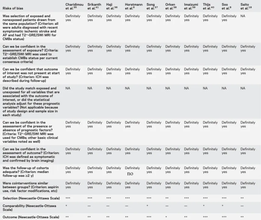
and was pooled with the rest of the cohorts in a post hoc sensitivity analysis. The risk of bias in the cohorts was moderate to low (table 2). Estimation of publication bias via the Egger test and the Begg test returned nonsignificant results (all p . 0.20). Follow-up was both retrospective and prospective, done face to face, and supplemented by telephone follow-up and patient record review as appropriate in all studies. All included
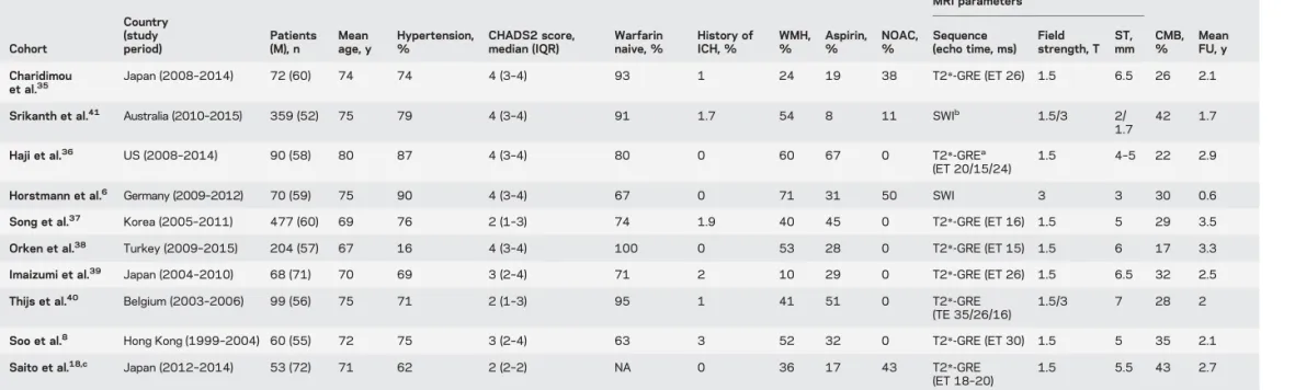
studies had.90% completeness of clinical follow-up. Cohorts had some variation in baseline characteristics and rates of concurrent aspirin use during follow-up (table 1). In 3 studies, a proportion of patients were using NOACs during follow-up; however, there was no difference in warfarin vs NOAC use based on CMBs. Seven studies used T2*-GRE MRI at 1.5T and 2 studies used SWI MRI at 1.5T or 3T to detect CMBs at baseline (table 1). The crude random-effects pooled prevalence of CMBs was 30% (95% CI 25%– 36%), and the pooled prevalence of$5 CMBs was 7% (95% CI 4%–10%).
A total of 22 patients had a symptomatic ICH and 81 patients had a recurrent ischemic stroke during follow-up. The pooled annual risk of ICH was 0.49% (95% CI 0.24%–0.745%) per year compared with a higher annual risk of ischemic stroke (2.22%, 95% CI 1.26%–3.18%) (p 5 0.001) but with mod-erate heterogeneity (figure 2). The pooled annual risk of ICH increased from 0.30% (95% CI 0.04%– 0.55%) per year among those without CMBs to 0.81% (95% CI 0.17%–1.45%) per year in patients with any CMBs (p 5 0.01) and 2.48% (95% CI 1.2%–6.2%) in patients with $5 CMBs (p 5 0.001). The annual pooled risk in patients with,5 CMBs was 0.49% (95% CI 0%–1.09%).
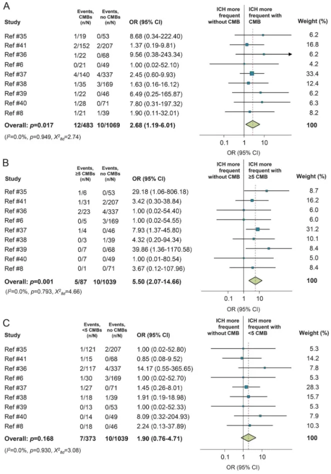
In the meta-analysis, CMB presence on MRI was associated with a higher risk of symptomatic ICH during follow-up compared with patients without CMBs (OR 2.68, 95% CI 1.19–6.01, p 5 0.017, figure 3A). This risk was particularly high for patients harboring $5 CMBs (OR 5.50, 95% CI 2.07– 14.66, p5 0.001), with some evidence of heteroge-neity across studies, but not for those with,5 CMBs (figure 3, B and C). There was no evidence for sub-stantial heterogeneity in any of the analyses. The pooled risk of recurrent ischemic stroke in the patient group with $5 CMBs was 0.29% (95% CI 0%– 2.77%) per year and for patients with ,5 CMBs was 0.02% (95% CI 0%–0.36%).
No significant heterogeneity was noted between studies for the main outcome (i.e., ICH) according to age, sex, hypertension, median CHADS2 score, ICH history, percentage of warfarin-naive patients, preva-lence of white matter hyperintensities, concurrent aspi-rin use, percentage of NOAC use, and MRI imaging parameters for CMB detection (all p. 0.1). Separate sensitivity analyses including only patients taking war-farin during follow-up were consistent and of an effect size similar to the main results presented here. The estimates did not change when the studies with zero cells in both groups were excluded post hoc.
Secondary analyses: CMBs distribution. In secondary analyses of CMBs distribution (vs no CMBs as the ref-erence group), strictly lobar CMBs (OR 2.88, 95% CI
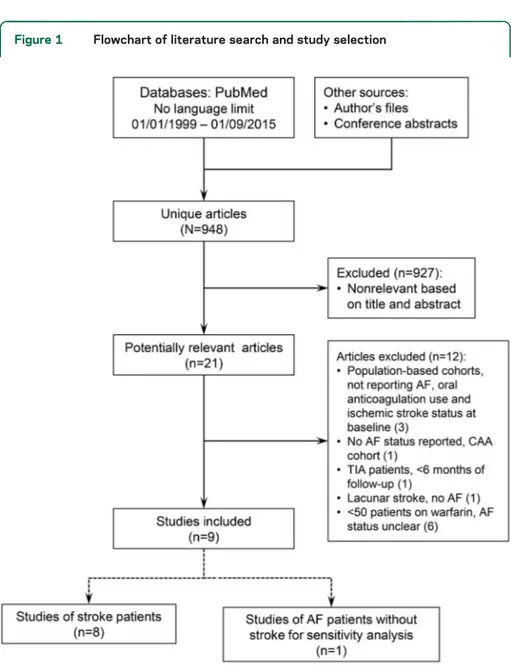
Figure 1 Flowchart of literature search and study selection
The search of electronic databases, authors’ own files, and abstract books from recent conferences yielded 948 publications. After initial screening, 21 reports were identified for full-text review. Of these reports that were reviewed in full text, 9 independent studies were identified as potentially eligible. Supplementary EMBASE searches over the same time period did not yield any extra articles. One included center also provided data on an addi-tional new cohort (n5 72), which was later published as a full article (Charidimou et al.35). AF
Table 1 Characteristics of included cohorts Cohort Country (study period) Patients (M), n Mean age, y Hypertension, % CHADS2 score, median (IQR) Warfarin naive, % History of ICH, % WMH, % Aspirin, % NOAC, % MRI parameters CMB, % Mean FU, y Sequence (echo time, ms) Field strength, T ST, mm Charidimou et al.35
Japan (2008–2014) 72 (60) 74 74 4 (3–4) 93 1 24 19 38 T2*-GRE (ET 26) 1.5 6.5 26 2.1
Srikanth et al.41 Australia (2010
–2015) 359 (52) 75 79 4 (3–4) 91 1.7 54 8 11 SWIb 1.5/3 2/ 1.7 42 1.7 Haji et al.36 US (2008 –2014) 90 (58) 80 87 4 (3–4) 80 0 60 67 0 T2*-GREa (ET 20/15/24) 1.5 4–5 22 2.9
Horstmann et al.6 Germany (2009–2012) 70 (59) 75 90 4 (3–4) 67 0 71 31 50 SWI 3 3 30 0.6
Song et al.37 Korea (2005–2011) 477 (60) 69 76 2 (1–3) 74 1.9 40 45 0 T2*-GRE (ET 16) 1.5 5 29 3.5
Orken et al.38 Turkey (2009–2015) 204 (57) 67 16 4 (3–4) 100 0 53 28 0 T2*-GRE (ET 15) 1.5 6 17 3.3
Imaizumi et al.39 Japan (2004–2010) 68 (71) 70 69 3 (2–4) 71 2 10 29 0 T2*-GRE (ET 26) 1.5 6.5 32 2.5
Thijs et al.40 Belgium (2003–2006) 99 (56) 75 71 2 (1–3) 95 1 41 51 0 T2*-GRE
(TE 35/26/16)
1.5/3 7 28 2
Soo et al.8 Hong Kong (1999–2004) 60 (55) 72 75 3 (2–4) 63 3 52 32 0 T2*-GRE (ET 30) 1.5 5 35 2.1
Saito et al.18,c Japan (2012
–2014) 53 (72) 71 62 2 (2–2) NA 0 36 17 43 T2*-GRE
(ET 18–20)
1.5 5.5 43 2.7
Abbreviations: CHADS25 congestive heart failure, hypertension, age $75 years, diabetes, stroke; CMB 5 cerebral microbleed; ET 5 echo time; FU 5 follow-up; GRE 5 gradient-recalled echo; ICH 5 intracerebral hemorrhage; IQR5 interquartile range; NA 5 not applicable; NOAC 5 non–vitamin K oral anticoagulant; ST 5 slice thickness; SWI 5 susceptibility-weighted imaging; WMH 5 white matter hyperintensities.
aMore than 90% of the cases.
bNinety-five percent patients had SWI; 20 patients had T2*-GRE (ET 15 milliseconds, slice thickness 7 mm). cStudy including patients with atrial fibrillation without ischemic stroke.
2320 Neurology 89 December 5, 2017
ª
2017
American
Academy
of
Neurology.
Unauthori
zed
reproduction
of
this
article
is
prohibited.
1.14–7.23, p 5 0.025) and mixed CMB pattern (OR 2.91, 95% CI 0.99–8.54, p 5 0.052), but not strictly deep CMBs (OR 2.43, 95% CI 0.83–7.14, p 5 0.107), were associated with the risk of ICH. CMB presence, burden, and distribution were not associated with recurrent ischemic stroke (all ORsz1.00). DISCUSSION This is a large-scale meta-analysis of the significance of CMBs in ischemic stroke pa-tients with AF treated with oral anticoagulation (mainly warfarin), a lingering question of clinical
relevance.9,19,20 In this study, we brought together
group-level data from .1,500 patients. The most important new finding is that the presence of $5 CMBs, regardless of their topographical distribution, identifies a patient group at particularly high risk for ICH. The risk of subsequent ICH in these patients of 2.8%/y may be high enough to be considered in the decision making for anticoagulation, at least in patients with characteristics similar to those of the included cohorts, after validation in international collaborative efforts.7,21
Table 2 Assessment of the 10 cohorts against each of 8 risks of bias of the Cochrane“Tool to Assess Risk of Bias in Cohort Studies”aand the
Newcastle–Ottawa Scaleb
Risks of bias
Charidimou
et al.35 Srikanthet al.41 Hajiet al.36 Horstmannet al.6 Songet al.37 Orkenet al.38 Imaizumiet al.39 Thijset al.40 Sooet al.8 Saitoet al.18
Was selection of exposed and nonexposed patients drawn from the same population? (Criterion: all were adults diagnosed with recent symptomatic ischemic stroke and AF and had T2*-GRE/SWI MRI for CMBs status) Definitely yes Definitely yes Definitely yes Definitely yes Definitely yes Definitely yes Definitely yes Definitely yes Definitely yes NA
Can we be confident in the assessment of exposure? (Criteria: T2*-GRE/SWI MRI was used to establish CMBs status per current consensus criteria) Definitely yes Definitely yes Definitely yes Definitely yes Definitely yes Definitely yes Definitely yes Definitely yes Definitely yes Definitely yes
Can we be confident that outcome of interest was not present at start of study? (Criterion: ICH was described during follow-up)
Definitely yes Definitely yes Definitely yes Definitely yes Definitely yes Definitely yes Definitely yes Definitely yes Definitely yes Definitely yes
Did the study match exposed and unexposed for all variables that are associated with the outcome of interest, or did the statistical analysis adjust for these prognostic variables? (Not applicable because of study design and sample size in each study)
NA NA NA NA NA NA NA NA NA NA
Can we be confident in the assessment of the presence or absence of prognostic factors? (Criteria: T2*-GRE/SWI MRI was used for CMBs; other basic clinical variables noted as well)
Definitely yes Definitely yes Definitely yes Definitely yes Definitely yes Definitely yes Definitely yes Definitely yes Definitely yes Definitely yes
Can we be confident in the assessment of outcome? (Criterion: ICH was defined as symptomatic and confirmed by brain imaging)
Definitely yes Definitely yes Definitely yes Definitely yes Definitely yes Definitely yes Definitely yes Definitely yes Definitely yes Definitely yes
Was the follow-up of cohorts adequate? (Criterion: median follow-up was‡2 y) Definitely yes Definitely yes Definitely yes Definitely
no
Definitelyyes Definitely yes Definitely yes Definitely yes Definitely yes Definitely yesWere cointerventions similar between groups? (Criterion: aspirin use, risk factor modifications, etc)
Definitely yes Definitely yes Definitely yes Definitely yes Definitely yes Definitely yes Definitely yes Definitely yes Definitely yes Definitely yes
Selection (Newcastle-Ottawa Scale) *** *** *** *** *** ** *** *** *** **
Comparability (Newcastle-Ottawa Scale)
* — — — * — — * * —
Outcome (Newcastle-Ottawa Scale) ** ** ** ** *** * ** *** *** **
Abbreviations: AF5 atrial fibrillation; CMB 5 cerebral microbleed; GRE 5 gradient-recalled echo; ICH 5 intracerebral hemorrhage; NA 5 not applicable; SWI5 susceptibility-weighted imaging.
aAn 8-item tool published by the Cochrane Methods Bias group: http://bmg.cochrane.org/sites/bmg.cochrane.org/files/uploads/Tool%20to%20Assess%
20Risk%20of%20Bias%20in%20Cohort%20Studies.pdf.
bRatings for the Newcastle-Ottawa Scale: a study can be awarded a maximum of 1 star for each numbered item within the selection (maximum 4 stars) and
outcome (maximum 3 stars) categories. A maximum of 2 stars can be given for comparability. For details, see Wells GA, Shea B, O’Connell D, et al. The Newcastle- Ottawa Scale (NOS) for assessing the quality of non-randomised studies in meta-analyses. Available at: http://www.ohri.ca/programs/clinical_ epidemiology/oxford.asp.
These results are largely in line with previous meta-analyses on CMBs in stroke.3,22,23 A
cross-sectional meta-analysis of stroke patients showed that CMBs are more common in warfarin- or antiplatelet-related ICH than spontaneous ICH without previous antithrombotic drug use.23A recent meta-analysis of
10 prospective ischemic stroke or TIA cohorts (n5
3,067) demonstrated that the presence of any CMB is associated with a higher risk of ICH.22These results
were extended by a more recent meta-analysis (n5 5,068) including similar cohorts, demonstrating that with increasing CMB burden (compared with no CMBs), the risk of ICH increases more steeply than that of ischemic stroke.3 However, the majority of
Figure 2 Annual absolute risk of stroke in included cohorts
Pooled annual rates of (A) incident symptomatic intracerebral hemorrhage and (B) recurrent ischemic stroke during follow-up in included studies. Weights are shown by the point estimate area. CI5 confidence interval.
2322 Neurology 89 December 5, 2017
these patients did not have AF and were treated with antiplatelet agents.
Our analysis shows that the overall annual risk for ICH in anticoagulant ischemic stroke patients with
AF is 0.24% to 0.74%, consistent with subgroup analyses for secondary prevention in randomized tri-als of vitamin K antagonist vs NOAC in stroke.24–26 In contrast, patients with$5 CMBs are at increased
Figure 3 Meta-analyses of CMBs and risk of future ICH in included studies
Forest plots of associations between (A) CMB presence, (B)$5 CMBs, and (C) ,5 CMBs and the risk of symptomatic ICH (main outcome) during follow-up. Plots show cohort-level and pooled estimates of these associations with no CMBs as the reference group in all analyses. The area of each shaded box is proportional to the weight of the cohort it represents. CI5 confidence interval; CMB5 cerebral microbleed; ICH 5 intracerebral hemorrhage; OR 5 odds ratio.
risk of ICH. Because of the devastating outcome of anticoagulation ICH and despite the effectiveness for preventing stroke due to AF, warfarin might carry more harm than benefit for those AF patients at great-est ICH risk.9,19A high burden of CMBs may thus tip
the balance in favor of risk rather than benefit for the decision to anticoagulate. In a hypothetical decision analysis model, an AF patient with typical risk for ischemic stroke (i.e., 4.5%/y if untreated) and pre-vious ICH should not be anticoagulated if the (untreated) risk for future recurrent ICH exceeds 1.4%/y.27 Of note, the ischemic stroke risk might
be even higher in patients with a previous ischemic stroke (similar to our meta-analysis population) if untreated. For this patient subgroup, greater empha-sis on the newer anticoagulants appears to be in order because of their generally lower risk of hemorrhagic side effects and ischemic outcomes.28 In the rapidly
evolving anticoagulation landscape, it is important to keep in mind that there are no data indicating whether the reduced ICH risk conferred by NOACs compared to warfarin extends to patients with multi-ple CMBs at high risk for future ICH or to patients with lobar ICH.29 Because of the class effect of all
NOACs in reducing ICH rates, these findings await replication in cohorts treated with NOACs.
In addition, no study to date has directly measured the additive effect of anticoagulation on ICH risk in CMB-positive patients.19 Percutaneous occlusion of
the left atrial appendage may be considered an another alternative stroke prevention therapy in pa-tients with$5 CMBs, but evidence for its safety in these patients with multiple CMBs or lobar ICH is very limited, and the effect of antithrombotic regi-mens required for the procedure remains to be defined.30,31 Removal or modification of additional
risk factors for ICH should include controlling blood pressure, stopping concurrent aspirin, replacing non-steroidal anti-inflammatory drugs with Cox inhibi-tors, and restricting alcohol intake as plausible strategies.19
Our study has notable strengths, including a large sample size from multiple cohorts and the homogene-ity of effect size direction across studies, all pointing to the validity of the results. We have included anal-yses based on a prespecified CMB cutoff based on previous data, presented secondary analyses based on CMBs distribution, and explored heterogeneity. Our meta-analysis has limitations that deserve careful consideration, especially methodological differences across studies. For example, studies had a small sam-ple size, variable and rather short follow-up (,3 years in general), and hence few outcome events (especially given the rarity of ICH as an outcome), leading to wide CIs around risk estimates. The MRI protocols for CMB detection were similar (including mainly
T2*-GRE at 1.5T, with echo times within a reason-able range) but not completely harmonized, poten-tially affecting the detection of CMBs.32In addition,
in our study, CMBs were determined by local inves-tigators and not by central readers, although all cen-ters used trained racen-ters and standardized definitions according to Standards for Reporting Vascular Changes on Neuroimaging criteria. Although treating clinicians in participating centers have not taken into account CMBs in routine clinical decision making for anticoagulation, this possibility cannot be completely excluded, especially for patients with very high CMB burden.
A potential important limitation of this study-level data approach is confounding of these estimates by other baseline variables related to future stroke risk in AF, including age, history of stroke, concurrent aspirin use, timing of starting oral anticoagulation after the baseline acute stroke, patient adherence to anticoagulation, and time in range. In addition, data on stroke severity and functional status at baseline were not available, although patients were fit enough to undergo MRI, were discharged on oral anticoagu-lation, and were to be followed up clinically. Despite our best efforts to adjust for certain available con-founding factors in meta-regression analyses, this is unlikely to have accounted for the full range of inter-actions between different variables. Hence, the pres-ent analyses do not adequately account for potpres-ential confounding by vascular risk factors or other varia-bles. None of the included cohorts were specifically designed to answer the specific questions explored in the present analysis. Moreover, all cohorts used clinical data, introducing some bias, and data were not collected with same accuracy or did not include the full range of covariates as research data. However, all studies showed a consistent direction of association between CMBs and ICH risk. Further stratified anal-yses are needed to explore the risk of future stroke and CMBs in Asian vs non-Asian patients and according to anticoagulation strategy, e.g., warfarin vs NOACs. Of relevance, the MRI substudy of A Phase III Study of Apixaban in Patients With Atrial Fibrillation (AVERROES) showed that patients on apixaban and patients on aspirin had a similar number of inci-dent CMBs.33
Our secondary analyses based on CMB distribution were not powerful enough to fully disentangle the role of different predominant small vessel disease patholo-gies in increasing the risk of ICH. This is due partly to confounding by CMB number across different topo-graphical categories. Determining patients’ underlying small vessel disease subtype on the basis of CMB dis-tribution would be a key next step for future analyses. Bleeding in other intracranial compartments (including subdural and rarely subarachnoid), especially in the
2324 Neurology 89 December 5, 2017
elderly, should also be investigated in further stud-ies and meta-analyses. There might be a selection bias because not all AF patients with stroke undergo an MRI with blood-sensitive MRI se-quences in clinical practice. Finally, although AF was defined by ECGs, either they were done at baseline stroke presentation or information was based on clinical history documentation of ECG findings. Presumed cardioembolic strokes (con-firmed on either MRI or CT) in the setting of AF were included in our analysis but with potential competing causes. Both can affect the generaliz-ability of our estimates.
Our data suggest that MRI could identify a specific subgroup of patients with ischemic stroke and AF (treated mainly with warfarin), namely those with $5 CMBs, who are at high risk for ICH events. Although MRI screening for CMBs is not needed before antithrombotic therapies initiation,34 in this
patient subgroup, the modification of anticoagulation strategy for stroke prevention might be relevant and in line with recent recommendations34and should be
explored in future studies and trials.9,20,34Given the
limitations discussed, these results can be generalized only to patient populations with overall characteris-tics similar to those of the patients included in our study, i.e., patients with generally mild strokes who are fit enough to undergo MRI at baseline and have CHADS2 scores between 2 and 4. Large interna-tional efforts in the field are underway to validate and expand these findings.7,21
AUTHOR CONTRIBUTIONS
Statistical analysis was conducted by Dr. A. Charidimou. A. Charidimou: study concept and design, systematic review, data analysis, write-up. Christopher Karayiannis, Tae-Jin Song, Dilek Necioglu Orken, Vincent Thijs, Robin Lemmens, Jinkwon Kim, Su Mei Goh, Thanh G. Phan, Cathy Soufan, Ronil V. Chandra, Lee-Anne Slater, Shamir Haji, Vincent Mok, Solveig Horstmann, Kam Tat Leung, Yuichiro Kawamura, Nobuyuki Saito, Naoyuki Hasebe, Tsukasa Saito, Lawrence K.S. Wong, Yannie Soo, Roland Veltkamp, Kelly D. Flemming, Toshio Imaizumi, Velandai Srikanth, and Ji Hoe Heo: data acquisition, critical revisions.
STUDY FUNDING No targeted funding reported.
DISCLOSURE
The authors report no disclosures relevant to the manuscript. Go to Neurology.org for full disclosures.
Received December 19, 2016. Accepted in final form September 18, 2017.
REFERENCES
1. Camm AJ, Kirchhof P, Lip GY, et al. Guidelines for the management of atrial fibrillation: the Task Force for the Management of Atrial Fibrillation of the European Society of Cardiology (ESC). Europace 2010;12:1360–1420.
2. Fang MC, Go AS, Chang Y, et al. Death and disability from warfarin-associated intracranial and extracranial hemorrhages. Am J Med 2007;120:700–705.
3. Wilson D, Charidimou A, Ambler G, et al. Recurrent stroke risk and cerebral microbleed burden in ischemic stroke and TIA: a meta-analysis. Neurology 2016;87:1501–1510. 4. Wang Z, Soo YO, Mok VC. Cerebral microbleeds: is
antithrombotic therapy safe to administer? Stroke 2014; 45:2811–2817.
5. Wardlaw JM, Smith EE, Biessels GJ, et al. Neuroimaging standards for research into small vessel disease and its con-tribution to ageing and neurodegeneration. Lancet Neurol 2013;12:822–838.
6. Horstmann S, Mohlenbruch M, Wegele C, et al. Prevalence of atrial fibrillation and association of previous antithrom-botic treatment in patients with cerebral microbleeds. Eur J Neurol 2015;22:1355–1362.
7. Charidimou A, Soo Y, Heo JH, Srikanth V; META-MI-CROBLEEDS Consortium. A call for researchers to join the META-MICROBLEEDS Consortium. Lancet Neurol 2016;15:900.
8. Soo YO, Yang SR, Lam WW, et al. Risk vs benefit of anti-thrombotic therapy in ischaemic stroke patients with cerebral microbleeds. J Neurol 2008;255:1679–1686.
9. Fisher M. MRI screening for chronic anticoagulation in atrial fibrillation. Front Neurol 2013;4:137.
10. Vanassche T, Lauw MN, Eikelboom JW, et al. Risk of ischaemic stroke according to pattern of atrial fibrillation: analysis of 6563 aspirin-treated patients in ACTIVE-A and AVERROES. Eur Heart J 2015;36:281–287a.
11. Fazekas F, Chawluk JB, Alavi A, Hurtig HI, Zimmerman RA. MR signal abnormalities at 1.5 T in Alzheimer’s dementia and normal aging. AJR Am J Roentgenol 1987;149:351–356.
12. Greenberg SM, Vernooij MW, Cordonnier C, et al. Cerebral microbleeds: a guide to detection and interpretation. Lancet Neurol 2009;8:165–174.
13. Gregoire SM, Chaudhary UJ, Brown MM, et al. The Micro-bleed Anatomical Rating Scale (MARS): reliability of a tool to map brain microbleeds. Neurology 2009;73:1759–1766. 14. Cordonnier C, Potter GM, Jackson CA, et al. Improving
interrater agreement about brain microbleeds: develop-ment of the Brain Observer MicroBleed Scale (BOMBS). Stroke 2009;40:94–99.
15. Greenberg SM, Eng JA, Ning M, Smith EE, Rosand J. Hemorrhage burden predicts recurrent intracerebral hemorrhage after lobar hemorrhage. Stroke 2004;35: 1415–1420.
16. Cochrane_Methods_Bias_Group. Tool to assess risk of bias in cohort studies. Available at: http://bmgcochraneorg/sites/ bmgcochraneorg/files/uploads/Tool to Assess Risk of20Bias in Cohort Studiespdf. Accessed August 1, 2015. 17. DerSimonian R, Laird N. Meta-analysis in clinical trials.
Controlled Clin Trials 1986;7:177–188.
18. Saito T, Kawamura Y, Sato N, et al. Non-vitamin K antagonist oral anticoagulants do not increase cerebral mi-crobleeds. J Stroke Cerebrovasc Dis 2015;24:1373–1377. 19. Diener HC, Selim MH, Molina CA, Greenberg SM. Embolic stroke, atrial fibrillation, and microbleeds: is there a role for anticoagulation? Stroke 2016;47:904–907. 20. Fisher M. Cerebral microbleeds: where are we now? Neurology
2014;83:1304–1305.
21. Microbleeds International Collaborative Network. Worldwide collaboration in the Microbleeds International Collaborative Network. Lancet Neurol 2016;15:1113–1114.
22. Charidimou A, Kakar P, Fox Z, Werring DJ. Cerebral microbleeds and recurrent stroke risk: systematic review and meta-analysis of prospective ischemic stroke and tran-sient ischemic attack cohorts. Stroke 2013;44:995–1001. 23. Lovelock CE, Cordonnier C, Naka H, et al. Antithrom-botic drug use, cerebral microbleeds, and intracerebral hemorrhage: a systematic review of published and unpub-lished studies. Stroke 2010;41:1222–1228.
24. Diener HC, Connolly SJ, Ezekowitz MD, et al. Dabiga-tran compared with warfarin in patients with atrial fibril-lation and previous transient ischaemic attack or stroke: a subgroup analysis of the RE-LY trial. Lancet Neurol 2010;9:1157–1163.
25. Hankey GJ, Patel MR, Stevens SR, et al. Rivaroxaban com-pared with warfarin in patients with atrial fibrillation and previous stroke or transient ischaemic attack: a subgroup analysis of ROCKET AF. Lancet Neurol 2012;11:315–322. 26. Easton JD, Lopes RD, Bahit MC, et al. Apixaban compared with warfarin in patients with atrial fibrillation and previous stroke or transient ischaemic attack: a subgroup analysis of the ARISTOTLE trial. Lancet Neurol 2012;11:503–511. 27. Eckman MH, Rosand J, Knudsen KA, Singer DE, Greenberg
SM. Can patients be anticoagulated after intracerebral hem-orrhage? A decision analysis. Stroke 2003;34:1710–1716. 28. Connolly SJ, Eikelboom J, Joyner C, et al. Apixaban in
patients with atrial fibrillation. N Engl J Med 2011;364: 806–817.
29. Ruff CT, Giugliano RP, Braunwald E, et al. Comparison of the efficacy and safety of new oral anticoagulants with warfarin in patients with atrial fibrillation: a meta-analysis of randomised trials. Lancet 2014;383:955–962. 30. Lewalter T, Kanagaratnam P, Schmidt B, et al. Ischaemic
stroke prevention in patients with atrial fibrillation and high bleeding risk: opportunities and challenges for percu-taneous left atrial appendage occlusion. Europace 2014;16: 626–630.
31. Horstmann S, Zugck C, Krumsdorf U, et al. Left atrial appendage occlusion in atrial fibrillation after intracranial hemorrhage. Neurology 2014;82:135–138.
32. Charidimou A, Werring DJ. Cerebral microbleeds: detec-tion, mechanisms and clinical challenges. Future Neurol 2011;6:587–611.
33. O’Donnell MJ, Eikelboom JW, Yusuf S, et al. Effect of apixaban on brain infarction and microbleeds: AVERROES-MRI assessment study. Am Heart J 2016; 178:145–150.
34. Smith EE, Saposnik G, Biessels GJ, et al. Prevention of stroke in patients with silent cerebrovascular disease: a sci-entific statement for healthcare professionals from the American Heart Association/American Stroke Association. Stroke 2017;48:e44–e71.
35. Charidimou A, Inamura S, Nomura T, Kanno A, Kim SN, Imaizumi T. Cerebral microbleeds and white matter hyperintensities in cardioembolic stroke patients due to atrial fibrillation: single-centre longitudinal study. J Neurol Sci 2016;369:263–267.
36. Haji S, Planchard R, Zubair A, et al. The clinical relevance of cerebral microbleeds in patients with cerebral ischemia and atrial fibrillation. J Neurol 2016;263:238–244. 37. Song TJ, Kim J, Song D, et al. Association of cerebral
microbleeds with mortality in stroke patients having atrial fibrillation. Neurology 2014;83:1308–1315.
38. Orken DN, Uysal E, Timer E, Kuloglu-Pazarci N, Mumcu S, Forta H. New cerebral microbleeds in ischemic stroke patients on warfarin treatment: two-year follow-up. Clin Neurol Neurosurg 2013;115:1682–1685.
39. Imaizumi T, Inamura S, Kohama I, Yoshifuji K, Nomura T, Komatsu K. Antithrombotic drug uses and deep intracerebral hemorrhages in stroke patients with deep cerebral microbleeds. J Stroke Cerebrovasc Dis 2013;22:869–875.
40. Thijs V, Lemmens R, Schoofs C, et al. Microbleeds and the risk of recurrent stroke. Stroke 2010;41: 2005–2009.
41. Karayiannis C, Soufan C, Chandra RV, et al. Preva-lence of brain MRI markers of hemorrhagic risk in patients with stroke and atrial fibrillation. Front Neu-rol 2016;7:151.
BrainPAC
BrainPAC is the American Academy of Neurology’s (AAN) federal political action committee. • Since its inception, more than 3,600 AAN members have contributed $2,000,000 to
BrainPAC.
• BrainPAC contributed more than $600,000 to individuals who ran for election in 2016, including several first-time candidates.
• During the 2016 congressional election, 92% of candidates supported by BrainPAC won election to the US Congress.
BrainPAC supports both Democrats and Republicans who support issues important to the practice of neurology and the care of patients with neurologic conditions. US AAN members are invited to learn more at BrainPAC.org.
2326 Neurology 89 December 5, 2017
DOI 10.1212/WNL.0000000000004704
2017;89;2317-2326 Published Online before print November 8, 2017
Neurology
Andreas Charidimou, Christopher Karayiannis, Tae-Jin Song, et al.
patients with AF
Brain microbleeds, anticoagulation, and hemorrhage risk: Meta-analysis in stroke
This information is current as of November 8, 2017
Services
Updated Information &
http://n.neurology.org/content/89/23/2317.full
including high resolution figures, can be found at:
References
http://n.neurology.org/content/89/23/2317.full#ref-list-1
This article cites 40 articles, 14 of which you can access for free at:
Citations
http://n.neurology.org/content/89/23/2317.full##otherarticles
This article has been cited by 1 HighWire-hosted articles:
Subspecialty Collections http://n.neurology.org/cgi/collection/intracerebral_hemorrhage Intracerebral hemorrhage http://n.neurology.org/cgi/collection/class_iii Class III e http://n.neurology.org/cgi/collection/all_cerebrovascular_disease_strok
All Cerebrovascular disease/Stroke
following collection(s):
This article, along with others on similar topics, appears in the
Permissions & Licensing
http://www.neurology.org/about/about_the_journal#permissions
its entirety can be found online at:
Information about reproducing this article in parts (figures,tables) or in
Reprints
http://n.neurology.org/subscribers/advertise
Information about ordering reprints can be found online:
rights reserved. Print ISSN: 0028-3878. Online ISSN: 1526-632X.
1951, it is now a weekly with 48 issues per year. Copyright © 2017 American Academy of Neurology. All ® is the official journal of the American Academy of Neurology. Published continuously since