2 0 1 3 년 2 월 박사학위논문
한국인 눈확위구멍과 눈확아래구멍의 형태계측학적 분석
조선대학교 대학원
치의학과
정 구 수
[UCI]I804:24011-200000263869
한국인 눈확위구멍과 눈확아래구멍의 형태계측학적 분석
Mor phomet r i canal ysi soft hesupr aor bi t alandi nf r aor bi t al f or ami nai n Kor eans
2013년 2월 25일
조선대학교 대학원
치의학과
정 구 수
한국인 눈확위구멍과 눈확아래구멍의 형태계측학적 분석
지도교수 김 흥 중
이 논문을 치의학 박사학위신청 논문으로 제출함
2012년 10월
조선대학교 대학원
치의학과
정구수의 박사학위 논문을 인준함
위원장 연세대학교 교 수 김 희 진 인
위 원 조선대학교 교 수 국 중 기 인
위 원 조선대학교 교 수 김 도 경 인
위 원 조선대학교 교 수 김 춘 성 인
위 원 조선대학교 교 수 김 흥 중 인
2012년 12월
조선대학교 대학원
목 차
ABSTRACT . . . . . . . . . . . . . . . . . . . . . . . . . . . . . . . . . . . . . . . . . . . . . . . . . . . . . . . . . . . . . . . . . . . . . . . . . . . . . . . . . . . . . . . . . . . . . . . . . . . . . . ⅳ
I .서론 . . . . . . . . . . . . . . . . . . . . . . . . . . . . . . . . . . . . . . . . . . . . . . . . . . . . . . . . . . . . . . . . . . . . . . . . . . . . . . . . . . . . . . . . . . . . . . . . . . . . . . . . . . . . . . . . . . . . 1
I I .재료 및 방법 . . . . . . . . . . . . . . . . . . . . . . . . . . . . . . . . . . . . . . . . . . . . . . . . . . . . . . . . . . . . . . . . . . . . . . . . . . . . . . . . . . . . . . . . . . . . . . . . . . 3
Ⅲ.결과. . . . . . . . . . . . . . . . . . . . . . . . . . . . . . . . . . . . . . . . . . . . . . . . . . . . . . . . . . . . . . . . . . . . . . . . . . . . . . . . . . . . . . . . . . . . . . . . . . . . . . . . . . . . . . . . . . . . 5
Ⅳ.고찰 . . . . . . . . . . . . . . . . . . . . . . . . . . . . . . . . . . . . . . . . . . . . . . . . . . . . . . . . . . . . . . . . . . . . . . . . . . . . . . . . . . . . . . . . . . . . . . . . . . . . . . . . . . . . . . . . . . 8
Ⅴ.결론 . . . . . . . . . . . . . . . . . . . . . . . . . . . . . . . . . . . . . . . . . . . . . . . . . . . . . . . . . . . . . . . . . . . . . . . . . . . . . . . . . . . . . . . . . . . . . . . . . . . . . . . . . . . . . . . . 1 1
참고문헌 . . . . . . . . . . . . . . . . . . . . . . . . . . . . . . . . . . . . . . . . . . . . . . . . . . . . . . . . . . . . . . . . . . . . . . . . . . . . . . . . . . . . . . . . . . . . . . . . . . . . . . . . . . . . . . . 1 3
표 목 차
Table 1. Distances from the supraorbital foramen, medial canthus, and infraorbitalforamentofacialmidline...5
Table 2.Location of the supraorbitalforamen with reference to the medial canthus...6
Table 3.Location of the infraorbital foramen with reference to the medial canthus...6
Table4.Diameterandshapeofinfraorbitalforamen...7
Table5.Comparison between stuideson therelation to diameterand shapeof theinfraorbitalforamen...10
도 목 차
Fig.1.The parameters ofthesupraorbitaland infraorbitalforamina in relation tomedialcanthus...4
ABSTRACT
Morphometricanalysisofthesupraorbitaland infraorbitalforaminain Koreans
Jeong,Goosoo
Advisor:Prof.Kim,Heung-Joong,Ph.D.
DepartmentofDentistry,
GraduateSchoolofChosun University
Knowledge of the location of the maxillo-facial foramina is essential for regionalnerveblocksand endoscopic surgicalproceduresto avoid nerveinjury passing through theseforamina.Thepurposesofthisstudy wereto determine the locations ofthe supraorbitalforamen (SOF) and the infraorbitalforamen (IOF)related to medialcanthus(MC),and to analyzethemorphology ofthese foramina.
Thirty-two embalmed cadavers (64 sides,mean age:64.1 years)and 33 dry skulls(66sides)wereused.Thedistancesfrom theSOF,IOF,andMC tofacial midline were directly measured on the cadavers using digitalVernier caliper. TheverticalandhorizontaldistancesoftheSOF andIOF relativetothemedial canthus were indirectly measured on the digital photographs using image analyzer software.The verticaland horizontaldiameters ofthe IOF,and its
locationinrelationtomaxillarytoothwereevaluatedonthedryskull.Statistical analysis was performed using one-way ANOVA with declaration ofsignificant differencewhenP< 0.05.
ThemeandistancesofSOF,MC,andIOF tothefacialmidlinewere24.13㎜, 15.00㎜,and29.11㎜,respectively.TheSOF waslocated18.99㎜ superiorand 9.05㎜ lateraltothemedialcanthus.Thedistancebetween themedialcanthus and the SOF was 22.67 ㎜,and the verticalangle (Angle 1)between these structureswas24.36˚ superolaterally.TheIOF waslocated26.69㎜ inferiorand 13.53㎜ lateraltothemedialcanthus.Thedistancebetween themedialcanthus andIOF was30.82㎜,andtheverticalangle(Angle2)betweenthesestructures was26.59˚ inferolaterally.In thethisstudy,spraorbitalnotch (SON)wasfound more frequently than the SOF.The mean verticaland horizontaldiameters of IOF were3.36㎜,3.45 ㎜,respectively.IOF wasmostcommonly found in the sameverticalplanewiththesecondupperpremolar.
Inconclusion,theseresultsareimportantforperforminglocalanesthetic,facial plastic surgery,and other invasive procedures in the forehead and periorbital region to prevent injury of neurovascular bundles passing through these foramina.
...
Key Words: supraorbital foramen, infraorbital foramen, medial canthus, soft-tissuelandmark
Ⅰ.서 론
악안면영역에서 시술부위 해부학적 구조물의 위치와 형태에 관한 정확한 지식은 국소마취와 얼굴성형,악안면 수술과정에서 신경혈관다발의 손상을 방지하기 위해 중요하다(Hwang과 Baik 1999,Aziz등 2000).특히,얼굴부위에 넓게 분포하는 감 각신경인 눈확위신경과 눈확아래신경이 나오는 눈확위구멍(패임)과 눈확아래구멍은 얼굴상처의 봉합,흉터재생,미용성형을 위한 다양한 시술 시 구조물 주위의 과도한 절개나 봉합으로 인하여 출혈이나 감각이상,신경통 등과 같은 부작용을 유발할 수 있기 때문에 고려되어야 할 구조물이다(Webster등 1986,Caputi와 Firetto 1997, Curight등 2003,Saylam 등 2003,Gupta2008).
눈확위구멍(패임)은 눈확위신경과 혈관의 통로로써 이마뼈의 눈확위모서리 안쪽 부위에 위치한다.눈확위신경은 눈시경의 가지인 이마신경의 연속가지이며,눈확위 패임 또는 눈확위구멍으로 나와 위 눈꺼풀,이마 및 머리덮개피부에 분포한다(Kim 2011).
눈확아래구멍은 위턱뼈 몸통의 앞면에서 눈확아래모서리로부터 약 8~10㎜ 아래 에 위치한다.눈확아래신경은 위턱신경의 가지로써 아래눈확틈새를 지나 눈확으로 들어간 후,눈확아래관의 연속인 눈확아래구멍을 통해 얼굴부위로 나와서 위턱굴 점막,위턱 앞니,작은어금니부위와 잇몸,아래눈꺼풀,코 바깥쪽,위 입술 등의 피 부와 점막에 분포한다(Moore와 Dalley2007).
계측점에 관련한 기존의 많은 연구에서 다양한 인종의 눈확위구멍과 눈확아래구 멍의 위치와 형태를 분석하였다(Canan 등 1999,Apinhasmit등 2006,Song 등 2007,Chrcanovic등 2011,Takahashi등 2011,Lee등 2012,Zheng 등 2012).최근 에는 주름제거술,안검미용성형 등의 확산으로 인하여 이들의 위치와 형태의 다양 성에 대한 연구가 진행되었다(Karakas등 2002,Huanmanop등 2007).
눈확위구멍과 눈확아래구멍의 위치와 형태에 관한 연구들은 머리뼈에서 경조직 계측점과 관련한 연구가 대부분이었으며,시신에서 연조직 계측점을 이용한 연구는 아직 미흡한 실정이다(Gupta2008,Liu등 2011,Zheng 등 2012).많은 연구의 결과 들로 인해 상당한 정확도를 가지고 눈확위구멍과 눈확아래구멍의 위치와 형태를 예 측할 수 있게 되었으나 경조직 계측점은 인체 내에서 쉽게 인지할 수 없으며,특히
눈확아래구멍은 눈확위구멍에 비해 그 형태와 위치가 다양하여 임상적용 시 더 많 은 어려움이 있다(Berge와 Bergman2001).따라서 이러한 경조직 계측점의 단점을 보완하면서 이들의 위치를 더 정확하게 설명할 수 있는 기준점이 필요하다.
본 연구에서는 시신에서 연조직 계측점인 안쪽 눈꼬리점을 이용하여 눈확위구멍 과 눈확아래구멍의 위치를 확인하고 더불어 머리뼈에서 이들의 형태를 분석하여 악 안면 수술과 얼굴성형 같은 임상시술 시 유용한 해부학적 자료를 제공하고자 한다.
Ⅱ.재료 및 방법
1.재료
본 연구에서는 조선대학교 의학전문대학원 해부학 교육용 시신 32구(64쪽;남자 24구,여자 8구)를 사용하였으며 사망 시 평균 연령은 64.1세(29~91세)였다.눈확위 구멍과 눈확아래구멍을 노출시키기 위해 골막을 포함하여 주변의 연조직을 모두 제 거한 후 디지털 카메라(D90;Nikon,Tokyo,Japan)를 사용하여 눈금자를 포함한 각 표본의 정면 사진을 촬영하였다.경조직에서의 분석을 위해 해부학 실습용 머리뼈 33개(66쪽)를 추가로 사용하였으며,사망 시 연령과 성별은 알 수 없었다.
2.측정
본 연구에서는 얼굴부위 연조직 계측점 중 하나인 안쪽 눈꼬리점(medialcorner ofthe eye;medialcanthus)을 기준으로 사용하였다.각 시신에서 디지털 캘리퍼 (CD-15CP,MitutoyoCo.,Kawasaki,Japan)를 사용하여 얼굴 정중선으로부터 눈확 위구멍,안쪽 눈꼬리점,눈확아래구멍까지의 거리를 0.01㎜ 수준까지 각각 측정하였 다.그리고 디지털 카메라로 촬영한 사진을 iSolution Capture(iMT,Vancouver, Canada)프로그램을 이용하여 안쪽 눈꼬리점을 기준으로 하여 눈확위구멍과 눈확 아래구멍의 위치를 측정하였다.모든 측정값들은 눈확위구멍과 눈확아래구멍의 중 심을 기준으로 측정하였다.
각 머리뼈에서 육안으로 눈확위구멍 또는 패임의 빈도를 확인하였고,디지털 캘 리퍼(CD-15CP,MitutoyoCo.,Kawasaki,Japan)를 사용하여 눈확아래구멍의 수직, 수평직경을 계측하여 형태를 분류하였다.눈확아래구멍과 위턱 치아와의 관계를 측 정하기 위해 눈확아래구멍의 중심을 지나며 얼굴 정중선과 평행한 수직선을 아래쪽 으로 연장하여 위턱 치아와 만나는 지점을 확인하였다.
3.통계 분석
모든 측정값의 통계분석은 SPSS 12.0(Chicago,IL,USA)프로그램을 이용하여 one-way ANOVA를 시행하였다.해부학적 지식이 있는 두 사람이 동일한 방법에 의하여 모든 표본을 반복 측정한 후,계측자간의 차이와 각 계측항목의 좌우 차이 를 분석하였다.분석 결과 계측자간의 유의적 차이가 나타나지 않아(P=0.843)두 명 의 계측자가 측정한 값의 평균을 최종 계측 자료로 사용하였다.또한,각 계측항목 의 좌우 역시 유의적 차이를 나타내지 않아 동일한 집단으로 간주하였다.본 연구 에서는 성별,연령간의 유의적 차이는 고려되지 않았다.모든 측정값들은 평균과 표 준편차로 나타내었으며 각 항목의 분석은 유의수준 0.05에서 이루어졌다.
Fig.1.The parameters ofthesupraorbitaland infraorbitalforamina in relation to medial canthus. FM, facial midline; SOF, supraorbital foramen; IOF, infraorbitalforamen;MC,medialcanthus;P1,cross pointbetween MC vertical line and supraorbital foramen horizontal line; P2,cross point between MC verticallineandinfraorbitalforamenhorizontalline;Angle1,verticalanglefrom MC to supraorbitalforamen; Angle 2,verticalangle from MC to infraorbital
Mean±SD (㎜)
Right Left Combined P SOF-FM 22.86±3.18 25.40±5.77 24.13±4.78 0.066
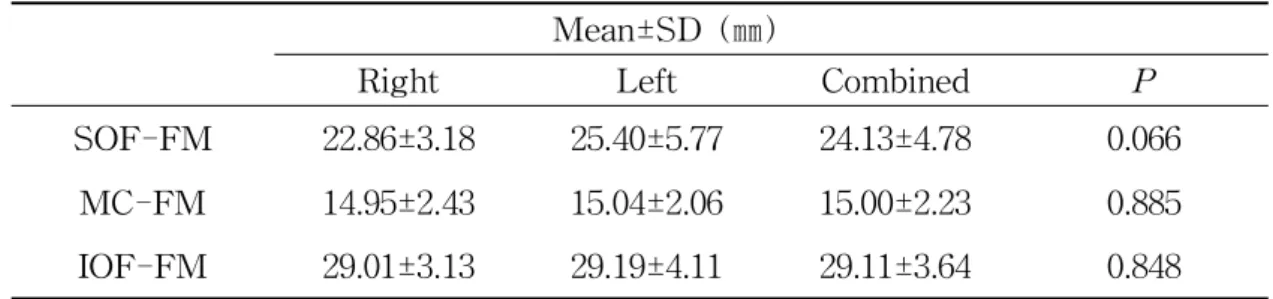
MC-FM 14.95±2.43 15.04±2.06 15.00±2.23 0.885 IOF-FM 29.01±3.13 29.19±4.11 29.11±3.64 0.848
Ⅲ.결 과
1.얼굴 정중선으로부터 계측점까지의 거리
얼굴 정중선과 눈확위구멍 사이의 평균거리는 24.13±4.78㎜로 얼굴 정중선과 눈확 아래구멍 사이의 평균거리인 29.11±3.64㎜보다 짧았다.얼굴 정중선과 안쪽 눈꼬리 점 사이의 평균거리는 15.00±2.23㎜였다.얼굴 정중선을 기준으로 양쪽 동일구조물 과 계측점 사이의 거리는 통계적 유의성을 나타내지 않았다(Table1).
Table1.Distancesto thesupraorbitalforamen,medialcanthus,and infraorbital foramenfrom facialmidline
Abbreviations;SOF,supraorbitalforamen;IOF,infraorbitalforamen;MC,medial canthus;FM,facialmidline.Thesignificancedifferencewasconsidered atP<
0.05.
2.안쪽 눈꼬리점을 기준으로 한 눈확위구멍의 위치
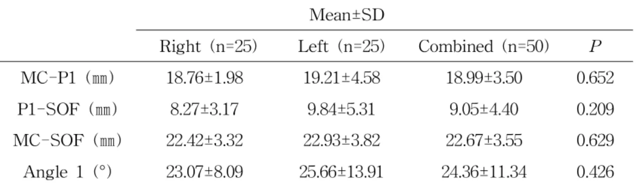
눈확위구멍은 안쪽 눈꼬리점에서 위쪽으로 18.99±3.50㎜,가쪽으로 9.05±4.40㎜에 위치하였다.안쪽 눈꼬리점과 눈확위구멍 사이의 거리는 22.67±3.55㎜이었으며,안쪽 눈꼬리점과 눈확위구멍 사이의 수직적 각도(angle 1)는 위가쪽으로 24.36±11.34°였 다(Table2).
Mean±SD
Right(n=25) Left(n=25) Combined(n=50) P MC-P1(㎜) 18.76±1.98 19.21±4.58 18.99±3.50 0.652 P1-SOF (㎜) 8.27±3.17 9.84±5.31 9.05±4.40 0.209 MC-SOF (㎜) 22.42±3.32 22.93±3.82 22.67±3.55 0.629 Angle1(°) 23.07±8.09 25.66±13.91 24.36±11.34 0.426
Mean±SD
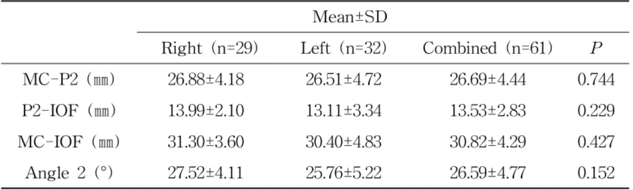
Right(n=29) Left(n=32) Combined(n=61) P MC-P2(㎜) 26.88±4.18 26.51±4.72 26.69±4.44 0.744 P2-IOF (㎜) 13.99±2.10 13.11±3.34 13.53±2.83 0.229 Table 2.Location of the supraorbitalforamen with reference to the medial canthus
Abbreviations;SOF,supraorbitalforamen;MC,medialcanthus;P1,cross point between medialcanthus verticalline and supraorbitalforamen horizontalline; Angle 1, vertical angle from medial canthus to supraorbital foramen. The significancedifferencewasconsideredatP< 0.05.
3.안쪽 눈꼬리점을 기준으로 한 눈확아래구멍의 위치
눈확아래구멍은 안쪽 눈꼬리점에서 아래쪽으로 26.69±4.44㎜,가쪽으로 13.53±2.83
㎜에 위치해 있었다.안쪽 눈꼬리점과 눈확아래구멍 사이의 거리는 30.82±4.29㎜이 었으며,안쪽 눈꼬리점과 눈확아래구멍 사이의 수직적 각도(angle2)는 아래가쪽으 로 26.59±4.77°였다(Table3).
Table 3.Location of the infraorbital foramen with reference to the medial canthus
Angle 2, vertical angle from medial canthus to infraorbital foramen. The significancedifferencewasconsideredatP< 0.05.
4.머리뼈에서 눈확위구멍과 눈확아래구멍의 형태
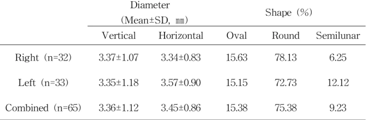
눈확위구멍의 형태는 눈확위패임이 56.60%,눈확위구멍이 43.40%로 패임이 더 많 았다.눈확아래구멍의 수직 직경은 3.36±1.12㎜,수평 직경은 3.45±0.86㎜였으며,형 태는 원형이 75.38%로 가장 많았고,그 다음으로 타원형(15.38%)과 반달형(9.23%) 을 보였다(Table4).눈확아래구멍은 위턱 둘째작은어금니와 같은 수직선상에 위치 하고 있는 경우가 90%로 가장 많은 빈도를 보였다.
Table4.Diameterandshapeofinfraorbitalforamen Diameter
(Mean±SD,㎜) Shape(%)
Vertical Horizontal Oval Round Semilunar Right(n=32) 3.37±1.07 3.34±0.83 15.63 78.13 6.25
Left(n=33) 3.35±1.18 3.57±0.90 15.15 72.73 12.12 Combined(n=65) 3.36±1.12 3.45±0.86 15.38 75.38 9.23
Ⅳ.고 찰
눈확위구멍(패임)과 눈확아래구멍은 다양한 악안면 수술 과정 중에 일반적으로 접하는 구조물이다.이 구멍들을 빠져나온 눈확위신경과 눈확아래신경은 얼굴과 이 마 등 넓은 부위에 분포하고 있기 때문에 국소마취와 수술과정에서 쉽게 손상을 받 을 수 있다(Moore와 Dalley 2007,Drake등 2010).최근 미용에 대한 인식이 높아 지면서 증가하고 있는 다양한 내시경 시술은 좁은 시야를 통해 이루어지므로 신경 혈관다발의 손상 방지를 위해 눈확위구멍과 눈확아래구멍의 위치와 형태를 정확하 게 인지하는 것이 중요하다(Curight등 2003,Suresh등 2006,Song등 2007).
대부분의 기존 연구들은 머리뼈에서 경조직 계측점을 이용하여 눈확위구멍과 눈 확아래구멍의 위치와 형태를 분석하였으나,이러한 연구 결과들은 임상에서 적용하 기에는 많은 어려움이 있다(Kazkayasi등 2001,Agthong 등 2005,Singh2011).이 러한 단점을 보완하고자 본 연구에서는 얼굴부위의 연조직 계측점 중 하나인 안쪽 눈꼬리점을 사용하여 그 위치를 계측하고,또한 머리뼈를 이용하여 이들의 형태를 분석하였다.
Liu 등(2011)이 시신에서 연조직 계측점인 안쪽 눈꼬리점을 기준으로 눈확위구멍 의 위치를 계측한 결과 눈확위구멍은 안쪽 눈꼬리점에서 위쪽으로 25.09㎜,가쪽으 로 7.86㎜,위가쪽으로 72.54°지점에 위치해 있었다.또한,Zheng 등(2012)이 동일 한 계측점을 이용하여 눈확위구멍의 위치를 확인한 결과 눈확위구멍은 안쪽 눈꼬리 점으로부터 위쪽으로 23.11㎜,가쪽으로 9.48㎜,위가쪽으로 72.54°지점에 위치해 있 었다.이와 동일한 계측점인 안쪽 눈꼬리점을 기준으로 한 본 연구에서 눈확위구멍 은 안쪽 눈꼬리점으로부터 위쪽으로 18.99㎜,가쪽으로 9.05㎜,위가쪽으로 24.36°지 점에 위치해 있으며,본 연구의 결과들은 Zheng 등(2012)의 연구와는 유사하였으나 Liu등(2011)에 의한 연구에 비해서는 전체적으로 약간 낮은 값을 나타내었다.
Song 등(2007)이 연조직 계측점인 콧방울점을 기준으로 눈확아래구멍의 위치를 확인한 결과 눈확아래구멍은 콧방울점에서 가쪽으로 1.6㎜,위쪽으로 14.1㎜,가위쪽
이 안쪽 눈꼬리점을 기준으로 눈확아래구멍의 위치를 확인한 결과 눈확아래구멍은 안쪽 눈꼬리점에서 아래쪽으로 25.75㎜,가쪽으로 10.67㎜,아래가쪽으로 66.77°지점 에 위치해 있었다.Zheng 등(2012)은 콧방울점과 안쪽 눈꼬리점을 기준점으로 사용 하여 눈확아래구멍의 위치를 계측하였는데,눈확아래구멍은 콧방울점에서 가쪽으로 6.09㎜,위쪽으로 11.22㎜,가위쪽으로 61.7°지점에 위치해 있었으며,안쪽 눈꼬리점 으로부터는 아래쪽으로 24.81㎜,가쪽으로 10.89㎜,아래가쪽으로 66.5°지점에 위치 해 있었다.안쪽 눈꼬리점을 기준점으로 이용한 본 연구에서 눈확아래구멍은 안쪽 눈꼬리점으로부터 아래쪽으로 26.69㎜,가쪽으로 13.53㎜,아래가쪽으로 26.59°지점 에 위치해 있으며,본 연구의 결과들은 동일한 계측점을 사용한 Liu 등(2011)과 Zheng등(2012)에 의한 연구와 비교하여 전체적으로 약간 높은 수치를 나타내었다.
한국인에서 눈확위구멍을 지나는 시상면을 기준으로 눈확아래구멍의 위치적 관 계를 분석한 결과 눈확아래구멍은 눈확위구멍보다 더 가쪽에 위치해 있었다.이는 위턱뼈의 일부분인 눈확아래구멍이 광대활의 가쪽 성장에 의해 영향을 받기 때문일 것이다(Chung 등 1995).Apinhasmit등(2006)의 연구에 의하면 머리뼈에서 눈확아 래구멍은 얼굴정중선에서 가쪽으로 28.43㎜ 떨어진 지점에 위치해 있다.본 연구에 서 얼굴정중선에서 눈확위구멍,안쪽 눈꼬리점,눈확아래구멍까지의 평균거리는 각 각 24.13㎜,15.00㎜,29.11㎜로 눈확아래구멍은 눈확위구멍과 안쪽 눈꼬리점보다 더 가쪽에 위치하고 있었다.
눈확위구멍과 눈확위패임의 발현 빈도에 관한 많은 연구가 이루어졌으며,그 양 상은 인종에 따라 다양하다.Turhan-Haktanir등(2008)은 눈확위패임이 눈확위구멍 보다 더 많이 나타난다고 보고하였으며,본 연구에서도 눈확위패임(56.60%)이 눈확 위구멍(43.40%)보다 더 많이 관찰되었다.이는 북서인도인 머리뼈에 관한 연구에서 눈확위패임(54.4%)이 눈확위구멍(45.6%)보다 더 많이 나타난다는 Gupta(2008)의 연 구 결과와 비슷하다.기존의 연구들에서 눈확위구멍의 발현률은 다른 인종에 비해 동북아시아인과 북미인에서 더 높다고 보고하였다(Hanihara와 Ishida 2001,Cheng 등 2006).눈확위신경혈관다발은 패임에 비해 상대적으로 눈확위구멍 위치에 고정되 어 있기 때문에 눈확위구멍의 발현률이 높은 인종의 경우 이마와 눈썹부위에서 절 개,봉합과 같은 외과적 수술시 더 큰 주의가 필요하다(Cheng등 2006).
본 연구에서 눈확아래구멍의 수직,수평 직경은 각각 3.36㎜,3.45㎜로 인도인 머 리뼈에서 측정한 Boopathi등(2010)에 의한 연구 결과에 비하여 그 크기가 더 크게
나타났다.눈확아래구멍의 형태는 원형이 75.38%로 가장 많이 나타났으며,타원형 (15.38%)과 반달형(9.23%)도 관찰되었다.이는 인도인 머리뼈에서 눈확아래구멍의 형태를 확인한 결과 타원형이 70.8%,원형이 29%였다는 Singh(2011)의 연구 결과 와 상반된다(Table5).
Aziz등(2000)이 눈확아래구멍은 위턱 치아와 관련하여 일반적으로 위턱 첫째작 은어금니와 같은 수직선상에 존재한다고 보고하였으나,본 연구에서는 둘째작은어 금니와 같은 수직선상에 위치해 있는 경우가 90%로 가장 높은 빈도를 보였다.한국 인에서 위턱뼈의 앞쪽벽이나 눈확아래모서리 부위의 골절로 인해 눈확아래구멍의 위치를 구분하기 힘든 경우 둘째작은어금니는 하나의 기준점으로 이용될 수 있을 것으로 생각된다.
본 연구에서 안쪽 눈꼬리점을 기준으로 측정한 눈확위구멍과 눈확아래구멍의 위 치에 대한 결과들은 경조직 계측점의 임상적용의 어려움을 보완해 줄 것이며,이들 의 크기 및 형태에 관한 결과들은 한국인에서 국소마취 시 유용한 해부학적 지표를 제공할 것이다.
Table5.Comparison between stuideson therelation to diameterand shapeof theinfraorbitalforamen
Study
Diameter
(Mean±SD,㎜) Shape(%)
Vertical Horizontal Oval Round Semilunar Azizetal,2000 4.50±1.10
Kazkayasietal,2001 34.3 38 27.1 Kazkayasietal,2003 30 40 30 Apinhasmitetal,2006 3.35±0.62 50 20.8 29.2 Gupta,2008 3.70±0.90
Ⅴ.결 론
악안면영역에서 해부학적 구조물의 위치에 관한 지식은 국소마취와 내시경을 이 용한 수술과정에서 신경손상을 피하기 위해 필수적이다.따라서 본 연구에서는 안 쪽 눈꼬리점과 관련하여 눈확위구멍과 눈확아래구멍의 해부학적 위치를 계측하고 이들의 형태를 분석하고자 하였다.
본 연구에서는 조선대학교 의학전문대학원 해부학교실에 교육용으로 기증된 시신 32구(64쪽;평균 연령 64.1세)와 해부학 실습용 머리뼈 33개를 사용하였다.눈확위구 멍과 눈확아래구멍을 노출시키기 위해 주변의 연조직을 모두 제거한 후 디지털 카 메라를 사용하여 눈금자를 포함하는 각 표본의 정면 사진을 촬영하였다.각 시신에 서 디지털 캘리퍼를 사용하여 얼굴 정중선으로부터 눈확위구멍,안쪽 눈꼬리점,눈 확아래구멍까지의 거리를 0.01㎜ 수준까지 각각 측정하였다.그리고 이미지 분석 프 로그램을 이용하여 디지털 카메라로 촬영한 사진 상에서 안쪽 눈꼬리점과 관련하여 눈확위구멍과 눈확아래구멍의 위치를 측정하였다.각 머리뼈에서 눈확위구멍과 눈 확아래구멍의 형태를 관찰하였으며 눈확아래구멍의 중심을 지나는 수직선을 아래쪽 으로 연장하여 이 연장선이 위턱 치아와 만나는 위치를 확인하였다.모든 측정값은 SPSS 12.0 프로그램을 이용하여 one-way ANOVA를 시행하여 다음과 같은 결과 를 얻었다.
1.얼굴 정중선과 눈확위구멍,안쪽 눈꼬리점,눈확아래구멍 사이의 평균거리는 각 각 24.13㎜,15.00㎜,29.11㎜로 눈확아래구멍은 눈확위구멍과 안쪽 눈꼬리점보다 가 쪽에 위치하고 있었다.
2.눈확위구멍은 안쪽 눈꼬리점에서 위쪽으로 18.99㎜,가쪽으로 9.05㎜,위가쪽으로 24.36°지점에 위치하였다.
3.눈확아래구멍은 안쪽 눈꼬리점에서 아래쪽으로 26.69㎜,가쪽으로 13.53㎜,아래 가쪽으로 26.59°지점에 위치하였다.
4.눈확위구멍의 형태는 눈확위패임이 56.60%,눈확위구멍이 43.40%로 패임이 더 많았다.
5.눈확아래구멍의 수직,수평 직경은 각각 3.36㎜,3.45㎜로 형태는 원형이 가장 많
았으며,눈확아래구멍은 대부분 위턱 둘째작은어금니 부위에 위치하고 있었다.
본 연구의 결과들은 국소마취,얼굴성형수술,그리고 이마와 눈 주위의 외과적 시 술 과정 시 눈확위구멍과 눈확아래구멍을 통과하는 신경혈관다발의 손상 방지를 위 해 중요한 자료를 제공할 것이다.
참고문헌
Agthong S, Huanmanop T, Chentanez V : Anatomical variations of the supraorbital,infraorbital,andmentalforaminarelated togenderand side.JOral MaxillofacSurg63:800-804,2005.
ApinhasmitW,Chompoopong S,MethathrathipD,SansukR,PhetphunphiphatW : Supraorbital Notch/Foramen,Infraorbital Foramen and Mental Foramen in Thais:anthropometricmeasurementsand surgicalrelevance.JMed AssocThai 89:675-682,2006.
Aziz SR,Marchena JM,Puran A :Anatomic characteristics ofthe infraorbital foramen:acadaverstudy.JOralMaxillofacSurg58:992-996,2000.
BergeJK,BergmanRA :Variationsinsizeandinsymmetryofforaminaofthe humanskull.ClinAnat14:406-413,2001.
BoopathiS,Chakravarthy Marx S,Dhalapathy SL,Anupa S :Anthropometric analysisoftheinfraorbitalforameninaSouthIndian population.SingaporeMed J51:730-735,2010.
Canan S,Asim OM,Okan B,Ozek C,AlperM :Anatomic variations ofthe infraorbitalforamen.AnnPlastSurg43:613-617,1999.
CaputiCA,FirettoV :Therapeuticblockadeofgreateroccipitalandsupraorbital nervesinmigrainepatients.Headache37:174-179,1997.
Cheng AC,Yuen HK,Lucas PW,Lam DS,So KF :Characterization and
localization ofthe supraorbitaland frontalexits of the supraorbitalnerve in Chinese:ananatomicstudy.OphthalPlastReconstrSurg22:209-213,2006.
Chrcanovic BR, Abreu MH, Custódio AL : A morphometric analysis of supraorbitalandinfraorbitalforaminarelativetosurgicallandmarks.Surg Radiol Anat33:329-335,2011.
Chung MS,Kim HJ,Kang HS,Chung IH :Locationalrelationship of the supraorbitalnotch orforamen and infraorbitaland mentalforamina in Koreans. ActaAnat(Basel)154:162-166,1995.
CurightB,Quillopa N,SchubertW :An anthropometric analysis ofthe key foraminaformaxillofacialsurgery.JOralMaxillofacSurg61:354-357,2003.
Drake RL,VoglAW,MitchellAWM :Gray,s Anatomy forstudents.2th ed., philadelphia,ChurchillLivingstone.pp.812-813,2010.
GuptaT :Localizationofimportantfacialforaminaencounteredinmaxillo-facial surgery.ClinAnat21:633-640,2008.
Hanihara T,Ishida H :Frequency variationsofdiscretecranialtraitsin major humanpopulations.IV.Vesselandnerverelatedvariations.JAnat199:273-287, 2001.
Huanmanop T,Agthong S,Chentanez V :Surgicalanatomy offissures and foramina in the orbits ofThaiadults.JMed Assoc Thai90:2383-2391,2007.
referencepointsin theorbitofmaleCaucasians.Surg RadiolAnat24:358-362, 2002.
KazkayasiM,Ergin A,Ersoy M,BengiO,Tekdemir I,Elhan A :Certain anatomical relations and the precise morphometry of the infraobital foramen-canalandgroove:ananatomicalandcephalometricstudy.Laryngoscope 111:609-614,2001.
Kim MK :Head & Neck anatomy.5th ed,Dental& MedicalPublishing,Seoul. pp.82-101,2011.
Lee YH,Lee MH,Yu SK,Jeong GS,Kim DK,Kim HJ :Localization ofthe mentaland infraorbitalforamen with related tothesoft-tissuelandmarks.IntJ OralBiol37:25-29,2012.
Liu DN, Guo JL,Luo Q,Tian Y, Xia CL,Li YQ, Su L : Location of supraorbitalforamen/notch and infraorbitalforamen with referenceto soft- and hard-tissuelandmarks.JCraniofacSurg22:293-296,2011.
MooreKL,Dalley AF :Clinically oriented anatomy (in Korean).5th ed.,Seoul, ShinHeungMediScience.pp.968-970,2007.
Saylam C,OzerMA,Ozek C,GurlerT :Anatomicalvariations ofthe frontal andsupraorbitaltranscranialpassages.JCraniofacSurg14:10-12,2003.
Singh R :Morphometric analysis ofinfraorbitalforamen in Indian dry skulls. AnatCellBiol44:79-83,2011.
Song WC,Kim SH,Paik DJ,Han SH,Hu KS,Kim HJ,Koh KS :Location of
theinfraorbitaland mentalforamen with referencetothesoft-tissuelandmarks. PlastReconstrSurg120:1343-1347,2007.
Suresh S, Voronov P, Curran J : Infraorbital nerve block in children: a computerized tomographic measurement of the location of the infraorbital foramen.RegAnesthPainMed31:211-214,2006.
TakahashiY,KakizakiH,NakanoT :Infraorbitalforamen:horizontallocationin relationtoalanasi.OphthalPlastReconstrSurg27:295-297,2011.
Turhan-Haktanir N, Ayçiçek A, Haktanir A, Demir Y : Variations of supraorbitalforamina in living subjects evaluated with multidetectorcomputed tomography.HeadNeck30:1211-1215,2008.
WebsterRC,GuantJM,Hamdan US,Fuleihan NS,Giandello PR,Smith RC : Supraorbitaland supratrochlearnotchesand foramina:anatomicalvariationsand surgicalrelevance.Laryngoscope96:311-315,1986.
Zheng WX,Guo JL,Song BX,Liu XL,Lv DL,Tian Y,LiYQ,Cheng FB : Location ofthesupraorbitaland infraorbitalforamen with referencestothesoft tissuelandmarksinaChinesepopulation.JCraniofacSurg23:1154-1155,2012.