저작자표시-비영리-변경금지 2.0 대한민국 이용자는 아래의 조건을 따르는 경우에 한하여 자유롭게
l 이 저작물을 복제, 배포, 전송, 전시, 공연 및 방송할 수 있습니다. 다음과 같은 조건을 따라야 합니다:
l 귀하는, 이 저작물의 재이용이나 배포의 경우, 이 저작물에 적용된 이용허락조건 을 명확하게 나타내어야 합니다.
l 저작권자로부터 별도의 허가를 받으면 이러한 조건들은 적용되지 않습니다.
저작권법에 따른 이용자의 권리는 위의 내용에 의하여 영향을 받지 않습니다. 이것은 이용허락규약(Legal Code)을 이해하기 쉽게 요약한 것입니다.
Disclaimer
저작자표시. 귀하는 원저작자를 표시하여야 합니다.
비영리. 귀하는 이 저작물을 영리 목적으로 이용할 수 없습니다.
변경금지. 귀하는 이 저작물을 개작, 변형 또는 가공할 수 없습니다.
의학석사 학위논문
The efficacy of intraoperative EEG to predict the occurrence of
emergence agitation after
sevoflurane anesthesia in children
세보플루레인 마취 후 각성 발작의 예측 인자로서의
수술 중 뇌파 검사의 유용성
2013년 8월 서울대학교 대학원 의학과 마취통증의학 전공
장 영 은
↑ 2cm
↓
The efficacy of intraoperative EEG to predict the occurrence of emergence agitation after sevoflurane anesthesia in children
↑ 2.5cm
↓
2 0 1 3 년
↑ 4cm
↓
↑ 3cm
↓
장 영 은
↑ 2cm
↓
의학석사 학위논문
The efficacy of intraoperative EEG to predict the occurrence of
emergence agitation after
sevoflurane anesthesia in children
세보플루레인 마취 후 각성 발작의 예측 인자로서의
수술 중 뇌파 검사의 유용성
2013 년 8 월 서울대학교 대학원 의학과 마취통증의학 전공
장 영 은
The efficacy of intraoperative EEG to predict the occurrence of emergence agitation after sevoflurane anesthesia
in children
지도 교수 김 희 수
이 논문을 의학석사 학위논문으로 제출함
2013년 4월
서울대학교 대학원
의학과 마취통증의학과
장 영 은
장영은의 의학석사 학위논문을 인준함
2013년 6월
위 원 장 (인)
부위원장 (인)
위 원 (인)
The efficacy of intraoperative EEG to predict the occurrence of emergence agitation after sevoflurane anesthesia
in children
by
Young Eun Jang
A thesis submitted to the Department of Anesthesiology and Pain Medicine in partial fulfillment of the requirements for the Degree of Master of Science in Medicine (Anesthesiology and
Pain Medicine) at Seoul National University College of Medicine
June 2013
Approved by Thesis Committee:
Professor Chairman
Professor Vice chairman
Professor
Abstract
Introduction:
Emergence agitation (EA) is common after sevoflurane anesthesia in children, but its appearance cannot be predicted. We investigated whether the intraoperative EEG during sevoflurane anesthesia can indicate the occurrence or degree of EA in children.Methods:
EEG-derived parameters (SEF95, beta, alpha, theta, and delta power) were measured at 2.0 vol% (during the maintenance of anesthesia) and 0.7 vol% (during emergence) end-tidal sevoflurane (EtSEVO) anesthesia in 29 pediatric patients (aged from 1 to 6 years).EA was evaluated using the emergence agitation score (EAS; 1-5) at the post-anesthetic care unit (PACU) on arrival (EAS 0) and at 15 and 30 minutes after the arrival (EAS 15 and EAS 30). The correlation between EEG-derived parameters and EA score was analyzed using Spearman correlation, and receiver operating characteristic (ROC) curve analysis was used to measure the predictability.
Results:
EA occurred in 11 patients. The alpha power at 0.7 vol% of EtSEVO was positively correlated with EAS 15 and EAS 30 (Spearman correlation coefficient; 0.392 and 0.566, p = 0.035 and 0.001, respectively). The theta/alpha ratio at 0.7 vol% of EtSEVO was negatively correlated with EAS 30 (Spearman correlation coefficient; - 0.478, p = 0.009). There were no significant differences in SEF95 at 2.0 vol% and 0.7 vol% of EtSEVO between patients with EA and those who without EA. The area under the ROC curve of the percentage of alpha bands at 0.7 vol% of EtSEVO and the occurrence of EA was0.672.
Conclusions:
We conclude that children showing high alpha powers and low theta powers (= low theta/alpha ratio) during emergence from sevoflurane anesthesia are at high risk of EA in PACU.(cris.nih.go.kr number, KCT0000652)
Keywords: Children, Emergence agitation, Intraoperative EEG, Sevoflurane
Student Number: 2011 - 23755
Contents
Abstract --- i
Contents --- iii
List of Tables --- iv
List of Figures --- v
Introduction --- 6
Methods --- 8
Results --- 12
Discussion --- 20
References --- 24
Korean Abstract--- 29
List of Tables
Table 1. Emergence agitation score. --- 11 Table 2. Patients' characteristics. --- 14 Table 3. Correlation analysis of EEG-derived parameters and EA score.
--- 15 Table 4. AUC of ROC curve of EEG-derived parameters for EA. -- 17
List of Figures
Figure 1. An example of time courses of percentages of theta and alpha bands of EEG during sevoflurane anesthesia. -- 18 Figure 2. ROC analysis of alpha power at 0.7 vol% of EtSEVO and the occurrence of EA. --- 19
Introduction
Sevoflurane is the most commonly used anesthetic agent. Its low blood/gas coefficient, non-pungency, and non-airway irritating properties made it as the anesthetic agent of choice for rapid induction and emergence in infants and children. However, emergence agitation (EA) after sevoflurane anesthesia is a concern for pediatric patient and caregivers. EA or emergence delirium (ED) is defined as an ‘acute and transient confusional state’,(1) and one of the most common side effects after general anesthesia with sevoflurane in children. The incidence reporting of EA is various from 10% to 50%, even up to 80%.(2-5)
EA itself is self-limited and does not show the severe sequelae, but it might cause difficulties in managing patients during post-anesthetic care unit (PACU) stay. Therefore, there were many literatures to prevent EA.(6-11) Also several risk factors of EA such as pre-school age(12), preoperative temperament and anxiety degree(13,14), sevoflurane(15,16), or surgical procedures(4) were suggested. However, there was no definite intraoperative predictive factor for EA.
There were several literatures reporting the abnormal findings of electroencephalography (EEG) in children during delirious status.(1,17,18) These findings suggested that the patients with EA showed the different EEG during general anesthesia. There was also one study that reported postoperative EA was associated with an increase in the portion of slow EEG rhythm at the lowest BIS value during the induction of anesthesia in children.(19) Therefore, EEG findings might have a correlation to the occurrence of EA in children.
We investigated whether the intraoperative changes of EEG during sevoflurane anesthesia are related with the occurrence or degree of
EA in children.
Methods
Patients and study design
This prospective double-blinded study was approved by the Institutional Review Board of Seoul National University Hospital (H- 1202-094-399; Apr 12, 2012, Seoul, Korea) and registered at cris.nih.go.kr (KCT0000652). After obtaining of informed consent from parents or guardians whose children were planned to get strabismus surgery, we recruited 31 patients aged from 1 to 6 years. They were classified as American Society of Anesthesiologists physical status I or II. Patients with an abnormal airway, reactive airway disease such as asthma, or a history of upper respiratory tract infection in the preceding 4 weeks, mental retardation, attention deficit hyperactivity disorder, or cerebral palsy were excluded.
The patients did not receive premedication. Upon arrival at the operating room, the patients were monitored with electrocardiography (ECG), pulse oximetry (SpO2), noninvasive arterial blood pressure, end-tidal CO2 (ETCO2), end-tidal sevoflurane concentration (EtSEVO), and single channel electroencephalographic monitor (EEG) (Solar 8000, GE, Milwaukee, WI, USA). All data from the patient monitor was recorded and stored in a personal computer.
Anesthesia was induced with 6 mg/kg of sodium thiopental and 0.02 mg/kg of atropine. After loss of consciousness, the patients were ventilated with 8.0 vol% of sevoflurane in 6 L/min of oxygen via a pediatric circle system. The patients were fully relaxed with 0.6 mg/kg of rocuronium and appropriate size of laryngeal mask airway (LMA) was inserted. The anesthesia was maintained around 2.0 vol% of sevoflurane (1 minimum alveolar concentration; 1MAC) in
approximately 50% oxygen in air with a total inflow of 2.5 L/min. The patients were ventilated with appropriate respiratory rate and tidal volume to keep 35-40 mmHg of ETCO2. The concentration of sevoflurane was maintained 2.0 vol% during surgery and adjusted by blood pressure or heart rate. At the end of surgery, the concentration of sevoflurane reduced and maintained 0.7 vol% (MAC-awake) for 5 minutes before turning the vaporizer off. If the patient could response the verbal comment, LMA was removed and the patient was transferred to the post-anesthetic care unit (PACU) when he/she was fully awakened. In the PACU, ECG, NIBP, SpO2, and the respiratory rate were checked.
EA was evaluated, using the emergence agitation score (EAS) (Table 1),(20) when the patient was first arrived at the PACU (EAS 0), 15 minutes after arrival (EAS 15), and 30 minutes after arrival (EAS 30) by one anesthesiologist at the 3 points. EA was defined when the children showed an EAS of 4 or 5 at least once, and 0.1 µg/kg of nalbuphine was administered intravenously to treat EA.
Analysis of EEG data
EEG was recorded continuously during anesthesia. Single monopolar channel was recorded from Ag-AgCl electrodes placed on the forehead.
The electrode impedance was checked automatically and maintained at less than 5 kΩ. The EEG was analyzed in the frequency domain automatically. Spectral edge frequency 95 (SEF95 = the frequency below which 95% of the EEG power is located), spectral bands of beta (13-30 Hz) , alpha (8-13 Hz), theta (4-8 Hz) and delta (0-4 Hz) were analyzed, calculated and expressed as a percentage of total spectral power. These EEG-derived parameters at 2.0 vol% (during the
maintenance of anesthesia) and 0.7 vol% (during emergence) of sevoflurane anesthesia were analyzed. For quality control of EEG data, EMG signal greater than 50Hz were excluded.
Statistical analysis
Patients' characteristics were compared between patients with EA and patients without EA using Mann-Whitney U-test. The correlation of EEG-derived parameters and EAS was analyzed using Spearman correlation. If there was meaningful correlation, simple regression analysis was performed. ROC curve of EEG parameters and overall EA (EAS ≥ 4) were analyzed to evaluate the predictability of them.
Statistical analysis was performed using SPSS 19.0 (IBM, Somers, NY, USA). P value < 0.05 was considered significant.
Table 1. Emergence agitation score.
Score Behavior
1 Sleeping
2 Awake, calm
3 Irritable, crying
4 Inconsolable crying
5 Severe restlessness, disorientation
Results
Thirty one patients were enrolled this study and completed the EEG recording. However, two patients were discarded because of the data artifacts of EEG.
Patients' characteristics, surgery, anesthetic, and PACU stay time, and depth of anesthesia as SEF95 were shown in table 2. The incidence of EA was 11 out of 29 patients (37.9%). Six patients experienced EA on the arrival at PACU. Four who developed EA at 15 minutes after the arrival had an EAS of 2 or 3 at the arrival. After these 10 patients had been given 0.1 mg/kg of nalbuphine, only three still showed EA at 30 minutes after the arrival. One patient suffered EA through the PACU stay (39 minutes). The use of nalbuphine was effective in 63.4% (7/11). However, subgroup analysis showed no difference between responders and non-responders to nalbuphine.
There was no significant adverse effect at PACU in any child.
Spearman correlation of EEG-derived parameters during sevoflurane anesthesia (2.0 and 0.7 vol% of EtSEVO) and EAS are shown in Table 3. The alpha power at 0.7 vol% of EtSEVO was positively correlated with EAS 15 and EAS 30 (Spearman correlation coefficient; 0.392 and 0.566, p = 0.035 and 0.001, respectively). The theta/alpha ratio at 0.7 vol% of EtSEVO was negatively correlated with EAS 30 (Spearman correlation coefficient; -0.478, p = 0.009). There were no significant differences in SEF95 at 2.0 vol% and 0.7 vol% of EtSEVO between patients with EA and patients without EA (Table 2.). An example of the different courses of theta and alpha powers during sevoflurane anesthesia in patients with and without EA is shown in Figure 1.
The area under the ROC curve of EEG-derived parameters for EA is
shown in Table 4. The area under the ROC curve of the percentage of alpha bands at 0.7 vol% of EtSEVO and the occurrence of EA was 0.672 (Figure 2.), and 29.3% of alpha bands at 0.7 vol% of EtSEVO showed a 72.7% sensitivity and 55.6% specificity. The positive predictive value (PPV) and negative predictive value (NPV) were 0.58 and 0.43.
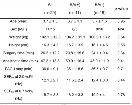
Table 2. Patients' characteristics.
All (n=29)
EA(+) (n=11)
EA(-)
(n=18) p value Age (year) 3.7 ± 1.5 3.7 ± 1.3 3.7 ± 1.6 0.95
Sex (M/F) 14/15 6/5 8/10 N/A
Weight (kg) 102.1 ± 12.3 104.2 ± 11.1 100.9 ± 13.2 0.64 Height (cm) 16.3 ± 4.3 16.7 ± 3.9 16.1 ± 4.6 0.55 Surgery time (min) 26.2 ± 12.2 29.8 ± 15.6 24.1 ± 9.4 0.34 Anesthetic time (min) 47.2 ± 13.8 50.9 ± 16.4 45.0 ± 11.9 0.41 PACU stay (min) 36.0 ± 9.1 35.1 ± 8.6 36.5 ± 9.7 0.71 SEF95 at 2.0 vol%
(Hz) 12.1 ± 2.7 11.6 ± 2.4 12.4 ± 3.0 0.44 SEF95 at 0.7 vol%
(Hz) 18.7 ± 3.8 18.2 ± 3.3 19.0 ± 4.1 0.78 Values are presented as mean ± SD.
Statistically significant, *P < 0.05
EA; Emergence agitation, EA(+); Patients with emergence agitation, EA(-); Patients without emergence agitation, PACU: Post-anesthetic care unit.
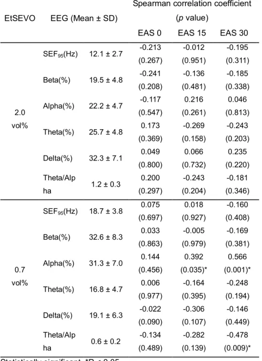
Table 3. Correlation analysis of EEG-derived parameters and EA score.
EtSEVO EEG (Mean ± SD)
Spearman correlation coefficient (p value)
EAS 0 EAS 15 EAS 30
2.0 vol%
SEF95(Hz) 12.1 ± 2.7 -0.213 (0.267)
-0.012 (0.951)
-0.195 (0.311) Beta(%) 19.5 ± 4.8 -0.241
(0.208)
-0.136 (0.481)
-0.185 (0.338) Alpha(%) 22.2 ± 4.7 -0.117
(0.547)
0.216 (0.261)
0.046 (0.813) Theta(%) 25.7 ± 4.8 0.173
(0.369)
-0.269 (0.158)
-0.243 (0.203) Delta(%) 32.3 ± 7.1 0.049
(0.800)
0.066 (0.732)
0.235 (0.220) Theta/Alp
ha 1.2 ± 0.3 0.200
(0.297)
-0.243 (0.204)
-0.181 (0.346)
0.7 vol%
SEF95(Hz) 18.7 ± 3.8 0.075 (0.697)
0.018 (0.927)
-0.160 (0.408) Beta(%) 32.6 ± 8.3 0.033
(0.863)
-0.005 (0.979)
-0.169 (0.381) Alpha(%) 31.3 ± 7.0 0.144
(0.456)
0.392 (0.035)*
0.566 (0.001)*
Theta(%) 16.8 ± 4.7 0.006 (0.977)
-0.164 (0.395)
-0.248 (0.194) Delta(%) 19.1 ± 6.3 -0.022
(0.090)
-0.306 (0.107)
-0.146 (0.449) Theta/Alp
ha 0.6 ± 0.2 -0.134
(0.489)
-0.282 (0.139)
-0.478 (0.009)*
Statistically significant, *P < 0.05
EA; Emergence agitation, EtSEVO; End-tidal sevoflurane concentration, EAS 0; EA score at the arrival on post-anesthetic care
unit, EAS 15; EA score 15 minutes after arrival, EAS 30; EA score 30 minutes after arrival, SEF95; Spectral edge frequency 95.
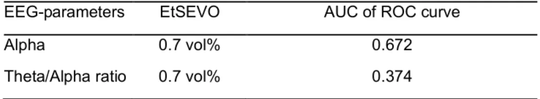
Table 4. AUC of ROC curve of EEG-derived parameters for EA.
EEG-parameters EtSEVO AUC of ROC curve
Alpha 0.7 vol% 0.672
Theta/Alpha ratio 0.7 vol% 0.374
EA; Emergence agitation, AUC; Area under curve.
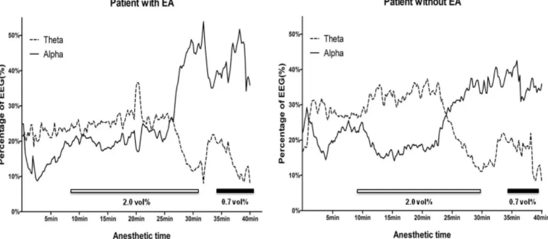
Figure 1. An example of time courses of percentages of theta and alpha bands of EEG during sevoflurane anesthesia.
3-year-old child with emergency agitation (EA) (left) showed high alpha power and low theta power during the maintenance of anesthesia (theta; 22.7, alpha; 23.5, theta/alpha ratio; 0.96, and SEF95; 16.6) and emergency (theta; 12.8, alpha; 46.2, theta/alpha ratio 0.28, and SEF95; 11.1), whereas 3-year-old child without EA (right) showed low alpha power and high theta power during the maintenance of anesthesia (theta; 34.0, alpha; 19.8, theta/alpha ratio; 1.71, and SEF95; 11.3) and emergency (theta; 17.1, alpha; 36.1, theta/alpha ratio; 0.47, and SEF95; 17.2). EAS of these children at 0, 15, and 30 minutes after post- anesthetic care unit arrival were '3 / 5 / 4' (left), and '2 / 1 / 2' (right), respectively. End-tidal sevoflurane concentrations are shown with bars.
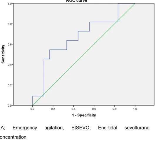
Figure 2. ROC analysis of alpha power at 0.7 vol%
of EtSEVO and the occurrence of EA. The area under ROC curve is 0.672.
EA; Emergency agitation, EtSEVO; End-tidal sevoflurane concentration
Discussion
Our data showed significant relationship between the percentage of EEG bands (high alpha powers) during emergence from sevoflurane anesthesia and the EAS in PACU.
The rhythmic activity in EEG is divided into several specific frequency bands; in the present study, the relative portion of beta, alpha, theta, and delta waves were monitored. Beta waves (13-30 Hz) are thought to be the result of sensory stimuli activating reticular activating system which desynchronizes the thalamic pacemaker cells. Alpha waves (8- 13 Hz) are usually seen in relaxed, awake patients with their eyes closed, and thought be related to the decrease of inhibitory activity of reticular nucleus on thalamic pacemaker cells. During alpha wave- predominant meditation or light sedation, thalamic pacemaker cells regulate and synchronize the thalamocortical activity. Theta waves (4-8 Hz) are normally seen during sleep and thought to be related to the inhibition of thalamic pacemaker cells by gamma-aminobutyric acid (GABA)-ergic action of reticular nucleus. During deep sleep, delta (0-3 Hz) waves are prominent and reflect extreme depression of thalamus.(21)
According to the thalamic theory, halogenated inhalation anesthetics cause unconsciousness by decreasing the neuronal activity of thalamocortical neurons (thalamic shunt).(22) Previous studies revealed this influence of inhalation anesthetics on EEG-derived parameters (SEF95 and the four frequency bands)(23,24); incremental concentration of inhalation anesthetics changes the EEG from fast (beta- and alpha-) waves during spontaneous arousal to slow (theta- and delta-) waves during anesthesia. SEF95 has been used to estimate the depth of
anesthesia and previous studies suggested SEF95 values for adequate anesthesia (10-14 Hz) and awaken state (15-20 Hz).(23,24) The values of SEF95 data in the present study during the maintenance of anesthesia (2.0 vol% of EtSEVO) and emergence (0.7 vol% of EtSEVO) were within these ranges, respectively (Table 2.). However, SEF95 value does not reflect the relative percentage of each spectral band (beta, alpha, theta, and delta-waves); it only reflects the sum of them. Therefore, it might be possible that there was a difference in alpha and theta bands without the difference in SEF95 values between patients with EA and without EA (Table 2.).
The previous study presented that children those who demonstrated agitation during induction showed slower EEG frequency during the second minute of induction compared with those who did not demonstrate agitation regardless of the kinds of premedication.(19) In the present study, induction of anesthesia was performed with high concentration of sevoflurane (8.0 vol%) with high fresh gas flow rate (6 L/min). Therefore, changes of EEG-derived parameters during induction were so rapid to observe. Instead, we obtained EEG-derived parameters at 0.7 vol% of EtSEVO (MAC-awake) for 5 minutes during emergence.
Higher alpha power and lower theta/alpha ratio during emergence from sevoflurane anesthesia were positively correlated to EAS in PACU. This trend means less GABA-ergic inhibition of thalamic pacemaker cells, and more thalamocortical activity during emergence from sevoflurane anesthesia.(22) Taken together, rapid recovery from EEG suppression of sevoflurane anesthesia was correlated with high EAS at PACU. There are two possible explanations for this phenomenon; patients with EA have high baseline EEG activity and/or
are more resistant to EEG suppression of sevoflurane. However, the PPV and NPV of alpha power at 0.7 EtSEVO were low and failed to show good predictablity of the occurrence of EA.
Smit DJ et al. (25) showed that individual differences in EEG spectral power reflect their genetic variance in brain development. Factors such as neuronal myelination, synaptic density, and dendritic outgrowth affect the volumes of gray and white matter and eventually results in different baseline theta and alpha spectral powers. Pediatric patients have large individual differences in the development of their central nervous system(26), and these differences can affect not only their baseline EEG activity and their EEG response to sevoflurane anesthesia, but also postoperative EA. However, no evidence exists regarding these issues.
We could not monitor postoperative EEG during PACU stay due to motion artifacts. There is no previous study about EEG monitoring in PACU. Previous studies conducted in intensive care unit or ward presented that the delirious patients showed significant lower alpha power and higher theta power, resulting in high theta/alpha ratio.(27-30) However, only one study was conducted in pediatric patients,(27) and only one study was conducted after general anesthesia. (30) Therefore, pediatric EEG data during EA after sevoflurane anesthesia could be hardly obtained.
There are several limitations in the present study. At first, it was a non-controlled, observational study of small score. Second, as mentioned before, we could not measure EEG at PACU, because of patients' discomfort and involuntary movement. Monitoring EEG at PACU, especially during EA, would have given more information about EA. Third, sevoflurane that remain in the body may played a role in
sedation, pain control, and EA at PACU. Forth, the present study was conducted under sevoflurane anesthesia for strabismus surgery.
Although adequate muscle relaxation was achieved and EMG was monitored simultaneously to exclude the effect of electric activity muscle, passive movement of extra-ocular muscle and eyelid would affect the EEG signal. Also, along with otolaryngeal surgery, strabismus surgery showed high incidence of EA than other pediatric surgery.(4)
In the present study, the authors found that the relative percentage of EEG bands during sevoflurane anesthesia showed significant relationship with postoperative EAS. Children showing high alpha powers and low theta/alpha ratio during emergence from sevoflurane anesthesia are at high risk of EA at 15 and 30 minutes after the arrival on PACU and need more careful attention.
Larger clinical trial with EEG monitoring at PACU and during EA should be needed in the future for an accurate assessment. Also, investigating the effect of alpha power-lowering at the emergence from sevoflurane anesthesia to prevent EA may clarify the relationship between intraoperative EEG and EA.
References
1. Prugh DG, Wagonfeld S, Metcalf D, Jordan K. A clinical study of delirium in children and adolescents. Psychosomatic medicine 1980;42:177-95.
2. Cravero J, Surgenor S, Whalen K. Emergence agitation in paediatric patients after sevoflurane anaesthesia and no surgery: a comparison with halothane. Paediatric anaesthesia 2000;10:419-24.
3. Galinkin JL, Fazi LM, Cuy RM, Chiavacci RM, Kurth CD, Shah UK, Jacobs IN, Watcha MF. Use of intranasal fentanyl in children undergoing myringotomy and tube placement during halothane and sevoflurane anesthesia. Anesthesiology 2000;93:1378-83.
4. Voepel-Lewis T, Malviya S, Tait AR. A prospective cohort study of emergence agitation in the pediatric postanesthesia care unit.
Anesthesia and analgesia 2003;96:1625-30, table of contents.
5. Welborn LG, Hannallah RS, Norden JM, Ruttimann UE, Callan CM. Comparison of emergence and recovery characteristics of sevoflurane, desflurane, and halothane in pediatric ambulatory patients. Anesthesia and analgesia 1996;83:917-20.
6. Dalens BJ, Pinard AM, Letourneau DR, Albert NT, Truchon RJ.
Prevention of emergence agitation after sevoflurane anesthesia for pediatric cerebral magnetic resonance imaging by small doses of ketamine or nalbuphine administered just before discontinuing anesthesia. Anesthesia and analgesia 2006;102:1056-61.
7. Hatzakorzian R, Shan WL, Cote AV, Schricker T, Backman SB.
The management of severe emergence agitation using droperidol. Anaesthesia 2006;61:1112-5.
8. Kararmaz A, Kaya S, Turhanoglu S, Ozyilmaz MA. Oral ketamine premedication can prevent emergence agitation in children after desflurane anaesthesia. Paediatric anaesthesia 2004;14:477-82.
9. Lankinen U, Avela R, Tarkkila P. The prevention of emergence agitation with tropisetron or clonidine after sevoflurane
anesthesia in small children undergoing adenoidectomy.
Anesthesia and analgesia 2006;102:1383-6.
10. Shukry M, Clyde MC, Kalarickal PL, Ramadhyani U. Does dexmedetomidine prevent emergence delirium in children after sevoflurane-based general anesthesia? Paediatric anaesthesia 2005;15:1098-104.
11. Sinha A, Levy N. Pre-emptive use of haloperidol in ICU to prevent emergence agitation. Anaesthesia 2007;62:753-4.
12. Aono J, Ueda W, Mamiya K, Takimoto E, Manabe M. Greater incidence of delirium during recovery from sevoflurane
anesthesia in preschool boys. Anesthesiology 1997;87:1298- 300.
13. Kain ZN, Caldwell-Andrews AA, Maranets I, McClain B, Gaal D, Mayes LC, Feng R, Zhang H. Preoperative anxiety and
emergence delirium and postoperative maladaptive behaviors.
Anesthesia and analgesia 2004;99:1648-54, table of contents.
14. Tripi PA, Palermo TM, Thomas S, Goldfinger MM, Florentino- Pineda I. Assessment of risk factors for emergence distress and postoperative behavioural changes in children following general anaesthesia. Paediatric anaesthesia 2004;14:235-40.
15. Cravero JP, Beach M, Dodge CP, Whalen K. Emergence characteristics of sevoflurane compared to halothane in pediatric patients undergoing bilateral pressure equalization tube insertion. Journal of clinical anesthesia 2000;12:397-401.
16. Oh AY, Seo KS, Kim SD, Kim CS, Kim HS. Delayed emergence process does not result in a lower incidence of emergence agitation after sevoflurane anesthesia in children.
Acta anaesthesiologica Scandinavica 2005;49:297-9.
17. Jacobson S, Jerrier H. EEG in delirium. Seminars in clinical neuropsychiatry 2000;5:86-92.
18. Koponen H, Partanen J, Paakkonen A, Mattila E, Riekkinen PJ.
EEG spectral analysis in delirium. Journal of neurology, neurosurgery, and psychiatry 1989;52:980-5.
19. Constant I, Leport Y, Richard P, Moutard ML, Murat I. Agitation and changes of Bispectral Index and electroencephalographic- derived variables during sevoflurane induction in children:
clonidine premedication reduces agitation compared with midazolam. British journal of anaesthesia 2004;92:504-11.
20. Cole JW, Murray DJ, McAllister JD, Hirshberg GE. Emergence behaviour in children: defining the incidence of excitement and agitation following anaesthesia. Paediatric anaesthesia
2002;12:442-7.
21. Jameson LC, Sloan TB. Using EEG to monitor anesthesia drug effects during surgery. Journal of clinical monitoring and
computing 2006;20:445-72.
22. Ries CR, Puil E. Mechanism of anesthesia revealed by shunting actions of isoflurane on thalamocortical neurons.
Journal of neurophysiology 1999;81:1795-801.
23. Schwender D, Daunderer M, Mulzer S, Klasing S, Finsterer U, Peter K. Spectral edge frequency of the electroencephalogram to monitor "depth" of anaesthesia with isoflurane or propofol.
British journal of anaesthesia 1996;77:179-84.
24. Schwender D, Daunderer M, Klasing S, Finsterer U, Peter K.
Power spectral analysis of the electroencephalogram during increasing end-expiratory concentrations of isoflurane, desflurane and sevoflurane. Anaesthesia 1998;53:335-42.
25. Smit DJ, Boomsma DI, Schnack HG, Hulshoff Pol HE, de Geus EJ. Individual differences in EEG spectral power reflect genetic variance in gray and white matter volumes. Twin research and human genetics : the official journal of the International Society for Twin Studies 2012;15:384-92.
26. Lenroot RK, Schmitt JE, Ordaz SJ, Wallace GL, Neale MC, Lerch JP, Kendler KS, Evans AC, Giedd JN. Differences in genetic and environmental influences on the human cerebral cortex associated with development during childhood and adolescence. Human brain mapping 2009;30:163-74.
27. Okumura A, Nakano T, Fukumoto Y, Higuchi K, Kamiya H, Watanabe K, Morishima T. Delirious behavior in children with influenza: its clinical features and EEG findings. Brain &
development 2005;27:271-4.
28. Jacobson SA, Leuchter AF, Walter DO. Conventional and quantitative EEG in the diagnosis of delirium among the elderly.
Journal of neurology, neurosurgery, and psychiatry 1993;56:153-8.
29. van der Kooi AW, Leijten FS, van der Wekken RJ, Slooter AJ.
What Are the Opportunities for EEG-Based Monitoring of
Delirium in the ICU? The Journal of neuropsychiatry and clinical neurosciences 2012;24:472-7.
30. Plaschke K, Fichtenkamm P, Schramm C, Hauth S, Martin E, Verch M, Karck M, Kopitz J. Early postoperative delirium after open-heart cardiac surgery is associated with decreased bispectral EEG and increased cortisol and interleukin-6.
Intensive care medicine 2010;36:2081-9.
국문 초록
서론:
마취 후 각성발작 (Emergence agitation; EA)은 소아 환자 의 세보플루레인 마취 후 흔히 나타나는 부작용이지만, 그 발생을예측하는 것은 어렵다. 본 연구는 수술 중 뇌파
(Electroencephalograpy; EEG) 측정을 통하여 수술 후 각성발작을 예측할 수 있는지 밝히기 위하여 진행되었다.
방법:
뇌파 측정은 29명의 소아 환자에서, 호기말 세보플루레인 농도(EtSEVO) 2.0 vol% (마취 유지 중)과 0.7 vol% (마취 회복 중) 일 때 이루어졌으며, SEF95, beta, alpha, theta, 및 delta power를 측정하였다. 각성 발작은 1점부터 5점으로 이루어진 각성발작점수 (Emergence agitation score; EAS)를 사용하여, 마취 회복실 도착 직후(EAS 0), 도착 15분 뒤(EAS 15), 도착 30분 뒤(EAS 30)에 측 정하였다. 뇌파 측정값들과 각정 발작 점수와의 관계는 Spearman correlation과 단순 회귀 분석을 사용하여 분석하였으며, 유의한 변 수의 진단적인 유용성을 알아보기 위해 receiver operating characteristic (ROC) 곡선을 그려보았다.결과:
마취 회복 중(0.7 vol% EtSEVO)에서의 alpha power는 EAS 15와 EAS 30과 양의 상관관계를 보였다 (순서대로, Pearson 상관 계수; 0.392 및 0.566, P=0.035 및 0.001). 마취 회복 중(0.7 vol%EtSEVO)의 theta/alpha 비율 또한 EAS 30과 음의 상관관계를 보 였다 (Pearson 상관계수; -0.478, P = 0.009). 마취 회복 중(0.7 vol% EtSEVO)의 alpha power와 각성발작의 발생여부로 작성한 ROC 곡선의 곡선하면적은 0.672이었다.
결론:
세보플루레인 마취를 받는 소아 환자에서 마취 회복 중에 높은 alpha power와 낮은 theta/alpha 비율을 보인 환자에서는 마 취 후 각성발작의 위험이 높다. (cris.nih.go.kr number, KCT0000652)