저작자표시-비영리-변경금지 2.0 대한민국 이용자는 아래의 조건을 따르는 경우에 한하여 자유롭게
l 이 저작물을 복제, 배포, 전송, 전시, 공연 및 방송할 수 있습니다. 다음과 같은 조건을 따라야 합니다:
l 귀하는, 이 저작물의 재이용이나 배포의 경우, 이 저작물에 적용된 이용허락조건 을 명확하게 나타내어야 합니다.
l 저작권자로부터 별도의 허가를 받으면 이러한 조건들은 적용되지 않습니다.
저작권법에 따른 이용자의 권리는 위의 내용에 의하여 영향을 받지 않습니다. 이것은 이용허락규약(Legal Code)을 이해하기 쉽게 요약한 것입니다.
Disclaimer
저작자표시. 귀하는 원저작자를 표시하여야 합니다.
비영리. 귀하는 이 저작물을 영리 목적으로 이용할 수 없습니다.
변경금지. 귀하는 이 저작물을 개작, 변형 또는 가공할 수 없습니다.
Master’s Thesis
Role of fibroblast growth factor 21 in obesity- induced hypothalamic inflammation
The Graduate School of the University of Ulsan
Department of Food Science and Nutrition Luthfiyyah Mutsnaini
[UCI]I804:48009-200000109071 [UCI]I804:48009-200000109071 [UCI]I804:48009-200000109071
Role of fibroblast growth factor 21 in obesity- induced hypothalamic inflammation
Supervisor: Dr. Rina Yu, Ph.D.
A Master’s Thesis
Submitted to
the Graduate School of the University of Ulsan In Partial Fulfillment of the Requirements
for the Degree of
Master of Science
by
Luthfiyyah Mutsnaini
Department of Food Science and Nutrition Ulsan, Korea
July, 2018
i ABSTRACT
Obesity-induced hypothalamic inflammation is associated with neuronal dysfunction and implicated in metabolic dysregulation. Fibroblast growth factor 21 (FGF21) is known to be an important metabolic regulator with anti-inflammatory properties. In this study, we investigated the effects of FGF21 deficiency on obesity-induced hypothalamic inflammation and thermogenic responses. FGF21- deficient mice or wild-type mice were fed a high-fat diet (HFD) or a regular diet (RD) for twelve weeks. The FGF21-deficient mice fed an HFD showed increased levels of inflammatory cytokines (TNFα and IL-1β) accompanied by elevated gliosis markers (Iba-1 and GFAP) expression levels when compared with HFD- fed control mice. The level of HSP72 expression, a neuronal damage marker, was increased in the FGF21-deficient obese mice, and hypothalamic neuronal markers, such as orexigenic (NPY), anorexigenic (POMC), anti-thermogenic (MCH), and thermogenic (TRH) markers, were altered. The levels of UCP1 expression in brown adipose tissue (BAT) were reduced in FGF21-deficient obese mice when compared with HFD-fed control mice. These findings suggest that FGF21 deficiency aggravates obesity-induced hypothalamic inflammation and reduces thermogenic responses and that this is associated with alteration of the hypothalamic neural circuits. FGF21 may be useful as a therapeutic target for controlling obesity-induced hypothalamic inflammation and metabolic derangement.
Keywords: Obesity, FGF21, Hypothalamic inflammation, Metabolism.
ii
ACKNOWLEGEMENT
First and foremost, I would like to express my sincere gratitude to our Almighty Allah SWT.
I would like to express my unfeigned thanks to my supervisor, Professor Rina Yu in every possible way. I am always grateful for her generous advice and encouragement during my research study. Her continuous guidance with invaluable and immense knowledge in which enhanced my understanding of the subject, to build my research skills and enabled me to accomplish this thesis.
Beside my advisor, I am gratefully obligated to the rest of my thesis’s examination committee: Dr. Suck-Young Choe and Dr. Jeong-Woo Park for their strenuous efforts to go through my thesis and for their insightful comments and feedback.
Moreover, I would like to thanks Dr. Chu-Sook Kim, who always provided help, motivation, and assistance in every step I had. Also many thanks to my fellow lab mates Jiye Kim, Jihyeon Yang, Hyunjung Choi, and Jeongheon Gong. I always appreciate their valuable support, opinions, and friendship throughout the years.
Furthermore, I am very much grateful to the National Institute for International Education (NIIED) for inviting and granting me a financial support through the Korean Government Scholarship Program (KGSP). Thanks to NIIED, I had an opportunity to study at higher educational institutions in Korea and learned many things about how wonderful Korea is.
Never enough thanks to my beloved father and mother, “Ibu, Bapak, I love you a lot.” I am ever grateful to my parents for giving me a blessing to continue my study abroad despite their extreme worry for me. Furthermore, I would like to express heartiest thanks to my twins sister upha and my younger brother Akil, who will always be there for me, even when I could not be for myself.
Last but not the least, I would like to thank my extended “WOW” family members and 2015 KGSP friends for their craziness, love, and console through the ups and downs this past three years. Thank you for surrounding me with a positive attitude!
“A thank you might seem underrated, but it is beyond every worth.”
July, 2018 Luthfiyyah Mutsnaini
iii
TABLE OF CONTENTS
ABSTRACT ... i
ACKNOWLEGEMENT ... ii
TABLE OF CONTENTS ... iii
1. Introduction... 1
1.1 Obesity ... 1
1.1.1 Obesity and metabolic disease ... 1
1.1.2 Obesity and peripheral inflammation ... 1
1.2 Obesity and brain ... 2
1.2.1 Obesity-induced hypothalamic inflammation ... 2
1.2.2 Hypothalamic inflammation and energy homeostasis ... 3
1.3 Fibroblast growth factor 21 (FGF21) ... 4
1.3.1 FGF21, metabolism, and inflammation ... 4
1.3.2 FGF21 and brain ... 5
2. Materials and Methods ... 7
2.1 Animal ... 7
2.2 Quantitative real-time PCR (qRT-PCR) ... 7
2.3 Western blot analysis ... 8
2.4 Statistical analysis ... 8
3. Results ... 9
3.1 FGF21 receptors were expressed in the hypothalamus ... 9
3.2 FGF21 deficiency altered brain weight changes in obese mice ... 9
3.3 FGF21 deficiency aggravated HFD-induced hypothalamic inflammation .. 9
3.4 FGF21 deficiency altered hypothalamic neuronal function ... 10
3.5 FGF21 deficiency abolished BAT thermogenesis function in obese mice 10 4. Discussion ... 16
5. Korean Summary... 22
References ... iv
Abbreviations ... x
1 1. Introduction
1.1 Obesity
1.1.1 Obesity and metabolic disease
Obesity, which has gradually become a major public health problem, has almost tripled since 1975. Based on statistical data from the world health organization (WHO), more than 650 million adults (18 years and older) are obese and 1.9 billion are overweight. In general, obesity is determined by the body mass index (BMI, with a BMI greater than or equal to 30 kg/m2 being considered obese. The WHO defines obesity as a condition in which fat is accumulated excessively in the body, to a degree that may cause health problems. An imbalance between food intake and energy disposal causes metabolic changes and leads to altered homeostasis in many organs. Thus, obesity is associated with several metabolic diseases (collectively known as metabolic syndrome), including insulin resistance, type 2 diabetes mellitus (T2DM), and cardiovascular diseases (Martyn, Kaneki and Yasuhara 2008, Shoelson, Herrero and Naaz 2007, Meshkani and Adeli 2009). Accordingly, it is very important to determine a method to prevent and treat obesity-induced metabolic diseases.
1.1.2 Obesity and peripheral inflammation
During obesity, the accumulation of excess body fat leads to the elevated plasma levels of free fatty acids (FFAs) (Boden 2008). The accumulation of FFAs in the peripheral systems drives adipose tissue to release a large amount of pro- inflammatory adipokines and cytokines, including leptin, tumor necrosis factor alpha (TNFα), interleukin-1β (IL-1β), interleukin-6 (IL-6), and monocyte chemoattractant protein-1 (MCP-1). Adipocytes have also been recognized as innate immunity regulators. Additionally, it had been demonstrated that toll-like receptors (TLR) might be activated by specific types of lipids. In a high-fat diet (HFD) fed obese animal models, saturated fatty acids manage the activation of TLR2 and TLR4 signaling to induce an inflammatory response. This might lead to the endoplasmic reticulum (ER) stress-mediated by cellular kinases, such as an inhibitor of NF-κB kinase (IKK) and c-Jun N-terminal kinase (JNK), as well as the production of reactive oxygen species (ROS) (Milanski et al. 2009). This condition is distinguished as a chronic low-grade systemic inflammation. Environmental factors may then increase the consequences of leptin and insulin resistance in
2
the obese state. Overall, many studies have indicated that obesity-induced inflammation is linked to the development of metabolic diseases.
1.2 Obesity and brain
1.2.1 Obesity-induced hypothalamic inflammation
Energy balance is a mechanism that is governed by the interplay between peripheral organs and the brain. Notably, the hypothalamus is an integral part of the brain responsible for the regulation of many fundamental physiological functions related to metabolism and maintenance of energy homeostasis (Le Thuc et al. 2017). Interestingly, growing evidence indicates that the hypothalamus is affected by obesity. During obesity, increased levels of circulating FFAs and pro-inflammatory cytokines can gain access to the hypothalamus via infiltration of the blood-brain barrier (BBB). Prolonged HFD for 14 weeks reportedly impaired BBB functions, as evidenced by the reduced BBB protein expression in the frontal cortices (Pepping et al. 2012). Upon sensing high-levels of FFAs, glial cells and neurons in the hypothalamus will be activated and transformed into their reactive form. This, in turn, elicits stress responses such as the production of ROS and activates pro-inflammatory NF-κB signaling (including TNFα and IL-1β), as well as elevates ionized calcium-binding adaptor molecule 1 (Iba-1; microglial marker) levels, which triggers the induction of hypothalamic inflammation (Zhang and Kaufman 2008, Zhang et al. 2008b).
Several studies have also suggested that activated glial cells have a prominent impact on hypothalamic inflammation and exacerbate metabolic dysregulation. The central nervous system (CNS) is a complex system composed of several cell types, including glial cells and neurons. Among glial cells, microglia and astrocytes are the most abundant in the brain (Argente-Arizón et al. 2017).
These cell types have both been implicated in the regulation of metabolic homeostasis in obesity pathogenesis (Douglass, Dorfman and Thaler 2017).
Astrocytes, which are large star-shaped glia, are only CNS cells capable of oxidizing fatty acids to ketones, which are necessary for neuronal survival (Argente-Arizón et al. 2017). Thus, astrocytes serve as neuronal support and enable communication (Argente-Arizón et al. 2017). Moreover, microglia are CNS macrophages that act as first responder cells that protect the CNS against infection, pathogens, and neuron injury (Douglass et al. 2017). Accordingly,
3
microglia and astrocytes are key cells involved in metabolic regulation of the hypothalamus in response to HFD or obesity. Morphological changes and activation of glial cells reportedly occur as early as 24 hours after HFD feeding (Thaler et al. 2012, Buckman et al. 2015). Indeed, many animal studies have shown evidence of the involvement of glial cells in HFD-induced hypothalamic inflammatory response. Notably, the accumulation of FFAs under obese conditions caused the release of inflammatory cytokines in astrocytes, which induced microglia migration and activation (Kwon et al. 2017). Furthermore, our study showed that the accumulation of lipid droplets in hypothalamic microglia mediates an inflammatory response, leading to neuronal damage in obese mice (Yang et al. 2017). Consequently, astrocytes and microglia are essential to neuronal activity and function. The activation of those glial cells will directly disrupt numerous physiological functions of neurons, including neurogenesis, neurotransmitter reuptake, and synaptic activity. These disruptions lead to neuronal dysregulation and neurodegenerative disorders under obese conditions.
1.2.2 Hypothalamic inflammation and energy homeostasis
Hypothalamic circuits consist of several small nuclei, including the arcuate nucleus (ARC), paraventricular nucleus (PVN), and lateral hypothalamus (LH), all of which directly regulate the activity of neurons that serve as metabolic sensors.
Those hypothalamic neurons project to autonomic sites through the sympathetic nervous system (SNS), where they regulate energy homeostasis in the peripheral system. The first-order neurons in the ARC, such as anorexigenic neurons that express proopiomelanocortin (POMC) and orexigenic neurons that express agouti-related peptide (AgRP) and neuropeptide Y (NPY), are responsible for sensing the integration of nutritional status between the peripheral and central system (Morton et al. 2006). The ARC also delivers nutritional signals directly or indirectly via the PVN and LH neurons, which are involved in the regulation of energy expenditure via thermoregulation. Moreover, those hypothalamic neurons can recognize and initiate adaptive responses to high FFAs levels (Morton et al.
2006). A previous study reported that low-grade hypothalamic inflammation causes significant reductions in the expression of the thermogenic neuronal markers thyrotropin-releasing hormone (TRH) and corticotropin-releasing hormone (CRH) in the PVN, as well as reduced expression of POMC and AgRP in the ARC, indicating defective hypothalamic neuronal circuit functions (Arruda
4
et al. 2011). Therefore, increasing the level of NPY in the ARC suppresses the expression of tyrosine hydroxylase (TH) in the PVN, which is an indicator of SNS outflow. Notably, reducing SNS outflow is associated with the reduction of uncoupling protein 1 (UCP1) expression in brown adipose tissue (BAT) (Shi et al.
2013). For instance, hypothalamic inflammation causes the disruption of energy homeostasis and body thermoregulation in the peripheral system. Moreover, blunted hypothalamic inflammation results in hypothalamic insulin resistance, brain volume decline, neuronal apoptosis, impaired neurogenesis, and brain synaptic remodeling (Taha et al. 2012, Jeon et al. 2012). These processes disrupt internal hypothalamic circuit sensing and alter hypothalamic outputs to other brain regions.
1.3 Fibroblast growth factor 21 (FGF21) 1.3.1 FGF21, metabolism, and inflammation
Fibroblast Growth Factor 21 (FGF21) is a member of the FGF family secreted by multiple organs, predominantly in liver, muscle and adipose tissue in response to diverse stresses or stimuli including starvation, ketogenic or high-fat diet, and the fibrate drugs (Kim and Lee 2015). The importance of metabolic functions of FGF21 has been reported in many human, non-human primate, and in vitro studies. The metabolic effects of FGF21 occur after FGF21 binds to its receptor (FGFR1) and co-receptor (β-Klotho) in each designated tissue and on the surface of cells. Binding of FGF21 to β-Klotho enables its interactions with the FGFR1, as well as leads to auto-phosphorylation and activation of its signaling pathway (Kharitonenkov et al. 2008). Moreover, there is growing evidence that exogenous FGF21 acts directly on the liver and adipose tissue to promote glucose uptake by the induction of glucose transporter-1 (GLUT-1) expression and decrease body weight mediated by induction of thermogenesis in BAT and browning of white adipose tissue (WAT). These changes are accompanied by increased leptin sensitivity in mice, decreased cholesterol/triglyceride (TG) levels, and improved insulin sensitivity (Ge et al. 2011, Véniant et al. 2012, Kleiner et al.
2012, Coskun et al. 2008). Moreover, FGF21 transgenic mice were shown to be resistant to weight gain and pharmacological treatment of FGF21 reduced adiposity and body weight in an obese mouse model (Coskun et al. 2008, Kharitonenkov et al. 2005). Notably, FGF21 has also been reported to increase in response to inflammatory stimuli. In the pancreas, FGF21 acts to inhibit islet
5
hyperplasia and prevent pancreatic inflammation in HFD-induced inflammation (Singhal et al. 2016). Moreover, treatment of recombinant murine FGF21 ameliorates obesity-related inflammation in rats with monosodium glutamate- induced obesity (Wang et al. 2015). Furthermore, FGF21 exerts an anti- inflammatory effect in macrophages in vitro by activating nuclear transcription factor-E2-related factor 2 (Nrf2) and suppressing the NF-κB signaling pathway (Yu et al. 2016). Taken together, the evidence has demonstrated the potential metabolic effects of FGF21 and its potential to correct obesity and mediate obesity-related metabolic diseases and inflammation.
1.3.2 FGF21 and brain
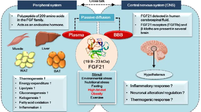
Although there are reports of the function of FGF21 in peripheral regulation, a recent study showed the beneficial effects of FGF21 in the brain.
Indeed, evidence suggests that FGF21 is present in human cerebrospinal fluid and brain tissues of rodents (Tan et al. 2011). However, it is not actually expressed in the central nervous system (CNS), although it can cross the BBB via simple diffusion (Hsuchou, Pan and Kastin 2007, Fon Tacer et al. 2010).
Moreover, a study demonstrated that FGF21 is produced by glial cells and expressed in specific regions of the midbrain, including the substantia nigra, striatum, hippocampus, and cortex (Mäkelä et al. 2014, Liang et al. 2014).
Figure 1. FGF21 and metabolism
6
Additionally, FGF21 receptors (FGFR1 and β-Klotho) have been found in several brain areas, including the hypothalamus (Fon Tacer et al. 2010, Bookout et al. 2013). It has also been shown that acute stimulation of hepatic gluconeogenesis by FGF21 is caused by activation of the hypothalamic pituitary adrenal (HPA) axis, which triggers adrenal corticosterone release (Liang et al.
2014). Moreover, FGF21 acts centrally to induce sympathetic nerve activity by enhancing energy expenditure through BAT thermogenesis in diet-induced obese (DIO) mice (Owen et al. 2014). A study of the CNS, documented that FGF21 protects against HFD-induced cognitive impairment via metabolic regulation and inhibition of neuro-inflammation and neurogenesis deficits in the hippocampus of obese mice (Wang et al. 2017b). These suggest that FGF21 elicits multiple metabolic functions and anti-inflammatory properties in the brain. Thus, we thought that FGF21 might have a potential to control hypothalamic inflammation in obese conditions.
However, a functional link and the underlying mechanisms of FGF21 action in obesity-induced hypothalamic inflammation have not fully understood. In this study, we investigated the role of FGF21 deficiency in obesity-induced hypothalamic inflammation inter-linked with metabolic derangements.
7 2. Materials and Methods
2.1 Animal
Six-week-old male C57BL/6 mice were purchased from Orient Bio Inc.
(Busan, Korea) and FGF21-deficient mice on the C57BL/6 background were established in the Immunomodulation Research Center of the University of Ulsan, South Korea. The mice were housed in plastic cages within a specific pathogen- free barrier animal facility that was maintained under a 12-h light/12-h dark cycle at 22±2°C. After one week of acclimatization, animals were randomly divided and fed either a regular diet or a high-fat diet (HFD, 60% calories from fat, Research Diets Inc., New Brunswick, NJ, USA) for 12 weeks, during which time they were given free access to food and water. Body weight and food intake were recorded every week. At the end of the feeding period, animals were food deprived for 4 hours, then euthanized by CO2 asphyxiation. The entire hypothalamus and hypothalamic specific nuclei regions (ARC, PVN, and LH) and BAT were subsequently collected and stored at -75°C. All animal care and procedures were conducted according to the protocols and guidelines approved by the animal ethics committee of the University of Ulsan and conformed to the National Institutes of Health guidelines (UOU-2015-Sang-005).
2.2 Quantitative real-time PCR (qRT-PCR)
One hypothalamic specific nuclei RNA sample was extracted from two combined animal tissues with Trizol-reagent (Invitrogen, Carlsbad, CA, USA). 2 µg aliquots of total RNA were reverse transcribed to cDNA using M-MLV reverse transcriptase (Promega, Madison, WI, USA). Next, qRT-PCR amplification of the cDNA was performed using SYBR premix Ex Taq (TaKaRa Bio Inc, Forster, CA, USA) in a Thermal Cycler Dice (TaKaRa Bio Inc., Japan). Additionally, a BAT RNA sample was extracted from one animal tissue sample, as described above.
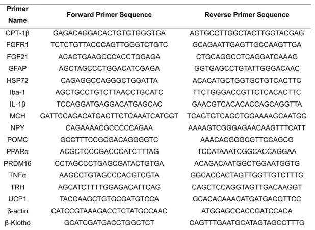
The results were analyzed with the Real-Time System TP800 software (TaKaRa), and all values were normalized to the levels of the house-keeping gene. The primers used in the analysis are listed in Table 1.
8
Table 1. Mouse primers used for quantitative real-time (qRT)-PCR.
Primer
Name Forward Primer Sequence Reverse Primer Sequence CPT-1β GAGACAGGACACTGTGTGGGTGA AGTGCCTTGGCTACTTGGTACGAG FGFR1 TCTCTGTTACCCAGTTGGGTCTGTC GCAGAATTGAGTTGCCAAGTTGA FGF21 ACACTGAAGCCCACCTGGAGA CTGCAGGCCTCAGGATCAAAG
GFAP AGCTAGCCCTGGACATCGAGA GGTGAGCCTGTATTGGGACAAC HSP72 CAGAGGCCAGGGCTGGATTA ACACATGCTGGTGCTGTCACTTC
Iba-1 AGCTGCCTGTCTTAACCTGCATC TTCTGGGACCGTTCTCACACTTC IL-1β TCCAGGATGAGGACATGAGCAC GAACGTCACACACCAGCAGGTTA MCH GATTCCAGACATGACTTCTCAAATCATGGT TCAGTGTCAGCTGGAAAAGCAATGG NPY CAGAAAACGCCCCCAGAA AAAAGTCGGGAGAACAAGTTTCATT POMC GCCTTTCCGCGACAGGGGTC AAACACGGGCGTTCCAGCG PPARα ACGCTCCCGACCCATCTTTAG TCCATAAATCGGCACCAGGAA PRDM16 CCTAGCCCTGAGCGATACTGTGA ACAGACAATGGCTGGAATGGTG
TNFα AAGCCTGTAGCCCACGTCGTA GGCACCACTAGTTGGTTGTCTTTG TRH AGCATCTTTTGGAGACATTCAG CAGCTCCAGGTAGTTGACAAGGT UCP1 TACCAAGCTGTGCGATGTCCA GCACACAAACATGATGACGTTCC β-actin CATCCGTAAAGACCTCTATGCCAAC ATGGAGCCACCGATCCACA β-Klotho GCATCGATGACCTGGCTCT CAGTTTGAATGCATAGTAGCCTTTG
2.3 Western blot analysis
Tissues were lysed in lysis buffer (150 mM NaCl, 50 mM Tris-HCl, 50 mM NaF, 10 mM Na4P2O7, 1 mM EDTA, 1% IGEPAL) supplemented with 0.25%
protease inhibitors cocktail (Sigma, St. Louis, MO, USA), and 1% phosphatase inhibitor cocktail (Sigma). Protein concentrations of the lysates were determined by BCA protein assay reagents (Pierce Biotechnology, Rockford, IL, USA). Equal amounts of protein (10-15 µg) were then subjected to western blot analysis using polyclonal antibodies to UCP1 (Abcam, ab10983, Cambridge, UK), PGC-1α (Abcam, ab54481), and alpha-tubulin (Abcam, ab7291). Protein bands were detected using an enhanced chemiluminescence western blotting detection kit (PerkinElmer, Waltham, MA, USA). Band intensities were quantified by densitometry using the Image J program.
2.4 Statistical analysis
All data are presented as the means ± SEM. Comparisons between the two groups were assessed by the Student’s t-test or one-way ANOVA (analysis of variance) for independent samples. Differences were considered to be significant at p < 0.05.
9 3. Results
3.1 FGF21 receptors were expressed in the hypothalamus
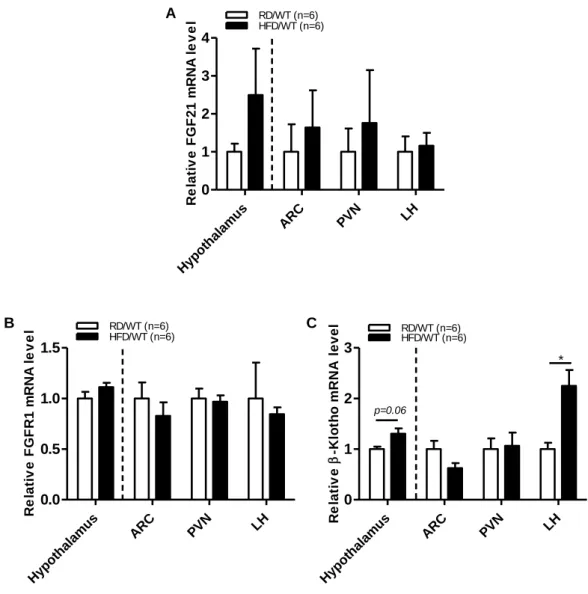
Growing evidence has shown that the metabolic actions of FGF21 require the cofactors β-Klotho and FGFR1 as primary receptors for signaling. To investigate the expression of FGFR1 and β-Klotho in the brain, we measured the mRNA expression within the hypothalamus region, specifically in the ARC, PVN, and LH, by real-time PCR. As shown in Fig. 2, FGF21, FGFR1 and β-Klotho were broadly expressed in the hypothalamic specific nuclei regions. When compared to RD-fed control mice, the expression levels of FGFR1 and β-Klotho were increased throughout the hypothalamus of HFD-fed mice. In contrast, the FGFR1 expression level was decreased in the ARC, PVN, and LH of HFD-fed mice. Moreover, the β-Klotho expression level in the PVN and LH of HFD-mice was upregulated when compared to control mice.
3.2 FGF21 deficiency altered brain weight changes in obese mice
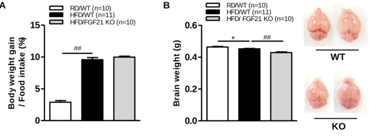
We examined whether FGF21 deficiency affects phenotypic changes in mice. Our experimental WT and FGF21 KO obese mouse groups did not show any significant differences in body weight changes and food intake (Fig. 3A).
However, the brain weight of FGF21 KO obese mice was smaller and lighter than that of WT obese mice (Fig. 3B). These findings demonstrate that FGF21 deficiency aggravates brain atrophy in HFD-fed obese mice.
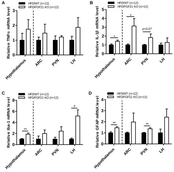
3.3 FGF21 deficiency aggravated HFD-induced hypothalamic inflammation Most studies conducted to date have revealed that chronic HFD feeding can mediate nutritional excess and low-grade inflammation in mice. Thus, these conditions might precede CNS hypothalamic inflammation. We next examined whether FGF21 plays a critical role in the regulation of inflammatory response in WT and FGF21 KO obese mice (Fig. 4). To accomplish this, we measured the expression of inflammatory cytokines (TNFα and IL-1β) and gliosis markers (Iba- 1 and GFAP) in the whole hypothalamus, ARC, PVN, and LH. The expression levels of inflammatory cytokines and gliosis markers mRNA was higher in FGF21 KO obese mice than control mice, demonstrating that FGF21 plays an important role in obese-induced hypothalamic inflammation.
10
3.4 FGF21 deficiency altered hypothalamic neuronal function
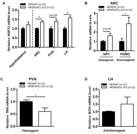
To further understand the metabolic effects of FGF21 in hypothalamic neuronal circuits, we first examined the expression of heat-shock protein 72 (HSP72), a critical protein that acts to protect neuronal damage. We found increased expression of HSP72 (Fig. 5A) in the whole hypothalamus, ARC, PVN, and LH of FGF21 KO obese mice relative to control obese mice. Moreover, the expression levels of anorexigenic marker (POMC), orexigenic marker (NPY) (Fig.
5B), and anti-thermogenic marker (MCH) (Fig. 5D) were increased in FGF21 KO obese mice. However, FGF21 deficiency caused suppression of the thermogenic marker (TRH) (Fig. 5C) in obese mice. These results show that FGF21 deficiency caused neuronal injury and altered hypothalamic neuronal circuits.
3.5 FGF21 deficiency abolished BAT thermogenesis function in obese mice
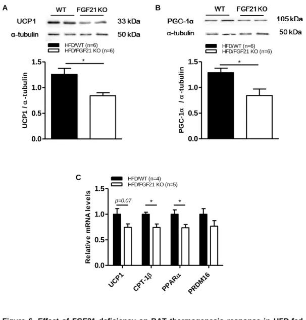
To establish whether alteration in hypothalamic neuronal circuits affects BAT thermogenesis, we measured the protein level of UCP1 and PGC-1α by western blot. Our findings showed that the ablation of FGF21 significantly reduced BAT thermogenesis function by reducing the protein level of UCP1 (Fig.
6A) and PGC-1α (Fig. 6B), as well as the mRNA levels of its transcription factors (Fig. 6C). These findings confirm that integration of all neuronal signals in the hypothalamus regulates the final output of BAT thermogenic response in the peripheral system.
11 A
B C
Figure 2. Expression of FGF21 and its receptors (FGFR1 and β-Klotho) in HFD-fed obese mice
The expression levels of (A) FGF21, (B) FGFR1, and (C) β-Klotho mRNA in the whole hypothalamus, ARC, PVN, and LH of 7-week-old male C57BL/6 mice fed a regular diet or HFD for 12 weeks as determined by real-time PCR analysis. All data are presented as the means ± SEM. * p < 0.05 significantly different from RD control. ARC, hypothalamic arcuate nucleus; FGFR1, fibroblast growth factor 21 receptor; FGF21, fibroblast growth factor 21; LH, lateral hypothalamus; PVN, hypothalamic paraventricular nucleus.
Hypo thala
mu s
ARC PVN LH
0 1 2 3
4 HFD/WT (n=6)
RD/WT (n=6)
Relative FGF21 mRNA level
Hypothala
mus ARC PVN LH
0.0 0.5 1.0
1.5 HFD/WT (n=6)RD/WT (n=6)
Relative FGFR1 mRNA level
Hypo thala
mu s
ARC PVN LH
0 1 2
3 HFD/WT (n=6)RD/WT (n=6)
Relative-Klotho mRNA level
*
p=0.06
12
A B
Figure 3. Body weight changes and food intake in FGF21 KO obese mice
7-week-old male C57BL/6 wild-type (WT) and FGF21-knockout (KO) mice were fed a regular diet or HFD for 12 weeks. (A) Body weight–food intake ratio and (B) brain weight.
All data are presented as the means ± SEM. * p < 0.05, ## p < 0.001 significantly different from RD control (RD/WT vs. HFD/WT), HFD control (HFD/WT vs. HFD/FGF21 KO).
WT
0 KO 5 10 15
RD/WT (n=10)
##
HFD/WT (n=11) HFD/FGF21 KO (n=10)
Body weight gain / Food intake (%)
0.0 0.2 0.4 0.6
RD/WT (n=10) HFD/WT (n=11) HFD/ FGF21 KO (n=10)
*
Brain weight (g) ##
13
A B
C D
Figure 4. Effect of FGF21 deficiency on inflammatory response in the hypothalamus of HFD-fed obese mice
Real-time PCR analysis of pro-inflammatory cytokines and glial cell markers; (A) TNFα, (B) IL-1β, (C) Iba-1, and (D) GFAP mRNA expression levels in the whole hypothalamus, ARC, PVN, and LH of 7-week-old male C57BL/6 WT and FGF21 KO mice fed an HFD for 12 weeks. All data are presented as the means ± SEM. * p < 0.05, ** p < 0.01, # p < 0.005 significantly different from WT control. ARC, hypothalamic arcuate nucleus; GFAP, glial fibrillary acidic protein; Iba-1, ionized calcium-binding adaptor molecule-1; IL-1β, interleukin-1-beta; LH, lateral hypothalamus; PVN, hypothalamic paraventricular nucleus;
TNFα, tumor necrosis factor alpha.
Hypo thala
mu s
ARC PVN LH
0 1 2 3
Relative TNF mRNA level HFD/FGF21 KO (n=12) HFD/WT (n=12)
Hypothala mus
ARC PVN LH
0 1 2 3 4 5
Relative IL-1 mRNA level HFD/FGF21 KO (n=12) HFD/WT (n=12)
*
p=0.07
*
Hypo thala
mu s
ARC PVN LH
0 2 4 6 8
HFD/WT (n=12)
Relative Iba-1 mRNA level HFD/FGF21 KO (n=12)
**
#
Hypo thala
mu s
ARC PVN LH
0 1 2 3 4
HFD/WT (n=12)
Relative GFAP mRNA level HFD/FGF21 KO (n=12)
** **
14
Orexigenic Anorexigenic
A B ARC
C PVN D LH
Figure 5. Effect of FGF21 deficiency on hypothalamic neuronal circuits in HFD-fed obese mice
Real-time PCR analysis of (A) neuronal damage marker (HSP72), (B) orexigenic marker (NPY), anorexigenic marker (POMC), (C) thermogenic marker (TRH), and (D) anti- thermogenic marker (MCH) mRNA expression levels in the whole hypothalamus, ARC, PVN, and LH of 7-week-old male C57BL/6J WT and FGF21 KO mice fed an HFD for 12 weeks. All data are presented as the means ± SEM. * p < 0.05, # p < 0.005 significantly different from WT control. ARC, hypothalamic arcuate nucleus; HSP72, heat-shock protein 72; LH, lateral hypothalamus; MCH, melanin-concentrating hormone; NPY, neuropeptide Y; POMC, proopiomelanocortin; PVN, hypothalamic paraventricular nucleus;
TRH, thyrotropin-releasing hormone.
Anti-thermogenic Hypo
thala mu
s
ARC PVN LH
0.0 0.5 1.0 1.5 2.0
Relative HSP72 mRNA level HFD/FGF21 KO (n=12) HFD/WT (n=12)
# p=0.05 #
NPY POMC
0 1 2 3 4
HFD/WT (n=12)
Relative mRNA levels
HFD/FGF21 KO (n=12)
p=0.06
*
0.0 0.5 1.0 1.5
HFD/WT (n=12) HFD/FGF21 KO (n=12)
Relative TRH mRNA level
*
0.0 0.5 1.0 1.5 2.0 2.5
HFD/WT (n=12) HFD/FGF21 KO (n=12)
Relative MCH mRNA level
Thermogenic
15
A B
C
Figure 6. Effect of FGF21 deficiency on BAT thermogenesis response in HFD-fed obese mice
Identification of (A) UCP1 protein level, (B) PGC-1α protein level, (C) thermogenic markers (UCP1, PPARα, and PRDM16), and mitochondrial β-oxidation gene marker (CPT-1β) in the BAT of 7-week-old male C57BL/6 WT and FGF21 KO mice fed a HFD for 12 weeks, as determined by western blot assay and RT-PCR. All data are represented as the means ± SEM. * p < 0.05 significantly different from WT control. CPT-1β, carnitine palmitoyltransferase-1-beta; PGC-1α, peroxisome proliferator-activated receptor gamma coactivator-1-alpha; PPARα, peroxisome proliferator-activated receptor alpha; PRDM16, PR domain containing 16; UCP1, uncoupling protein 1.
0.0 0.5 1.0 1.5
HFD/WT (n=6)
UCP1 /-tubulin
HFD/FGF21 KO (n=6)
*
0.0 0.5 1.0 1.5
HFD/WT (n=6)
PGC-1 /-tubulin
HFD/FGF21 KO (n=6)
*
UCP1
CP
T-1
PPAR
PRDM1 6 0.0
0.5 1.0 1.5
HFD/WT (n=4)
Relative mRNA levels
HFD/FGF21 KO (n=5)
p=0.07 * *
16 4. Discussion
FGF21 is known as a potent metabolic regulator and has emerged as an interesting new candidate for obesity treatment (Kharitonenkov et al. 2005, Sonoda, Chen and Baruch 2017). Studies have shown that the administration of FGF21 improves insulin sensitivity, glucose uptake, and lipid metabolism under obese conditions (Markan et al. 2014, Staiger et al. 2017, Kharitonenkov et al.
2005). Moreover, the potential effects of FGF21 in the brain have recently been reported (Sa-nguanmoo, Chattipakorn and Chattipakorn 2016). For example, FGF21 stimulates sympathetic nerve activity to regulate whole-body energy expenditure via thermogenesis (Sarruf et al. 2010, Douris et al. 2015). FGF21 also plays an important role in the hypothalamic-pituitary-adrenal (HPA) and hypothalamic-pituitary-gonadal (HPG) axis (Bookout et al. 2013, Liang et al. 2014, Fon Tacer et al. 2010). In addition, like other FGFs family, FGF21 and its receptors are broadly expressed in the brain, such as midbrain and hypothalamus (Mäkelä et al. 2014, Fon Tacer et al. 2010). However, a study of FGF21 functions in the hypothalamic specific nuclei regions is limited. Here, in the present study, we found for the first time that FGF21 and its receptors (FGFR1 and β-Klotho) were expressed in the hypothalamic specific nuclei regions (ARC, PVN, and LH) of mice, and that its expression was modulated by providing a high-fat diet. These findings indicate that FGF21 might act directly on the hypothalamus specific nuclei regions by binding to its complex receptors and regulating hypothalamic functions.
There is growing evidence that FGF21 exerts an anti-inflammatory function (Luo et al. 2017). Specifically, FGF21 protects against obesity-related inflammation and inhibits inflammation by suppressing IL-1β and TNFα production (Wang et al. 2015, Yu et al. 2016, Singhal et al. 2016, Li et al. 2018).
Interestingly, FGF21 was also found to increase in response to inflammatory stimuli and HFD feeding (Feingold et al. 2012, Zhang et al. 2008a). Moreover, a study of the CNS revealed that FGF21 protected against HFD-induced cognitive impairment by inhibiting neuro-inflammation (Wang et al. 2017b). Hence, the major concern of the present study was whether FGF21 has the ability to mediate obesity-induced hypothalamic inflammation. In this study, we used an FGF21 KO HFD-induced obese mouse model, to investigate whether FGF21 ablation aggravates obesity-induced hypothalamic inflammation. We found that FGF21
17
deficiency results in increased hypothalamic inflammatory cytokines (TNFα and IL-1β) expression levels in HFD-fed obese mice compared to control mice.
Hypothalamic inflammation is closely associated with gliosis, which referred to as the activation of astrocytes and microglia (Thaler et al. 2012, Dorfman and Thaler 2015). Obese conditions and HFD-feeding have been shown to induce the activation of hypothalamic astrocytes and microglia (Gao et al. 2014, Berkseth et al. 2014). Under obese conditions, upon sensing the FFAs-rich conditions, the hypothalamic astrocytes accumulate lipid droplets and induces microglia migration and activation, leading to the production of pro-inflammatory cytokines (Gorina et al. 2011, Masson et al. 2015, Kwon et al. 2017). This indicates that the activation of these glial cells, such as astrocytes and microglia contribute to the escalation of hypothalamic inflammation in obese conditions. The increased gliosis is considered as an important marker of hypothalamic inflammation in addition to inflammatory cytokines (Dorfman and Thaler 2015). In this study, we observed that gliosis markers (Iba-1 and GFAP) in FGF21 KO obese mice were upregulated. Together, our findings suggest that FGF21 ablation mediates the activation of hypothalamic glial cells, and this aggravates obesity-induced hypothalamic inflammation.
Inflammation has been shown to drive progressive neurotoxicity and glial cells activation, resulting in neuronal damage (Block, Zecca and Hong 2007).
Among hypothalamic glial cells, astrocytes are able to provide a nutrient that is necessary for neuronal survival, while microglia act as first responder cells that react to protect CNS against infection and neuronal injury (Douglass et al. 2017, Argente-Arizón et al. 2017). The initial response of activated glial cells is to neutralize threats and to maintain neuroprotective functions in response to neuronal injury (Ransohoff and Cardona 2010, Douglass et al. 2017). However, under obese conditions, the accumulation of lipid droplets in hypothalamic astrocytes and microglia promote the inflammatory response, leading to neuronal damage (Yang et al. 2017, Kwon et al. 2017). As a compensatory response after stress to promote cell survival and further prevent or limit injury, neuronal cells express heat-shock protein 72 (HSP72). HSP72 is a chaperone protein that protects cells from oxidative stress and it has been used as a neuronal injury marker (Turturici, Sconzo and Geraci 2011). It should be noted that one of the functions of FGF21 is to protect neuronal injury (Leng et al. 2015). In the present
18
study, we found the expression of HSP72 was increased in FGF21 KO obese mice. Given that gliosis markers were upregulated in FGF21 KO obese mice, the neuronal injury observed in the hypothalamus of FGF21 KO obese may result from the activated glial cells accompanied by increased inflammatory mediators.
Therefore, FGF21 might become a valuable target in order to protect neuron injury induced by hypothalamic inflammation or gliosis in obese conditions.
Notably, studies have documented the impact of obesity on hypothalamic neural network signaling. Feeding a high-fat diet induces inflammation in the hypothalamus, resulting in dysregulation of hypothalamic neuronal circuits, which leads to altered appetite and food intake, thus aggravating metabolic imbalance (Dalvi et al. 2017, Guillemot-Legris and Muccioli 2017). Therefore, we investigated whether FGF21 ablation alters hypothalamic neuronal functions. We found that FGF21 deficiency in the hypothalamic ARC nucleus of HFD-obese mice caused an increase in orexigenic marker (NPY), which is an appetite- regulating neuropeptide involved in body weight regulation and energy balance (Thaler et al. 2013). Interestingly, NPY hypothalamic expression increased in FGF21 KO obese mice, mice were not hyperphagic and did not gain more weight compared to control obese mice. It is not known why NPY failed to execute its function in FGF21 KO obese mice; therefore, further study is needed to explore this issue. However, the increased level of NPY was also accompanied by increased anorexigenic marker (POMC) expression level. We thought that the elevation of POMC was to compensate for the neuronal activity that suppressed the appetite signal, resulting in identical food intake between WT and FGF21 KO mice.
Studies have shown that FGF21 KO mice gained more weight compared to WT mice, but did not show metabolic abnormalities when fed a standard chow diet (Singhal et al. 2016, Badman et al. 2009, Hotta et al. 2009). However, we found that WT and FGF21 KO obese mice did not show any significant differences in body weight changes and food intake. This phenomenon is consistent with the results reported by Chui et al., who found that obese FGF21 KO mice were phenotypically similar to WT mice (Chui et al. 2010). They suggested that, despite the increased level of FGF21 in HFD-fed WT animals, FGF21 fails to function as a metabolic regulator. As a result, WT animals would
19
“obtain” the phenotype of mice lacking FGF21, suggesting that FGF21 resistance manifests during the obese state (Chui et al. 2010). Strikingly, FGF21 primarily occurs in the liver, where it has multiple metabolic roles, such as enhancing hepatic fatty acid oxidation (Kim and Lee 2015, Fisher et al. 2014). In accordance with the study by Liu et al., FGF21 deficiency exacerbates hepatic steatosis and injury in mice exposed to chronic alcohol (Liu et al. 2016). Moreover, FGF21 ablation induces hepatomegaly and excessive fat accumulation in the livers of obese mice, which was also observed in our mice (Badman et al. 2009).
Interestingly, we found that the outspread of adipose tissue in FGF21 KO mice was slightly smaller than that in WT mice. In particular, lipodystrophy was associated with hepatic injury, in which a lack of adipocytes or adipose tissue disorders caused excess FFAs to be directed into other ectopic storage areas, such as the liver (Garg 2006). Therefore, we considered the similar body weight between FGF21 KO and WT obese mice to have developed as a result of lipodystrophy, which resembles an extreme version of obesity-associated metabolic syndrome (Zadeh et al. 2013).
Regarding the impact of hypothalamic inflammation on the first order neuron (NPY and POMC) in the ARC, one can speculate that other integrated neuronal circuits may be disrupted. The first-order neurons in the ARC participate in regulation of nutritional status between the peripheral and central system (Morton et al. 2006). They also project the nutritional signals to other nearby hypothalamic neurons, such as PVN and LH (Morton et al. 2006). In the present study, we found that the expression levels of the anti-thermogenic marker (MCH) were upregulated in the LH and accompanied by a reduction of the thermogenic marker (TRH) in the PVN of FGF21 KO obese mice. Excessive HFD feeding likely results in alterations to these neuronal activities, where neurons in the LH and PVN initially played a role in the excitation of adipose tissue thermogenesis regulation (Contreras et al. 2015). Additionally, low-grade hypothalamic TNFα led to reductions in POMC, TRH, and CRH, all of which are neurotransmitters with important thermogenic function (Arruda et al. 2011). In this study, we thought that inflammation-induced hypothalamic neuronal circuit alteration may disrupt the regulation of energy expenditure in obese mice. We found that FGF21 deficiency significantly reduced the protein levels of UCP1 and PGC-1α in BAT, which was accompanied by a highly significant decrease in the gene expression of brown
20
fat-specific transcription factors (UCP1, CPT-1β, PPARα, and PRDM16). These findings indicate that FGF21 deficiency decreases BAT thermogenesis, which could be associated with neuronal dysregulation under an obesity-induced inflammatory condition in the hypothalamus.
In addition to being associated with metabolism and energy balance, the hypothalamus is inter-related with the functions of other brain regions. For instance, brain size is associated with neuronal system organization, where the numbers of inter-neuronal circuits are adjusted to the brain volume size (Butler and Hodos). The correlation between BMI and brain volume alterations has been associated with brain structural deficits, in which higher body adiposity may have deleterious consequences on brain atrophy (Raji et al. 2010, Wang et al. 2017a).
Interestingly, we found that brain size in FGF21 KO obese mice was smaller and lighter than in WT obese mice; thus, we suspect that the dysregulation of neuronal hypothalamic outputs in FGF21 KO obese mice is caused by inflammation-induced neuronal deficits, but exacerbated by brain atrophy.
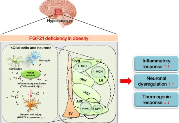
Figure 7. FGF21 deficiency in obesity-induced hypothalamic inflammation
21
In conclusion, FGF21 deficiency aggravates obesity-induced hypothalamic inflammation, leading to the alteration of hypothalamic neural circuits accompanied via reduction of the thermogenic response. Thus, FGF21 may be useful as a therapeutic target for controlling obesity-induced hypothalamic inflammation and another metabolic derangement.
22 5. Korean Summary
비만으로 유도된 시상하부 염증은 신경 세포 기능 장애 및 대사 조절 장애와 관련이 있다. Fibroblast growth factor 21 (FGF21)은 항염증 활성이 있으며, 중요한 대사 조절자로 알려져있다. 본 연구에서는 FGF21 결핍이 비만성 시상하부 염증 및 열 생성 반응에 미치는 영향을 조사하였다. FGF21이 결핍된 쥐 (FGF21 KO) 또는 대조군 쥐 (WT)에게 12 주간 고지방식 (HFD)이나 일반식 (RD)이 투여했다. FGF21 KO 비만 쥐는 WT 비만 쥐와 비교하여 gliosis 마커 (Iba-1 및 GFAP)와 염증성 사이토카인 (TNFα 및 IL-1β) 발현 수준이 증가하였다. FGF21 KO 비만 쥐에서 신경 손상 마커인 HSP72 발현 수준이 증가했으며, 시상하부 신경 마커의 식욕 촉진 (NPY)/ 식욕 부진 (POMC), 열 생성 억제 (MCH)/ 열 생성 촉진 (TRH) 마커들의 변화가 관찰되었다. 갈색 지방 조직 (BAT)에서의 UCP1 발현 수준은 FGF21 KO 비만 쥐에서 WT 비만 쥐 보다 감소하였다. 이러한 결과들은
FGF21 결핍이 비만으로 유도된 시상하부 염증을 악화시키고 열 생성 반응을
감소시킬 수 있으며, 이러한 변화는 시상하부 대사 조절능을 가진 신경회로의 변화와 관련이 있음을 시사한다. 따라서 FGF21은 비만성 시상하부 염증 및 대사 장애를 조절하기 위한 치료 표적으로서 유용할 것으로 사료된다.
iv References
Argente-Arizón, P., S. Guerra-Cantera, L. M. Garcia-Segura, J. Argente & J. A.
Chowen (2017) Glial cells and energy balance. Journal of molecular endocrinology, 58, R59-R71.
Arruda, A. P., M. Milanski, A. Coope, A. S. Torsoni, E. Ropelle, D. P. Carvalho, J.
B. Carvalheira & L. A. Velloso (2011) Low-grade hypothalamic inflammation leads to defective thermogenesis, insulin resistance, and impaired insulin secretion. Endocrinology, 152, 1314-1326.
Badman, M. K., A. Koester, J. S. Flier, A. Kharitonenkov & E. Maratos-Flier (2009) Fibroblast growth factor 21-deficient mice demonstrate impaired adaptation to ketosis. Endocrinology, 150, 4931-4940.
Berkseth, K. E., S. J. Guyenet, S. J. Melhorn, D. Lee, J. P. Thaler, E. A. Schur &
M. W. Schwartz (2014) Hypothalamic gliosis associated with high-fat diet feeding is reversible in mice: a combined immunohistochemical and magnetic resonance imaging study. Endocrinology, 155, 2858-2867.
Block, M. L., L. Zecca & J.-S. Hong (2007) Microglia-mediated neurotoxicity:
uncovering the molecular mechanisms. Nature Reviews Neuroscience, 8, 57.
Boden, G. (2008) Obesity and free fatty acids. Endocrinology and metabolism clinics of North America, 37, 635-646.
Bookout, A. L., M. H. De Groot, B. M. Owen, S. Lee, L. Gautron, H. L. Lawrence, X. Ding, J. K. Elmquist, J. S. Takahashi & D. J. Mangelsdorf (2013) FGF21 regulates metabolism and circadian behavior by acting on the nervous system. Nature medicine, 19, 1147-1152.
Buckman, L. B., M. M. Thompson, R. N. Lippert, T. S. Blackwell, F. E. Yull & K. L.
Ellacott (2015) Evidence for a novel functional role of astrocytes in the acute homeostatic response to high-fat diet intake in mice. Molecular metabolism, 4, 58-63.
Butler, A. B. & W. Hodos Evolution and Adaptation of the Brain, Behavior, and Intelligence. Comparative Vertebrate Neuroanatomy: Evolution and Adaptation, Second Edition, 93-111.
Chui, P. C., P. J. Antonellis, H. A. Bina, A. Kharitonenkov, J. S. Flier & E.
Maratos-Flier (2010) Obesity is a fibroblast growth factor 21 (FGF21)- resistant state. Diabetes, 59, 2781-2789.
Contreras, C., F. Gonzalez, J. Fernø, C. Diéguez, K. Rahmouni, R. Nogueiras &
M. López (2015) The brain and brown fat. Annals of medicine, 47, 150- 168.
Coskun, T., H. A. Bina, M. A. Schneider, J. D. Dunbar, C. C. Hu, Y. Chen, D. E.
Moller & A. Kharitonenkov (2008) Fibroblast growth factor 21 corrects obesity in mice. Endocrinology, 149, 6018-6027.
v
Dalvi, P., J. Chalmers, V. Luo, D.-Y. Han, L. Wellhauser, Y. Liu, D. Tran, J.
Castel, S. Luquet & M. Wheeler (2017) High fat induces acute and chronic inflammation in the hypothalamus: effect of high-fat diet, palmitate and TNF-α on appetite-regulating NPY neurons. International Journal of Obesity, 41, 149-158.
Dorfman, M. D. & J. P. Thaler (2015) Hypothalamic inflammation and gliosis in obesity. Current opinion in endocrinology, diabetes, and obesity, 22, 325.
Douglass, J. D., M. D. Dorfman & J. P. Thaler (2017) Glia: silent partners in energy homeostasis and obesity pathogenesis. Diabetologia, 60, 226-236.
Douris, N., D. M. Stevanovic, F. M. Fisher, T. I. Cisu, M. J. Chee, N. L. Nguyen, E.
Zarebidaki, A. C. Adams, A. Kharitonenkov & J. S. Flier (2015) Central fibroblast growth factor 21 browns white fat via sympathetic action in male mice. Endocrinology, 156, 2470-2481.
Feingold, K. R., C. Grunfeld, J. G. Heuer, A. Gupta, M. Cramer, T. Zhang, J. K.
Shigenaga, S. M. Patzek, Z. W. Chan & A. Moser (2012) FGF21 is increased by inflammatory stimuli and protects leptin-deficient ob/ob mice from the toxicity of sepsis. Endocrinology, 153, 2689-2700.
Fisher, F. M., P. C. Chui, I. A. Nasser, Y. Popov, J. C. Cunniff, T. Lundasen, A.
Kharitonenkov, D. Schuppan, J. S. Flier & E. Maratos-Flier (2014) Fibroblast growth factor 21 limits lipotoxicity by promoting hepatic fatty acid activation in mice on methionine and choline-deficient diets.
Gastroenterology, 147, 1073-1083. e6.
Fon Tacer, K., A. L. Bookout, X. Ding, H. Kurosu, G. B. John, L. Wang, R. Goetz, M. Mohammadi, M. Kuro-o & D. J. Mangelsdorf (2010) Research resource:
comprehensive expression atlas of the fibroblast growth factor system in adult mouse. Molecular endocrinology, 24, 2050-2064.
Gao, Y., N. Ottaway, S. C. Schriever, B. Legutko, C. García‐Cáceres, E. de la Fuente, C. Mergen, S. Bour, J. P. Thaler & R. J. Seeley (2014) Hormones and diet, but not body weight, control hypothalamic microglial activity. Glia, 62, 17-25.
Garg, A. (2006) Adipose tissue dysfunction in obesity and lipodystrophy. Clinical cornerstone, 8, S7-S13.
Ge, X., C. Chen, X. Hui, Y. Wang, K. S. Lam & A. Xu (2011) Fibroblast growth factor 21 induces glucose transporter-1 expression through activation of the serum response factor/Ets-like protein-1 in adipocytes. Journal of Biological Chemistry, 286, 34533-34541.
Gorina, R., M. Font‐Nieves, L. Márquez‐Kisinousky, T. Santalucia & A. M. Planas (2011) Astrocyte TLR4 activation induces a proinflammatory environment through the interplay between MyD88‐dependent NFκB signaling, MAPK, and Jak1/Stat1 pathways. Glia, 59, 242-255.
Guillemot-Legris, O. & G. G. Muccioli (2017) Obesity-Induced Neuroinflammation:
Beyond the Hypothalamus. Trends in Neurosciences.
vi
Hotta, Y., H. Nakamura, M. Konishi, Y. Murata, H. Takagi, S. Matsumura, K.
Inoue, T. Fushiki & N. Itoh (2009) Fibroblast growth factor 21 regulates lipolysis in white adipose tissue but is not required for ketogenesis and triglyceride clearance in liver. Endocrinology, 150, 4625-4633.
Hsuchou, H., W. Pan & A. J. Kastin (2007) The fasting polypeptide FGF21 can enter brain from blood. Peptides, 28, 2382-2386.
Jeon, B. T., E. A. Jeong, H. J. Shin, Y. Lee, D. H. Lee, H. J. Kim, S. S. Kang, G. J.
Cho, W. S. Choi & G. S. Roh (2012) Resveratrol attenuates obesity- associated peripheral and central inflammation and improves memory deficit in mice fed a high-fat diet. Diabetes, 61, 1444-1454.
Kharitonenkov, A., J. D. Dunbar, H. A. Bina, S. Bright, J. S. Moyers, C. Zhang, L.
Ding, R. Micanovic, S. F. Mehrbod & M. D. Knierman (2008) FGF‐
21/FGF‐21 receptor interaction and activation is determined by βKlotho.
Journal of cellular physiology, 215, 1-7.
Kharitonenkov, A., T. L. Shiyanova, A. Koester, A. M. Ford, R. Micanovic, E. J.
Galbreath, G. E. Sandusky, L. J. Hammond, J. S. Moyers & R. A. Owens (2005) FGF-21 as a novel metabolic regulator. Journal of Clinical Investigation, 115, 1627.
Kim, K. H. & M.-S. Lee (2015) FGF21 as a mediator of adaptive responses to stress and metabolic benefits of anti-diabetic drugs. Journal of Endocrinology, 226, R1-R16.
Kleiner, S., N. Douris, E. C. Fox, R. J. Mepani, F. Verdeguer, J. Wu, A.
Kharitonenkov, J. S. Flier, E. Maratos-Flier & B. M. Spiegelman (2012) FGF21 regulates PGC-1α and browning of white adipose tissues in adaptive thermogenesis. Genes & development, 26, 271-281.
Kwon, Y. H., J. Kim, C. S. Kim, T. H. Tu, M. S. Kim, K. Suk, D. H. Kim, B. J. Lee, H. S. Choi & T. Park (2017) Hypothalamic lipid‐laden astrocytes induce microglia migration and activation. FEBS letters, 591, 1742-1751.
Le Thuc, O., K. Stobbe, C. Cansell, J.-L. Nahon, N. Blondeau & C. Rovère (2017) Hypothalamic Inflammation and Energy Balance Disruptions: Spotlight on Chemokines. Frontiers in endocrinology, 8, 197.
Leng, Y., Z. Wang, L.-K. Tsai, P. Leeds, E. B. Fessler, J. Wang & D.-M. Chuang (2015) FGF-21, a novel metabolic regulator, has a robust neuroprotective role and is markedly elevated in neurons by mood stabilizers. Molecular psychiatry, 20, 215.
Li, J.-y., N. Wang, M. H. Khoso, C.-b. Shen, M.-z. Guo, X.-x. Pang, D.-s. Li & W.-f.
Wang (2018) FGF-21 Elevated IL-10 Production to Correct LPS-Induced Inflammation. Inflammation, 41, 751-759.
Liang, Q., L. Zhong, J. Zhang, Y. Wang, S. R. Bornstein, C. R. Triggle, H. Ding, K.
S. Lam & A. Xu (2014) FGF21 maintains glucose homeostasis by mediating the cross talk between liver and brain during prolonged fasting.
Diabetes, 63, 4064-4075.