J Vet Sci 2019, 20(1), 79-86ㆍhttps://doi.org/10.4142/jvs.2019.20.1.79 JVS
Received 10 Sep. 2018, Revised 8 Nov. 2018, Accepted 18 Nov. 2018
*Corresponding author: Tel: +82-2-880-1269; Fax: +82-2-873-1269; E-mail: [email protected]
Journal of Veterinary Scienceㆍⓒ 2019 The Korean Society of Veterinary Science. All Rights Reserved.
This is an Open Access article distributed under the terms of the Creative Commons Attribution Non-Commercial License (http://creativecommons.org/licenses/
pISSN 1229-845X eISSN 1976-555X
Iodixanol supplementation during sperm cryopreservation improves protamine level and reduces reactive oxygen species of canine sperm
Dimas A. Abdillah, Erif M. N. Setyawan, Hyun Ju Oh, Kihae Ra, Seok Hee Lee, Min Jung Kim, Byeong Chun Lee*
Department of Theriogenology and Biotechnology, College of Veterinary Medicine, Seoul National University, Seoul 08826, Korea
The objective of this study was to analyze the protective effects of iodixanol on dog spermatozoa during cryopreservation. The optimal concentration of iodixanol, 1.5%, was determined using fresh spermatozoa and was applied in the following experiments. The 1.5% iodixanol group showed significantly increased sperm motility from that in the control (p < 0.05). Lower mitochondrial reactive oxygen species (ROS) modulator (ROMO1) and pro-apoptotic gene (BAX) expressions, together with higher expressions of protamine-2 (PRM2), protamine-3 (PRM3), anti-apoptotic B-cell lymphoma-2 (BCL2), and sperm acrosome associated-3 (SPACA3) genes were detected in the iodixanol-treated group. In addition, decreased protamine deficiency and cryocapacitation were observed in the treatment group. Our results show that supplementation with 1.5% iodixanol is ideal for reducing production of ROS and preventing detrimental effects during the canine sperm cryopreservation process, effects manifested as increased motility and reduced cryocapacitation in frozen-thawed spermatozoa.
Keywords: Dogs; Sperm; Cryopreservation; Reactive oxygen species; Protamine
Introduction
Sperm cryopreservation is an essential step for artificial insemination (AI), which is the most widely used assisted reproductive technology in Canidae. The main goal of sperm cryopreservation is to conserve the fertility of high genetic value organisms or to preserve endangered species [7]. In addition, sperm cryopreservation for AI can prevent sexually transmitted diseases such as brucellosis and herpes virus infections [10]. In wild animals, AI with cryopreserved spermatozoa has had a considerable effect in conservation management of African wild dogs [5]. Moreover, AI using cryopreserved spermatozoa provides a number of potential advantages including reduction of stress associated with transportation of animals, avoiding resistance to copulation due to behavioral issues (female aggressiveness), and overcoming quarantine restrictions placed on live animals [8].
During the cryopreservation process, osmotic pressure and ice formation can cause cryoinjury and loss of viability and sperm function post-thawing [25]. Osmotic stress following cell dehydration induces destabilization of sperm membranes by reducing their fluidity, and this is exacerbated by excessive
production of endogenous and exogenous reactive oxygen species (ROS) [1,13]. An imbalance between ROS production during cryopreservation and antioxidant defenses of spermatozoa result in oxidative stress [3] and lipid peroxidation of plasma membranes that contain large amounts of polyunsaturated fatty acids [35]. Consequently, important cell components such as DNA are damaged by cryopreservation [16]. On the other hand, complete elimination of ROS is also detrimental because some ROS are required for normal reproductive events [15]. Thus, maintaining appropriate oxidative stress and ROS levels is necessary during sperm cryopreservation and thawing.
Protamine binding of large segments of DNA is stronger than
histone binding [6] and results in the construction of toroids,
which are condensed DNA strands that protect sperm
chromatin from oxidative damage [28]. After a freeze-thaw
cycle, bonding of disulfide bridges in protamine is disturbed,
and DNA damage is increased [12]. In addition, less
compaction of DNA and more susceptibility to damage has
been observed in frozen-thawed canine spermatozoa [4], and a
higher level of sperm DNA damage reduces the quality of the
embryo [33]. Insufficient sperm chromatin content is also
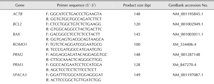
Table 1. Primer sequences used for gene expression analysis in spermatozoa treated with iodixanol during cryopreservation
Gene Primer sequence (5′–3′) Product size (bp) GenBank accession No.
ACTB F: GGCATCCTGACCCTGAAGTA 148 NM_001195845.1
R: GGTGTGGTGCCAGATCTTCT
BCL2 F: CTCCTGGCTGTCTCTGAAGG 120 NM_001002949.1
R: GTGGCAGGCCTACTGACTTC
BAX F: GACGGCCTCCTCTCCTACTT 145 NM_001003011.1
R: GGTGAGTGACGCAGTAAGCA
ROMO1 F: TGTCTCAGGATCGGAATGCG 100 XM_534406.4
R: TCCCGATGGCCATGAATGTG
PRM2 F: AGGAGGAGATACAGGAGGTGC 148 NM_001287148
R: CTTGCAAACTCAGGGCTTGG
PRM3 F: GGCCACGAATCCTCCATGGA 128 XM_847270.4
R: AGCTCCTCCTCTTCCTCCT
SPACA3 F: GGATTTCGGCATGGAGGGAT 149 NM_001197087.1
R: ACTTCCGGCTGTTGATCTGG F, forward; R, reverse.
Real-Time PCR System (Applied Biosystems, USA) and each target gene’s expression was quantified relative to that of the internal gene (ACTB) using the equation, R = 2
–[ΔCt sample – ΔCt
control]
[17].
Frozen-thawed canine sperm smears were fixed in methanol/glacial acetic acid (3:1) at 4
oC for 5 min. Control and treatment group slides were treated for 20 min with 100 L CMA
3
solution. The CMA
3
solution contained 0.25 mg/mL CMA
3
in McIlvane’s buffer (pH 7.0) supplemented with 10 mM MgCl
2
. Slides were then rinsed in McIlvain's buffer and air dried. Microscopic analysis of slides was performed by measuring fluorescence with a Zeiss microscope at 1,000×
magnification. A total of 200 spermatozoa were randomly evaluated on each slide. Evaluation of CMA
3
was completed by identifying two types of staining patterns: bright green fluorescence of the sperm head (CMA
3
positive/abnormal chromatin packaging) and dull green staining (CMA
3
negative/normal chromatin packaging) of the sperm head.
Sperm capacitation test
The contents of one cryopreserved straw were divided into 2 aliquots after thawing in a water bath at 37
oC for 30 sec.
Cryocapacitation was analyzed using the first aliquot, while capacitation ability after thawing was analyzed using the second aliquot.
The first aliquot was diluted by adding 1 part semen to 9 parts 0.9% NaCl then an equal volume of trypan blue 0.27% (v/v) was added and the combination was mixed on a slide. Sperm smears were then fixed in a 37% formaldehyde solution for 2 min, then rinsed with distilled water. The Giemsa stock solution was freshly made by adding the stain to distilled water. Slides were dipped in 7.5% (v/v) of Giemsa stain, air-dried in a vertical
position, then cover-slipped. Slides were examined by examining 200 sperm cells with 5 independent replications. Assessment included counting the following: live spermatozoa with intact acrosomes (LSIA), live spermatozoa with damaged acrosomes (LSDA), dead spermatozoa with intact acrosomes (DSIA), and dead spermatozoa with damaged acrosomes (DSDA). The posterior part of the sperm head was dark blue in dead spermatozoa and sky-blue in live spermatozoa. Based on the color of anterior part sperm head, spermatozoa were divided into three groups, spermatozoa with intact acrosomes (purple), damage acrosomes (lavender), and those without acrosomes (pale gray). A canine capacitation medium (CCM) supplemented with 1.0 mM MgCl
2
and 10 mM progesterone was used for incubating the second aliquot for 4 h, following incubation the second aliquot was stained to determine acrosomal status as described for the first aliquot [30].
Mucus penetration test
Modified synthetic oviduct fluid, as a surrogate mucus, was loaded into marked flat capillary tubes (10 cm long, 3 mm deep;
Camlab, UK) that were sealed on one end. The filled capillary tubes were left standing vertically with the sealed end on top to allow removal of bubbles and check seal tightness. Then, the capillary tube was inserted into an Eppendorf tube containing a 100 L sperm suspension and laid horizontally for 2 h at room temperature. The spermatozoa reaching the 1 cm and 3 cm markers in the capillary tube were then counted.
Statistical analysis
All values are presented as mean ± SEM values, and a p value <
0.05 was used to indicate statistical significance. For multiple
comparisons among treatments and control groups, one-way