Catheter Ablation of Multiple Accessory Pathways in Duchenne Muscular Dystrophy
4
0
0
전체 글
(2) 116 WPW-Syndrome in Duchenne Muscular Dystrophy. cessfully terminated with adenosine intravenously. Blood pressure was 110/55 mm Hg. Echocardiography was normal. After resolution of the acute rhythm abnormality, ECG showed a delta-wave over all precordial recordings (Fig. 1), leading to the diagnosis of WPW-syndrome. Ajmaline was started and catheter ablation was scheduled. Ablation of three accessory pathways (right anterior-septal with anterograde and retrograde conduction, right posterior-septal with exclusively retrograde conduction, and right posterior-lateral with exclusively retrograde conduction) (Figs. 2 and 3) was successfully carried out one week after starting ajmaline without complications (Fig. 4). One day after ablation, however, a relapse of the supraventricu-. A . lar tachycardia occurred, which could be again relieved with ajmaline intravenously. Six days after ablation, a re-entry tachycardia recurred, which responded favorably to adenosine intravenously, but could be finally stopped only with ajmaline intravenously. On ECG, 12 days after ablation, delta-waves had disappeared, but small Qwaves over V 2-6 were recorded (Fig. 1). To prevent tachycardia, verapamil (240 mg/d) was additionally given with success. Unfortunately, the delta-wave and re-entry tachycardias recurred four months after the ablation. After switching to propafenone, neither the delta-wave nor re-entry tachycardia recurred.. B . Fig. 1. ECG on admission showing shortening of the PQ-interval and a delta-wave over all chest wall recordings (A). ECG 12 days after the ablation shows incomplete right bundle branch block and small Q-waves over V 2-6, but no longer pre-excitation (B). ECG: electrocardiography.. A . B . C . D . Fig. 2. Surface ECG before ablation with delta-wave (A) and after ablation without delta-wave (B). Intracardiac ECG before ablation (C) and after bump of the accessory pathway (D). The left lower panel shows change of the retrograde activation pattern during energy delivery (3rd beat from the right) with a shift to the second exclusively retrograde conducting right postero-septal accessory pathway. The right lower panel shows the retrograde activation pattern after ablation with exclusive conduction via the retrograde conducting AV-node. ECG: electrocardiography, AV: atrioventricular.. http://dx.doi.org/10.4070/kcj.2013.43.2.115. www.e-kcj.org.
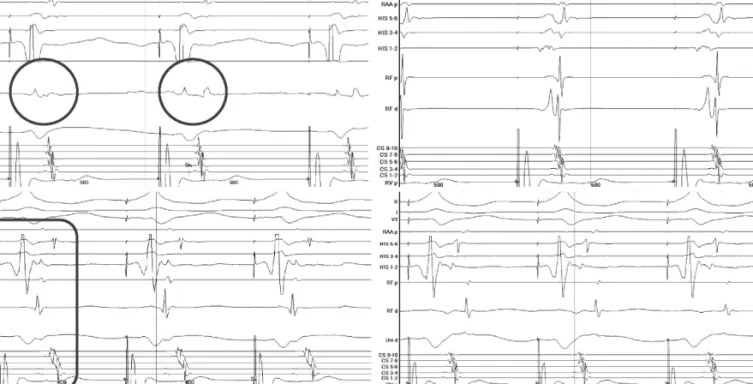
(3) Josef Finsterer, et al.. 117. Fig. 3. The left upper panel shows change of the retrograde activation pattern during energy delivery with a shift from the right anterior septal pathway (circle) to the right postero-septal pathway (circle). The right upper panel shows the target signal for the right postero-septal pathway. The right anterolateral accessory pathway with exclusively retrograde conduction (encircled) is shown in the left lower panel. Retrograde conduction via the AV-node after ablation is shown in the right lower panel. The ablation of the antero-lateral pathway was achieved by energy delivery via an Agilis long sheath during sinus rhythm. AV: atrioventricular.. A . B . C . D . Fig. 4. Catheter position during ablation of the 1. accessory, anterograde and retrograde conducting, right antero-septal pathway in LAO 45° (A) and in RAO 30° (B), of the 2. accessory exclusively retrograde conducting right posterior-septal pathway in LAO 45° (C), and during ablation of the 3. accessory, exclusively retrograde conducting right posterior-lateral pathway in LAO 45° (D). A stable position at the tricuspid anulus was achieved only with the steerable introducer Agilis NxT (St. Jude Medical). RV: right ventricle, HIS: HIS bundle, RAA: right atrial appendage, CS: coronary sinus, Abl: ablation catheter, LAO: left anterior oblique, RAO: right anterior oblique.. Discussion Cardiac involvement develops in the majority of the DMD-pawww.e-kcj.org. tients if they survive long enough,1) and is usually first recognized during the second decade,1)6) although a single case developed clinical or subclinical cardiac involvement already earlier.1) Cardiac involvement in DMD usually manifests as cardiomyopathy or rhythm abnormalities. WPW-syndrome has been only rarely reported as a manifestation of cardiac involvement in DMD.2) WPW-syndrome is a cardiac conduction abnormality, which is due to accessory conductive pathways between the atrium and the ventricle bypassing the atrioventricular (AV)-node.7) ECG typically shows PR-shortening, a delta wave, and QRS-widening, and patients with WPW-syndrome develop supraventricular re-entry tachycardias or atrial fibrillation. WPW-syndrome may be associated with sudden cardiac death, of which, the risk is estimated to be 0.02%/patient/y.8) The risk of sudden cardiac death is further increased in patients who additionally suffer from atrial fibrillation,8) or rapidly anterogradely conducting pathways with short effective refractory period. WPW-syndrome may respond to drug therapy with adenosine9) or ajmaline. The best and most effective and safe treatment of symptomatic WPW-syndrome, however, is ablation of the accessory pathways by radio-frequency ablation.10) After ablation, high activation recovery interval-dispersion may be observed in the persisting pre-excited rhythm, but may gradually return to normal within weeks.11) Also, in the presented patient ablation resulted in transient resolution of palpitations and disappearance of delta-waves. http://dx.doi.org/10.4070/kcj.2013.43.2.115.
(4) 118 WPW-Syndrome in Duchenne Muscular Dystrophy. Why the described DMD-patient presented with three accessory pathways remains speculative. WPW-syndrome is widely believed to result from incomplete regression of fetal accessory atrio-ventricular pathways, a procedure which may occur in neuromuscular disorders or independently from the muscle disease. Arguments for sporadic accessory pathway formation in the presented patient are that he never complained about palpitations before age 19 years and that an ECG at age 19 years did not show a delta-wave. Possibly, the atrio-ventricular connections developed due to abnormal enlargement of cardiomyocytes, since swelling of cardiomyocytes brings atrial and ventricular cardiomyocytes together, as has been described in patients with PRKAG2 syndrome.12) Absence of a deltawave on the previous ECG at age 19 years could be explained with intermittent occurrence of pre-excitation. Arguments for WPW-syndrome as a cardiac manifestation of DMD are that cardiac conduction abnormalities are a frequent finding in these patients, and that with improved survival, also previously non-reported cardiac conduction abnormalities may be seen. WPW-syndrome could also be unrelated to DMD, but clinical manifestation not earlier than at age 19 years argue against such an assumption. Why reentry tachycardias recurred after ablation is most likely due to an incomplete ablation of the right anterior-septal pathway located in the proximity of the AV-node and the HIS-bundle. This case shows that WPW-syndrome may occur in DMD-patients carrying a dystrophin mutation. The causal relation between WPWsyndrome and mutation, however, remains speculative. Repeated radio-frequency catheter ablation of accessory pathways may be necessary to completely block the re-entry mechanism in these patients.. References 1. Finsterer J, Stöllberger C. The heart in human dystrophinopathies. Cardiology 2003;99:1-19. 2. Agarwal RK, Misra DN, Verma RK. Wolff-Parkinson-White syndrome. http://dx.doi.org/10.4070/kcj.2013.43.2.115. with paroxysmal atrial fibrillation in pseudohypertrophic muscular dystrophy (Duchenne type). Indian Heart J 1973;25:346-8. 3. Finsterer J, Stöllberger C, Quasthoff S. Wolff-Parkinson-White syndrome as initial manifestation of Becker muscular dystrophy. Herz 2008;33:307-10. 4. Symons AL, Townsend GC, Hughes TE. Dental characteristics of patients with Duchenne muscular dystrophy. ASDC J Dent Child 2002;69: 277-83, 234. 5. Weiss C, Jakubiczka S, Huebner A, et al. Tandem duplication of DMD exon 18 associated with epilepsy, macroglossia, and endocrinologic abnormalities. Muscle Nerve 2007;35:396-401. 6. Kirchmann C, Kececioglu D, Korinthenberg R, Dittrich S. Echocardiographic and electrocardiographic findings of cardiomyopathy in Duchenne and Becker-Kiener muscular dystrophies. Pediatr Cardiol 2005; 26:66-72. 7. Sethi KK, Dhall A, Chadha DS, Garg S, Malani SK, Mathew OP. WPW and preexcitation syndromes. J Assoc Physicians India 2007;55 Suppl:10-5. 8. Cay S, Topaloglu S, Aras D. Percutenous catheter ablation of the accessory pathway in a patient with wolff-Parkinson-white syndrome associated with familial atrial fibrillation. Indian Pacing Electrophysiol J 2008;8:141-5. 9. Bodalski R, Maryniak A, Walczak F, Szumowski L, Jedynak Z. [Glass of water or ablation?-episodes of malignant atrial tachyarrhythmias during swimming in a lake in a woman with overt Wolff-Parkinson-White syndrome and benign palpitations for several decades of life]. Kardiol Pol 2008;66:1346-9. 10. Tischenko A, Fox DJ, Yee R, et al. When should we recommend catheter ablation for patients with the Wolff-Parkinson-White syndrome? Curr Opin Cardiol 2008;23:32-7. 11. Ghosh S, Rhee EK, Avari JN, Woodard PK, Rudy Y. Cardiac memory in patients with Wolff-Parkinson-White syndrome: noninvasive imaging of activation and repolarization before and after catheter ablation. Circulation 2008;118:907-15. 12. Light PE. Familial Wolff-Parkinson-White Syndrome: a disease of glycogen storage or ion channel dysfunction? J Cardiovasc Electrophysiol 2006;17 Suppl 1:S158-61.. www.e-kcj.org.
(5)
수치

관련 문서
