Veterinary Science
http://dx.doi.org/10.4142/jvs.2012.13.3.235
Received: 16 Aug. 2011, Revised: 31 Oct. 2011, Accepted: 19 Apr. 2012
Original Article
*Corresponding authors: Tel: +82-43-261-2868; Fax: +82-43-272-1603; E-mails: [email protected], [email protected]
†The first two authors contributed equally to this work.
ⓒ 2012 The Korean Society of Veterinary Science.
This is an Open Access article distributed under the terms of the Creative Commons Attribution Non-Commercial License (http://creativecommons.org/licenses/by-nc/3.0) which permits
unrestricted non-commercial use, distribution, and reproduction in any medium, provided the original work is properly cited.
Activation of Akt/protein kinase B mediates the protective effects of mechanical stretching against myocardial ischemia-reperfusion injury
Chan-Hyung Kim
2,*
,†, Jia Hao
1,†, Hee-Yul Ahn
2, Si Wook Kim
3,*
1
Department of Public Health, Chengdu Medical College, Chengdu, Sichuan 610083, China
Departments of
2Pharmacology, and
3Thoracic and Cardiovascular Surgery, College of Medicine, Chungbuk National University, Cheongju 361-763, Korea
Akt/protein kinase B is a well-known cell survival factor and activated by many stimuli including mechanical stretching.
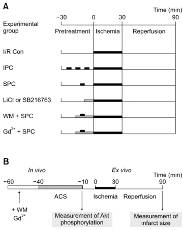
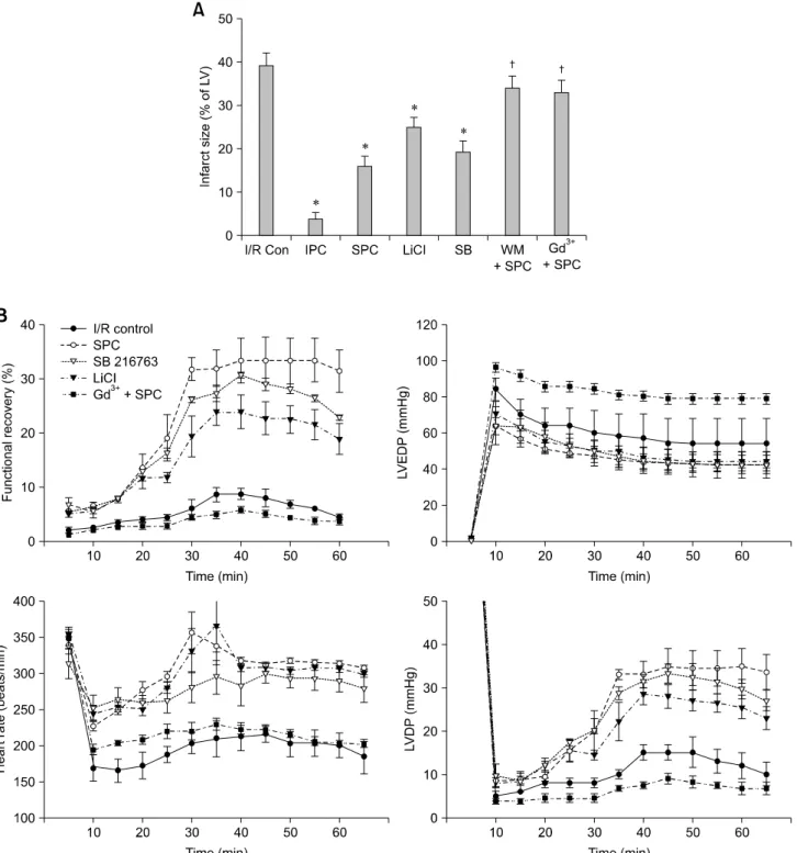
Therefore, we evaluated the cardioprotective effect of a brief mechanical stretching of rat hearts and determined whether activation of Akt through phosphatidylinositol 3-kinase (PI3K) is involved in stretch-induced cardioprotection (SIC). Stretch preconditioning reduced infarct size and improved post- ischemic cardiac function compared to the control group.
Phosphorylation of Akt and its downstream substrate, GSK-3β, was increased by mechanical stretching and completely blocked by wortmannin, a PI3K inhibitor. Treatment with lithium or SB216763 (GSK-3β inhibitors) before ischemia induction mimicked the protective effects of SIC on rat heart. Gadolinium (Gd
3+), a blocker of stretch-activated ion channels (SACs), inhibited the stretch-induced phosphorylation of Akt and GSK-3β. Furthermore, SIC was abrogated by wortmannin and Gd
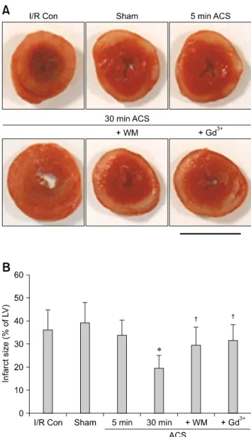
3+. In vivo stretching induced by an aorto-caval shunt increased Akt phosphorylation and reduced myocardial infarction; these effects were diminished by wortmannin and Gd
3+ pretreatment. Our results showed that mechanical stretching can provide cardioprotection against ischemia- reperfusion injury. Additionally, the activation of Akt, which might be regulated by SACs and the PI3K pathway, plays an important role in SIC.
Keywords: Akt/protein kinase B, cardioprotection, ischemia- reperfusion injury, mechanical stretching
Introduction
Ischemic heart disease is due to deficient coronary circulation. This pathologic condition is usually associated
with myocardial damage caused by cardiac ischemia- reperfusion (I/R) and has been the leading cause of death worldwide [35]. Ischemic preconditioning (IPC) achieved by repeated short episodes of ischemia protects the heart against a subsequent sustained period of ischemia, and consequently reduces post-ischemic arrhythmia, abnormal contractility, and the extent of myocardial infarction [5].
IPC also can be mimicked by pharmacological pretreatment [20], heat shock [16], and mechanical stretching of the heart [28]. Many studies have shown that stretch preconditioning (SPC) enables the myocardium to better tolerate a subsequent ischemic insult. Ovize et al. [28] showed that brief stretching of the myocardium significantly reduces myocardial infarction extent in canines, and that this protective effect is blocked by gadolinium (Gd
3+), a potent inhibitor of stretch-activated ion channels (SACs).
Additionally, temporary myocardial stretching induced by increased diastolic pressure in the left ventricle results in significantly decreased infarct size and improves post-ischemic contractile function in isolated rat hearts [25,27]. However, details about the signaling pathways involved in stretch-induced cardioprotection (SIC) have not been previously reported.
Mechanical stretching directly affects cardiac functions
and rapidly activates multiple signal transduction factors in
cardiac myocytes such as the c-Jun N-terminal kinase,
mitogen-activated protein kinases, protein kinase C, and
Janus kinase/signal transducers and activators of
transcription pathways [6,29]. The serine/threonine-
specific protein kinase Akt (also known as protein kinase
B), is a potent survival factor [7] and one of the primary
kinases activated by phosphatidylinositol-3-kinase (PI3K)
which is activated by several myocardial protective