359
ORIGINAL ARTICLEDOI 10.4070 / kcj.2009.39.9.359
Print ISSN 1738-5520 / On-line ISSN 1738-5555 Copyright ⓒ 2009 The Korean Society of Cardiology
Cardioprotective Effects of Alpha-Lipoic Acid on Myocardial Reperfusion Injury: Suppression of Reactive Oxygen Species Generation and Activation of Mitogen-Activated Protein Kinase
Seok Kyu Oh, MD
1, Kyeong Ho Yun, MD
1, Nam Jin Yoo, MD
1, Nam-Ho Kim, MD
1, Min-Sun Kim, MD
2, Byung-Rim Park, MD
2and Jin-Won Jeong, MD
11Division of Cardiology, Departments of Internal Medicine and 2Physiology, Wonkwang University School of Medicine, Iksan, Korea
ABSTRACT
Background and Objectives:
Reactive oxygen species (ROS) and mitogen-activated protein (MAP) kinase play an important role in the development of myocardial reperfusion injury. In this study, we examined whether treatment with alpha-lipoic acid (ALA) before reperfusion could prevent myocardial reperfusion injury in vivo.
Materials and Methods:Sprague-Dawley rats were subjected to a 45-minute left anterior descending coronary artery ligation followed by 45- or 10-minute reperfusion. ALA was administered 10 minutes prior to reperfusion. The infarct size ratio of the infarct area to the ischemic area at risk, was measured based on 10, 25, 50, and 100 mg/kg of ALA, with propidium iodide (PI) fluorescence. Apoptosis was evaluated by TdT-mediated dUDP nick end labeling (TUNEL) staining. The generation of intracellular ROS was evaluated using the fluorogenic probe, dichlorodihy- drofluorescein diacetate (CM-H
2DCFDA). Western blot analysis was performed for MAP kinase (pERK 1/2 and pJNK 1/2) activity.
Results:The infarct size, according to ALA dose, was significantly suppressed 29.1% with ALA 25 mg/kg (p<0.0001), 41.5% with 50 mg/kg (p<0.05), and 41.4% with 100 mg/kg (p<0.05) compared to the con- trols (54.3%). However, the results were not significantly different with 47.2% of the ALA 10 mg/kg (p=0.192). A few apoptotic nucleoli were detected in the ALA 25 mg/kg group, but were frequently detected in the control group.
The ROS generation was significantly suppressed (p<0.0001), the activity of pERK 1/2 was significantly increased (p<0.05) and the activity of pJNK 1/2 was significantly decreased (p<0.05) in the ALA 25 mg/kg group com- pared to the controls.
Conclusion:The results of this study suggested that adequate doses of ALA before reper- fusion was effective for the prevention of myocardial reperfusion injury in vivo. This cardioprotective activity of ALA might be associated with an anti-apoptotic effect of ALA via suppression of ROS generation, increase of pERK 1/2 and decrease of pJNK 1/2 activity.
(Korean Circ J 2009;39:359-366)KEY WORDS:
Myocardial reperfusion injury; Alpha-lipoic acid; Apoptosis; Reactive oxygen species; Mitogen- activated protein kinase.
Introduction
Following acute myocardial infarction, re-establishing coronary blood flow with the rapid use of reperfusion strategies such as thrombolysis or primary coronary intervention is essential to salvage viable myocardium.
However, paradoxically, the process of reperfusion can itself result in myocyte death, a phenomenon referred to as “lethal reperfusion injury”.
1)Apoptosis and necrosis
are the two major distinct types of cell death of cardio- myocytes that have been associated with ischemia and reperfusion. Particularly, apoptosis-induced myocyte dea- th is thought to play an important role during the early stages of reperfusion.
2-4)The exact mechanisms underlying myocardial reperfu- sion injury are not known. However, reactive oxygen spe- cies (ROS) are formed in excessive amounts within the first few minutes following reperfusion and are considered important to myocardial reperfusion injury.
5)In addition, excessive ROS during the early stages of reperfusion in- creases the activity of mitogen-activated protein (MAP) kinase and induces apoptosis following reperfusion.
6-8)Anti-oxidant treatments have been reported to prevent myocardial reperfusion injury.
9)10)Among the anti-oxi- dants, alpha-lipoic acid (ALA) thiol antioxidant, has
Received: October 22, 2008 Revision Received: January 28, 2009 Accepted: February 11, 2009
Correspondence: Seok Kyu Oh, MD,Division of Cardiology, Department of Internal Medicine, Wonkwang University School of Medicine, 344-2 Sin- yong-dong, Iksan 570-711, Korea
Tel: 82-63-859-2522, Fax: 82-63-852-8480 E-mail: [email protected]
360
·Alpha-Lipoic Acid on Myocardial Reperfusion Injurybeen shown to preserve cardiac function during ische- mia-reperfusion while acting as a co-factor for mito- chondrial dehydrogenase.
11-13)Several authors have re- ported that low-dose ALA reduces myocardial reperfu- sion injury in vitro.
13)14)To date, however, few studies have reported on the effectiveness of ALA in preventing apoptotic myocardial reperfusion injury in vivo.
We examined whether the administration of ALA prior to reperfusion therapy would reduce apoptotic re- perfusion injury of myocytes in an ischemia-reperfusion rat model.
Materials and Methods
Experimental animals
Healthy male Sprague-Dawley rats weighed 250-300 g were divided into an ALA treatment group and a control group. The rats in both groups received 45-minutes of ischemia. The infarct size of the myocardium was assessed 45 minutes following reperfusion. Apoptosis staining, ROS synthesis and the activity of MAP kinase were assessed 10 minutes following reperfusion. To confirm the dose of ALA, at which the infarct size was suppressed most effectively, ALA 10, 25, 50 and 100 mg/kg was administered to the right ventricle 10 minutes prior to the reperfusion. The infarct size was smallest in the ALA 25 mg/kg group. Following this, apoptosis ROS syn- thesis and MAP kinase activity were measured in the ALA 25 mg/kg group.
To induce cardiac ischemia, chloral hydrate (100 mg/
kg) (Merck, Germany) was injected intra-abdominally for anesthesia. In the supine position, the rats were fixed onto an experimental plate. An endotracheal tube was placed using a 16-G tube and ventilation provided. The sternum was incised and the heart exposed. The proxi- mal part of the left anterior descending artery was ligated using No. 6 silk and maintained for 45 minutes. Con- firmation of myocardial infarction was based on decreased movement of the left ventricle and change of the coloring to blue. After the indicated time, the ligated suture was released. Reperfusion was confirmed by the return to normal coloring of the left ventricle. Thereafter, the rats were sacrificed according to schedule. During all experi- mental procedures, the temperature of the rats was main- tained at 37℃ with a white lamp.
The measurement of the infarct size
To measure the infarct size based on the dose of ALA in all groups including: ALA 10 mg/kg (n=5), 25 mg/
kg (n=5), 50 mg/kg (n=5), 100 mg/kg (n=5) and the control group (saline 0.5 mL) (n=5), propidium iodide (PI) 1 mg (Sigma Chemical Co.) was injected into the right ventricle over 30 minutes following reperfusion. PI, 1 mg was diluted in saline 0.5 mL. Forty five minutes following reperfusion, the left anterior descending artery
was ligated again. Then, in order to differentiate between the ischemic and normal areas, polystyrene/divinylben- zene fluorescence (Duke Scientific) was injected into the proximal part of the aorta.
15)The rats were then sacrific- ed and stored in a -70℃ refrigerator. Thereafter, the samples were fixated in 4% paraformaldehyde for two hours. Using a vibrotom, the left ventricle was vertically sectioned at a thickness of 400 μm. Following this, the basal, middle and apical areas were selected and the in- farct size of myocardium was measured using a polarized microscope. On polarized microscopy, the normal area was observed as a green color, the ischemic area was black and the infarct area was a red color. The infarct size was defined as the ratio of the infarct area relative to the is- chemic area of the myocardium.
The measurement of apoptosis
To measure apoptosis at the myocardial infarction site, terminal deoxynucleotidyl transferase mediated biotin- dUTP nick-end labeling (TUNEL) staining was perform- ed in the ALA 25 mg/kg group (n=5) and the control group (n=5), using an in situ apoptosis detection kit (TaKaRa Shuzo Co.). The cardiac tissue was attached to a glass slide and rinsed with 0.01 M phosphate buffered saline (PBS) (pH 7.4), and then treated with proteinase K (20 g/mL) at room temperature for 20 minutes. The sample was again rinsed with PBS. To suppress peroxi- dase activity, intrinsic peroxidase activity was blocked using 3% H
2O
2at room temperature for 5 minutes. Fol- lowing the rinsing, the sample was treated with a perme- ability buffer on ice for 2-5 minutes. Fluorescein-dUTP (TdT+labeling safe buffer) was used for the labeling performed at 37℃ in an incubator for 90 minutes. Fol- lowing rinsing, the anti-FITC HRP conjugate underwent antibody reaction using a 37℃ incubator. Then, the color was developed using DAB (5 mg/mL in Tris buf- fer, pH 7.0) at room temperature for 10-15 minutes. The degree of apoptosis was then observed using a light mi- croscope. On light microscopy, with the use of a digital camera, the microscopic images were converted to a digital format and then saved in a computer. An auto- matic system for the imaging analysis was used to meas- ure the number of cells that were positive for TUNEL staining.
The measurement of reactive oxygen species ge- neration
To measure ROS generation, 5-(and-6)-chloromethyl- 2’,7’-dichlorodihydro-fluorescein diacetate (CM-H
2DC- FDA) (Molecular Probes Inc., Eugene, USA) 100 μg was directly injected into the right ventricle 10 minutes prior to the ligation of the left anterior descending ar- tery in the ALA 25 mg/kg (n=5) and control groups (n=
5). Ten minutes prior to reperfusion, ALA or saline was
administered to the right ventricle. Ten minutes following
Seok Kyu Oh, et al.·
361
reperfusion, the rats were sacrificed and then stored in a -70℃ refrigerator. Thereafter, with the use of a freezing microtom, the left ventricle was sectioned at a thickness of 40 μm in the vertical direction of the long axis. Fol- lowing this, basal, middle and apical areas were selected.
Then, using a polarized microscope, the fluorescent de- velopment of a clear green color was noted, which re- flected the ROS.
16)17)The amount of ROS synthesis was analyzed using image pro plus in the areas of ischemic injury. The images of the original green-colored samples were converted to an 8 bit gray color. In the areas of is- chemic injury, the values of the gray scale were averaged and the expression of ROS defined. The amount of ROS generation that was calculated in the control group was summed. Then, the average was defined as the amount of ROS expression, which was considered 100% com- pared to the ALA 25 mg/kg group. The amount of ROS generation in the ALA 25 mg/kg group was obtained by calculating the average value of the gray scale in the areas of injury in each experimental animal. This value was calculated as a percentile compared to the values for the control group.
The measurement of mitogen-activated protein ki- nase activity
Among the MAP kinases, pERK 1/2 and pJNK 1/2 were measured in the rats after they were sacrificed in both the ALA 25 mg/kg (n=5) and the control groups (n=5) 10 minutes following reperfusion. The infarction area of the left ventricle was extracted and then frozen using liquid nitrogen. The sample was stored in a -70℃
refrigerator. To isolate the protein from cardiac muscle, the cardiac muscles were converted to a powder using liquid nitrogen and a grinder. The powder of cardiac muscle was diluted in a histolysis buffer solution (20 mM Tris-HCl pH 8.0, 1% NP-40, 150 mM EDTA, 10% gly- cerol, 0.1 % beta-mercaptoethanol, 0.5 mM DTT) at a ratio of 1 : 5 of weight/volume, and maintained in an ice bath for more than an hour. Centrifugation of the sample was performed at 4℃ at 12,000×g for 15 minutes. Then, the supernatant protein was harvested.
Thereafter, using the Bradford method,
18)the protein concentration was quantified using a spectrophotometer (Beckman Co., USA).
Protein electrophoresis was performed using sodium dodecyl sulfate polyacrylamide gel electrophoresis (SDS- PAGE). The protein was mixed with a sample buffer (0.125 M Tris pH 6.8, 2% SDS, 25% glycerol, bromo- phenol blue, 2-mercaptoethanol) at 100℃. The protein content (80 μg), which was mixed with a histolysis buf- fer solution, underwent electrophoresis with a 12%- and 15%-gradient. The electrophoresis was performed using a mini gel electrophoremeter (SE 600 Hoefer Sci. Ins), at 90 V for two hours. Following electrophoresis, the gel was stained using Coomassie brilliant blue R-250. Des-
taining was carried out with destaining solution (10%
acetic acid and 10% methanol) and the protein bands were confirmed.
With the use of protein transfer equipment (Hoefer Semiphor, Phamacia Bio.), the protein gels were trans- ferred to polyvinylidene difluoride (PVDF) membranes of 0.45 μm in thickness (Millipore Co., USA) for two hours. To block the non-specific binding of primary an- tibodies, this PVDF membrane was placed in a block- ing buffer where Tris buffer saline (TBS) (pH 7.6) was dissolved in a 3% bovine serum albumin and then the reaction was performed for an hour. The primary anti- body, pERK 1/2 (1:500) (Cell Signaling Technology, USA), was reacted overnight at 4℃ and then rinsed with 0.05% Tween-20-TBS (TBST) three times. There- after, the secondary antibody, goat rabbit immunoglo- bulin G (IgG) conjugated AP (Santa Cruz, USA), was diluted at a ratio of 1 : 2,000 and the reaction was per- formed for one hour. Following rinsing with TBST, rin- sing with alkaline phosphates (0.1 M Tris, 0.1 M NaCl, 0.01 M MgCl
2) was performed. Thus, the color was de- veloped with NBT and BCIP. In addition, with the same conditions, as the controlled experiment for pERK 1/2, the inactive form of ERK 1/2 protein expression was ob- served. The expression of pJNK 1/2 and JNK 1/2 were also observed in the same manner as ERK.
To analyze the degree of the expression of pERK 1/2 and pJNK 1/2, on Western blot analysis, with the use of a scanner (HP4P), the pattern of the proteins’ color was developed on the PVDF membrane and then converted to a digital format. Then, the data was saved in a personal computer. With the use of an imaging analysis program (UN-SCAN-IT gel, USA), the area and density of each protein expression were measured. Then, these two pa- rameters were multiplied and the volume of each protein was thereby calculated. The expression of active proteins, pERK 1/2 and pJNK 1/2, were defined based on the fi- nal amount of expression compared to the values of ERK 1/2 and JNK 1/2. In addition, the relative ratio of expression was calculated based on the amount of ex- pression of the active forms noted in the sham surgery group (n=2).
Statistical analysis
The experimental results were expressed as the mean
±standard deviation (SD). The inter-group analysis of the statistical data was performed using the Kruskall- Wallis test. A value of p<0.05 was considered statistically significant.
Results
The effect of alpha-lipoic acid on the infarct size
The infarct size based on the dose of ALA was signifi-
cantly smaller in the ALA 25 mg/kg (29.1±4.8%, p<
362
·Alpha-Lipoic Acid on Myocardial Reperfusion Injury0.0001), ALA 50 mg/kg (41.5±9.5%, p<0.05), and ALA 100 mg/kg groups (41.4±7.9%, p<0.05) compared to the controls (54.3±8.7%). However, the ALA 10 mg/
kg group (47.2±9.5%, p=0.192) did not differ signifi- cantly from the control group. The ALA 25 mg/kg treat- ment group had the smallest infarct size (Fig. 1) (Table 1).
The effect of alpha-lipoic acid on apoptosis Following TUNEL staining, on light microscopy, apo- ptotic nucleoli, which were stained as a dark brown color, indicating reperfusion injury due to apoptosis, were fre- quently detected in the control group. By contrast, this finding was rarely seen in the ALA 25 mg/kg group (Fig. 2).
The effect of alpha-lipoic acid on reactive oxygen species generation
In the ALA 25 mg/kg group, the color development indicating the synthesis of ROS was only observed in 41.6% (p<0.0001) compared to 100% in the control gro- up (Fig. 3).
The effect of alpha-lipoic acid on the activation of mitogen-activated protein kinases
In regard to the pattern of the expression of pERK 1/2 with regard to anti-apoptotic effects and pJNK 1/2
with regard to apoptotic effects, the relative ratio of ex- pression was calculated compared to the sham surgery group in the controls and the ALA 25 mg/kg group (Fig.
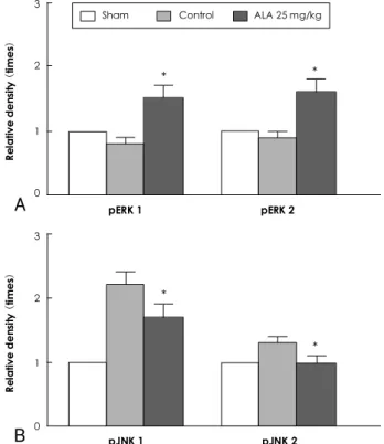
4). The expression of pERK 1/2 was significantly incre- ased in the ALA 25 mg/kg group compared to the control group as shown with pERK 1 (0.8±0.1 times vs. 1.5±
0.2 times, p<0.05) and pERK 2 (0.9±0.1 times vs. 1.6
±0.2 times, p<0.05) (Fig. 5A) (Table 2). In addition, the expression of pJNK 1/2 was significantly decreased in the ALA 25 mg/kg group compared to the control group as shown with pJNK 1 (2.2±0.2 times vs. 1.7±0.2 times, p<0.05) and pJNK 2 (1.3±0.1 times vs. 1.0±0.1 times, p<0.05) (Fig. 5B) (Table 2).
Discussion
In the current study, the administration of ALA prior to reperfusion significantly reduced the infarct size in rats after myocardial ischemia-reperfusion. The admini- stration of ALA 25 mg/kg prior to reperfusion significan- tly reduced apoptotic cell death in the myocardium.
This anti-apoptotic effect of ALA was observed to be closely associated with the inhibition of ROS generation, the increased activity of pERK 1/2 and the decreased activity of pJNK 1/2.
Prompt reperfusion is essential for the survival of car- diac muscle after an ischemic event. In addition to the cardiac injury caused by ischemia, the reperfusion itself can promote cardiac injury and make it worse. This phe-
Table 1. Ischemic area, infarct area, and infarct size according to ALA dose by propidium iodide fluorescence
Ischemic area (mm2)
Infarct area (mm2)
Infarct size*
(%) Control 0.97±0.24 0.53±0.190 54.3±8.70 ALA 10 (mg/kg) 0.95±0.27 0.45±0.200 47.2±9.50 ALA 25 (mg/kg) 0.94±0.29 0.28±0.12† 29.1±4.8† ALA 50 (mg/kg) 0.98±0.33 0.41±0.19‡ 41.5±9.5‡ ALA 100 (mg/kg) 0.97±0.29 0.40±0.22‡ 41.4±7.9‡
*Infarct size: ratio of infarct area to ischemic area, †p<0.0001, ‡ p<0.05 compared with control. ALA: alpha-lipoic acid
Infarct area/ischemic area at risk (%) 100 80
60
40
20
0
p=0.192
p<0.0001
p<0.05 p<0.05
Control ALA 10 ALA 25 ALA 50 ALA 100 mg/kg mg/kg mg/kg mg/kg
47.2%
29.1%
41.5% 41.4%
54.3%
Fig. 1. This graph shows the infarct size (ratio of infarct to ische- mic area) according to ALA dose at 45 minutes after reperfusion by propidium iodide fluorescence. The ALA at doses of 25 mg/
kg (p<0.0001), 50 mg/kg (p<0.05), and 100 mg/kg (p<0.05) sig- nificantly reduced the infarct size compared to the control group, but not the ALA 10 mg/kg (p=0.192) group. The ALA 25 mg/kg group had the smallest infarct size. ALA: alpha-lipoic acid.
A
B
Fig. 2. Light microscopic findings of TUNEL staining in the infarct area at 10 minutes after reperfusion in the control (A) and ALA 25 mg/kg (B) groups. Apoptotic nucleoli (brown spot) were frequently detected in the control group (A). By contrast, only a few apop- totic nucleoli were detected in the ALA 25 mg/kg group (B). TUNEL:
terminal deoxynucleotidyl transferase-mediated biotin-dUDP nick end labeling, ALA: alpha-lipoic acid.
Seok Kyu Oh, et al.·
363
nomenon is referred to as reperfusion injury.
1)In myo- cardial ischemia-reperfusion, the myocardial injury is caused by necrosis and apoptosis. Kajstura et al.
19)and Fliss et al.
20)reported that myocardial injury caused by apoptosis was significantly more prevalent than injury due to necrosis in a 2-hour ischemia model and a 45- minute ischemia and a 1-hour reperfusion model. Sev- eral investigators have reported that apoptosis was a more
critical factor than necrosis with regard to myocardial injury in ischemia-reperfusion animal models, and that apoptosis was significantly increased by reperfusion.
2-4)The exact mechanisms underlying myocardial reper- fusion injury is not known. However, ROS are formed in excessive amounts within the first few minutes fol- lowing reperfusion and are considered a major factor involved in myocardial reperfusion injury.
5)21-23)To mi- nimize reperfusion injury, powerful antioxidants have been administered to reduce ROS generation and the- reby decrease myocardial injury.
17)24)25)Among the antio- xidants, ALA, a thiol compound, is a cofactor for mito- chondrial dehydration enzymes. In the tissue mitochon- dria, it is reduced to dithiol dihydrolipoic acid (DHLA), an effective form. DHLA has been reported to play a
Table 2. The relative expression of pERK 1/2 and pJNK 1/2 in the infarct area by Western blot and densitometry analysis
Sham Control ALA 25 mg/kg
pERK 1 1 0.8±0.1 1.5±0.2*
pERK 2 1 0.9±0.1 1.6±0.2*
pJNK 1 1 2.2±0.2 1.7±0.2*
pJNK 2 1 1.3±0.1 1.0±0.1*
*p<0.05 compared with control. ALA: alpha-lipoic acid, ERK: ex- tracellular signal-regulated kinases, JNK: c-Jun-NH2-terminal kinases
A B
p<0.0001
Relative ROS generation (%) 100%
41.6%
120
100
80
60
40
20
0
Control ALA 25 mg/kg
C
Fig. 3. Confocal laser microscopic findings of fluorescence on intracellular ROS generation detected by CM-H2DCFDA at 10 mi- nutes after reperfusion in the control (A) and ALA 25 mg/kg (B) groups. The fluorescence expression (bright green color) was more frequently detected in the control group (A) than in the ALA 25 mg/kg group (B). The relative ROS generation and the relative le- vel of CM-H2DCFDA fluorescence by confocal microscopy, was significantly suppressed in the ALA 25 mg/kg group compared to the control group (C). ROS: reactive oxygen species, CM-H2DC- FDA: 5-(and-6)-chloromethyl-2’,7’-dichlorodihydro-fluorescein dia- cetate, ALA: alpha-lipoic acid.
Sham Control ALA 25 mg/kg
ERK 1: 44k Da ERK 2: 42k Da
pERK1: 44k Da pERK2: 42k Da
JNK1: 57k Da JNK2: 46.5k Da
pJNK1: 57k Da pJNK2: 46.5k Da
Fig. 4. The expression of mitogen-activated protein (MAP) kinase by Western blot analysis pERK 1/2 expression was increased more in the ALA 25 mg/kg group compared to controls. By contrast, pJNK 1/2 expression was decreased more in the ALA 25 mg/kg group than in controls. ALA: alpha-lipoic acid, ERK: extracellular signal-regulated kinase, JNK: c-Jun-NH2-terminal kinase.
Fig. 5. This graph shows the relative expression of pERK 1/2 (A) and pJNK 1/2 (B) in the infarct area by Western blot and den- sitometry analysis. The relative density, of the Western blot analy- sis by densitometry, was compared with the sham density. The relative density of pERK 1/2 was significantly increased (A), and for pJNK 1/2 it was significantly decreased (B) in the ALA 25 mg/kg group compared to controls. *p<0.05 compared with control. ALA:
alpha-lipoic acid, ERK: extracellular signal-regulated kinase, JNK:
c-Jun-NH2-terminal kinase.
Relative density (times)
Sham Control ALA 25 mg/kg 3
2
1
0
pERK 1 pERK 2
* *
A
Relative density (times)
3
2
1
0
pJNK 1 pJNK 2
*
*
B
364
·Alpha-Lipoic Acid on Myocardial Reperfusion Injuryrole in preserving myocardium function by normaliza- tion of the intracellular pH, increased mitochondrial ATP synthesis and decreased ATP hydrolysis in the sett- ing of myocardial ischemia-reperfusion.
12)13)According to Schonheit et al.
13)a low-dose of ALA improved cardiac function following reperfusion in an in vitro study. By contrast, a high-dose of ALA increased cardiac damage following reperfusion. Based on these findings, the ef- fect of ALA treatment on the prevention of reperfusion injury was thought to be dose-dependent. According to Cao et al.
14)a minimal dose of ALA increased intrinsic antioxidants and the concentration of phase 2 enzymes with defense against oxidation, in an experiment using cultured cardiac cells from rats. Thus, ALA has been re- ported to reduce myocardial injury caused by ROS.
ALA has been reported to prevent ischemia-reperfu- sion injury in a dose-dependent manner in vitro. To date, however, few studies have examined the effects of ALA on the prevention of myocardial ischemia-reperfu- sion injury in vivo. In the current study, the infarct size of the myocardium was differentially suppressed depend- ing on the dose of ALA administered 10 minutes prior to reperfusion. Compared to the control group, the ALA 25 mg/kg group (p<0.0001) had the smallest infarct size.
In addition, the infarct size was also suppressed in the 50 mg/kg (p<0.05) and the 100 mg/kg groups (p<0.05).
However, the infarct size was not significantly sup- pressed in the ALA 10 mg/kg group. Furthermore, in the ALA 25 mg/kg group compared to the control group, apoptosis was significantly suppressed. These results in- dicate that ALA suppressed apoptosis and reduced myo- cardial injury during reperfusion.
Little is known about the mechanisms that cause myo- cardial injury due to apoptosis as a result of ischemia- reperfusion in the myocardium. Immediately after reper- fusion, however, an excessive degree of ROS generation and the activation of MAP kinase might be involved.
ROS generation reached the highest levels within a mi- nute following reperfusion and thereafter persisted. An excessive degree of ROS generation is known to play a crucial role in the pathogenesis of myocardial injury due to apoptosis immediately after reperfusion.
21-23)The ROS can be synthesized from endothelial cells, inflammatory cells and cardiac cells through a variety of enzyme activity.
Particularly in the early stage of reperfusion, an excessive amount of ROS generation in the endothelial cells and cardiac cells stimulates inflammatory cells and the acti- vated inflammatory cells release ROS. Thus, ROS plays a key role in the early and late stages of apoptosis.
22)26)27)ROS generation reached maximum levels within a min- ute following reperfusion. Therefore, to minimize the reperfusion injury, antioxidant treatment must be per- formed prior to reperfusion.
Bolli et al.
23)assessed myocardial contractility in groups where treatment was performed 15 and 1 minute prior
to reperfusion and 1 minute following reperfusion with a control group in a canine myocardial ischemia-reper- fusion model. In the groups where the treatment was performed 15 and 1 minute prior to reperfusion, com- pared to the control group, the myocardial contractility was significantly improved. In the group where the treat- ment was performed 1 minute following reperfusion, however, there was no significant difference between the treated and control groups. The investigators concluded that treatment must be performed prior to reperfusion to prevent reperfusion injury. In the current study, ALA was administered 10 minutes prior to reperfusion. On polarized light microscopy, using a detector for active oxygen, CM-H
2DCFDA, in the ALA 25 mg/kg group compared to the control group, showed that the genera- tion of ROS was significantly suppressed at the site of myocardial infarction (p<0.0001). These findings suggest that the cardioprotective effects of ALA might be closely associated with the suppression of apoptosis due to dec- reased generation of ROS.
It is not known whether ROS is involved in the acti- vation of the protein kinase pathway, which is associated with the induction of apoptosis following myocardial is- chemia and reperfusion. An excessively produced ROS during the early stages of reperfusion activates the in- flammatory cells and augments the intracellular calcium levels. In addition, it increases the secretion of pro-apo- ptotic genes and activates MAP kinase, nuclear factor- κB (NF-κB) and tumor necrosis factor-α (TNF-α), and thereby induces apoptosis. Thus, it is involved in triggering myocardial injury.
28)Protein kinase pathways that are activated by ischemiareperfusion of the myocar- dium include MAP kinase, PI-3 kinase/Akt and tyrosine kinase. MAP kinases can be divided into three types in- cluding the extracellular signal-regulated kinase (ERK 1/2), c-Jun-NH2-terminal kinase 1/2 (JNK 1/2) and the p38 α/β kinase.
7)MAP kinase belongs to a class of serinethreonine protein kinases that are activated in response to the various stimuli. ERK 1/2 is involved in the survival of cells and JNK 1/2 is involved in apop- tosis; thus, it is involved in maintaining the balance bet- ween survival and apoptosis.
29)30)According to Yue et al.,
29)in a myocardial ischemia-reperfusion rat model, the activation of ERK 1/2 reached the highest levels 10 minutes following ischemia and in 10 minutes following reperfusion. When PD98059 selectively blocked the ERK 1/2 pathway, the degree of apoptosis in the myo- cardium was significantly higher compared to controls.
Thus, it was demonstrated that pERK 1/2 has anti-
apoptotic effects in myocardial ischemia-reperfusion. In
addition, Ferrandi et al.
30)reported that the size of the
infarction site was decreased and the apoptosis of cardiac
muscles was markedly reduced after activation of the JNK
1/2 pathway was blocked following the use of AS601245,
a JNK 1/2 inhibitor, in a myocardial ischemia-reperfu-
Seok Kyu Oh, et al.·
365
sion rat model. Thus, it was demonstrated that pJNK 1/2 was involved in the apoptosis of myocardial ische- mia-reperfusion. These findings suggest novel treatment modalities that increase pERK 1/2 involved in cell sur- vival and suppress pJNK 1/2 involved in apoptosis; mo- dulation of these factors might prevent myocardial re- perfusion injury due to apoptosis. In the current study, the administration of ALA 25 mg/kg prior to reperfusion resulted in significantly increased pERK 1/2 activity 10 minutes following reperfusion (p<0.05) compared to controls. In addition, the activity of pJNK 1/2 was sig- nificantly decreased (p<0.05). These findings suggest that the cardioprotective effects of ALA treatment prior to reperfusion were closely associated with the suppression of apoptosis due to increased pERK 1/2 and decreased pJNK 1/2.
In conclusion, the results of this study show that ALA therapy prior to reperfusion significantly reduced the in- farct size in a myocardial ischemiareperfusion rat model.
The cardioprotective effects of ALA might be associated with the anti-apoptotic effects of ALA. The anti-apopto- tic effects of ALA were closely associated with the in- hibition of ROS generation, increased activity of pERK 1/2 and decreased activity of pJNK 1/2. The cardiopro- tective effects of ALA with regard to reperfusion injury
in vivo suggest that ALA therapy might be a novel treat-ment modality for minimizing reperfusion injury in pa- tients with acute myocardial infarction in the clinical setting. Additional studies are needed to examine the ef- fects of ALA administration in a large series of patients with acute myocardial infarction.
Acknowledgments
This work was financially supported by the grants of The Korean Society of Cardiology in 2004.
REFERENCES
1) Braunwald E, Kloner RA. Myocardial reperfusion: a double-edg- ed sword? J Clin Invest 1985;76:1713-9.
2) Eefting F, Rensing B, Wigman J, et al. Role of apoptosis in re- perfusion injury. Cardiovasc Res 2004;61:414-26.
3) Zhao ZQ, Nakamura M, Wang NP, et al. Reperfusion induces myo- cardial apoptotic cell death. Cardiovasc Res 2000;45:651-60.
4) Anversa P, Cheng W, Liu Y, Leri A, Redaelli G, Kajstura J. Apo- ptosis and myocardial infarction. Basic Res Cardiol 1998;93 (Suppl 3):8-12.
5) Zweier JL. Measurement of superoxide-derived free radicals in the reperfused heart: evidence for a free radical mechanism of reperfusion injury. J Biol Chem 1988;263:1353-7.
6) Cicconi S, Ventura N, Pastore D, et al. Characterization of apop- tosis signal transduction pathways in HL-5 cardiomyocytes ex- posed to ischemia/reperfusion oxidative stress model. J Cell Phy- siol 2003;195:27-37.
7) Abe J, Baines CP, Berk BC. Role of mitogen-activated protein kinases in ischemia and reperfusion injury: the good and the bad.
Circ Res 2000;86:607-9.
8) Omura T, Yoshiyama M, Shimada T, et al. Activation of mitogen- activated protein kinases in in vivo ischemia/reperfused myocar-
dium in rats. J Mol Cell Cardiol 1999;31:1269-79.
9) Dhalla NS, Elmoselhi AB, Hata T, Makino N. Status of myocar- dial antioxidants in ischemia-reperfusion injury. Cardiovasc Res 2000;47:446-56.
10) Marczin N, El-Habashi N, Hoare GS, Bundy RE, Yacoub M. An- tioxidants in myocardial ischemia-reperfusion injury: therapeutic potential and basic mechanisms. Arch Biochem Biophys 2003;
420:222-36.
11) Biewenga GP, Haenen GR, Bast A. The pharmacology of the an- tioxidant lipoic acid. Gen Pharmacol 1997;29:315-31.
12) Freisleben HJ. Lipoic acid reduces ischemia-reperfusion injury in animal models. Toxicology 2000;148:159-71.
13) Schonheit K, Gille L, Nohl H. Effect of alpha-lipoic acid and dihydrolipoic acid on ischemia/reperfusion injury of the heart and heart mitochondria. Biochem Biophys Acta 1995;1271:335-42.
14) Cao Z, Tsang M, Zhao H, Li Y. Induction of endogenous anti- oxidants and phase 2 enzymes by alpha-lipoic acid in rat cardiac H9C2 cells: protection against oxidative injury. Biochem Bio- phys Res Commun 2003;310:979-85.
15) Wolff RA, Chien GL, van Winkle DM. Propidium iodide com- pares favorably with histology and triphenyl tetrazolium chloride in the assessment of experimentally-induced infarct size. J Mol Cell Cardiol 2000;32:225-32.
16) Doi K, Suzuki Y, Nakao A, Fujita T, Noiri E. Radical scavenger edaravone developed for clinical use ameliorates ischemia/re- perfusion injury in rat kidney. Kidney Int 2004;65:1714-23.
17) Kim H, Park CK, Kim KS. Antioxidants inhibit smooth muscle cell proliferation in vitro and neointimal hyperplasia in vivo af- ter carotid artery injury in the rat. Korean Circ J 2004;34:69-75.
18) Bradford MM. A rapid and sensitive method for the quantitation of microgram quantities of protein utilizing the principle of pro- tein-dye binding. Anal Biochem 1976;72:248-54.
19) Kajstura J, Chen W, Reiss K, et al. Apoptotic and necrotic myo- cyte cell deaths are independent contributing variables of infarct size in rats. Lab Invest 1996;74:86-107.
20) Fliss H, Gattinger D. Apoptosis in ischemic and reperfused rat myocardium. Circ Res 1996;79:949-56.
21) Ambrosio G, Zweier JL, Flaherty JT. The relationship between oxygen radical generation and impairment of myocardial energy metabolism following post-ischemic reperfusion. J Mol Cell Car- diol 1991;23:1359-74.
22) Duilio C, Ambrosio G, Kuppusamy P, Dipaula A, Becker LC, Zweier JL. Neutrophils are primary source of O2 radicals during reperfusion after prolonged myocardial ischemia. Am J Physiol Heart Circ Physiol 2001;280:H2649-57.
23) Bolli R, Jeroudi MO, Patel BS, et al. Marked reduction of free radial generation and contractile dysfunction by antioxidant the- rapy begun at the time of reperfusion: evidence that myocardial stunning is a manifestation of reperfusion injury. Circ Res 1989;
65:607-22.
24) Kim TY, Chung HM, Park HJ, et al. The cardioprotective effects of resveratrol via anti-apoptosis in hypoxic injury of myocardial cells. Korean Circ J 2007;37:408-13.
25) Koh JH, Ryu KH, Lim SH, Hong KS, Choi YJ, Park SW. Effect of antioxidants on myocardial damage in streptozotocin-induced diabetic rats. Korean Circ J 2006;36:261-71.
26) Scarabelli T, Stephanou A, Rayment N, et al. Apoptosis of endo- thelial cells precedes myocyte cell apoptosis in ischemia/reperfu- sion injury. Circulation 2001;104:253-6.
27) Dun Y, Zhi JM, Sun HY, Zhao RR, Zhao ZQ. Activated polymor- phonuclear leukocytes induce cardiomyocyte apoptosis and the protective effects of carvedilol. Methods Find Exp Clin Pharmacol 2002;24:403-12.
366
·Alpha-Lipoic Acid on Myocardial Reperfusion Injury28) Zhao ZQ. Oxidative stress-elicited myocardial apoptosis during reperfusion. Curr Opin Pharmocol 2004;4:159-65.
29) Yue TL, Wang C, Gu JL, et al. Inhibition of extracellular signal- regulated kinase enhances Ischemia/Reoxygenation-induced apo- ptosis in cultured cardiac myocytes and exaggerates reperfusion
injury in isolated perfused heart. Circ Res 2000;86:692-9.
30) Ferrandi C, Ballerio R, Gaillard P, et al. Inhibition of c-Jun N- terminal kinase decreases cardiomyocyte apoptosis and infarct size after myocardial ischemia and reperfusion in anaesthetized rats. Br J Pharmacol 2004;142:953-60.