415
The Current Concept of Cell Therapy for Heart Failure
Sang Hong Baek, M.D.
Division of Cardiovascular Medicine, Kangnam St. Mary’s Hospital, The Catholic University of Korea College of Medicine, Seoul, Korea
ABSTRACT
Cardiac repair is a dream in the field of medical science. The biological limitations to human cardiac regenerative growth require the creation of new strategies for cardiac regeneration using cells, genes and protein. Recent experimental studies and early-phase clinical trials showed stem cells have the potential to enhance myocardial perfusion and contractile performance in patients with acute myocardial infarction, advanced coronary artery di- sease and chronic heart failure. Overall clinical experience also suggests that stem cell therapy can be safely per- formed if the right cell protocol is used within the correct clinical setting. Some experimental data have shown stable stem cell engraftment due to fusion or transdifferentiation into cardiomyocyte or vascular cell lineages, which could be likely explanations for these beneficial effects. Others have proposed that transient cell retention may be sufficient to promote functional effects, e.g., by release of paracrine mediators. We should proceed cau- tiously with carefully designed clinical trials, and concern for patient safety must remain the key issue. The trans- lational basic research will be required to elucidate the mechanism of stem cell therapy. (Korean Circulation J 2005;35:415-423)
KEY WORDS:Heart failure, congestive;Stem cells.
Introduction
Heart failure continues to be a major medical problem, despite advances in medical management and device therapy. Heart failure is a clinical syndrome, which can result from any structural or functional cardiac disorder that impairs the ability of the ventricle to fill with or pump blood. These include intrinsic defects of the con- tractility of cardiac muscle due to the altered expression of calcium-cycling proteins, components of the sarco- mere and enzymes for cardiac energy production; ex- trinsic defects to cardiac muscle cells, such as interstitial fibrosis, affecting the compliance of the heart; and myocyte loss, unmatched by myocyte replacement. Car- diac regeneration is robust in certain organisms, such as the newt and zebrafish, where total replacement can transpire, even for an amputated limb, fin or tail, via the production of an undifferentiated cell mass, called the blastema.
1)The degree of regeneration might also be dependent on the retention of proliferate potential in
a subset of adult cardiomyocytes, which is impossible in mammals under normal, unassisted biological circum- stances. Several complementary strategies can be helpful in potentially aiding this process: overriding cell-cycle checkpoints that constrain the reactive proliferation of ventricular myocytes
2); supplementing the naturally oc- curring cytoprotective mechanisms, or inhibiting pro- death pathways
3); supplementing the naturally occurring angiogenic mechanisms using defined growth factors or arteriole-forming cells
4); or providing exogenous cells as a surrogate or precursor for cardiac muscle itself.
5)Among these conceptual possibilities, cell transplantation, in various forms, is the first strategy that has been trans- lated from the bench to the bedside. The possibility of tissue repair by autologous adult stem and progenitor cells, suggested by the auspicious findings in experimen- tal studies of various cell sources, immediately captured the attention of clinicians confronted with patients who suffer from disabling, life-threatening heart failure in acute or chronic ischemic heart disease.
Within only the past three years, more than a half- dozen preliminary clinical studies have been published, ranging from case reports to formal trials, deploying a range of differing cell-based therapies, with the shared objective of improving cardiac repair. In general, these small initial human trials have pointed toward a func-
Correspondence:Sang Hong Baek, M.D.,Division of Cardiovascular Me- dicine, Kangnam St. Mary’s Hospital, The Catholic University of Korea College of Medicine, 505 Banpo-dong, Seocho-gu, Seoul 137-040, Korea
Tel: 82-2-590-2075, Fax: 82-2-591-1075 E-mail: [email protected]
tional improvement, despite their different strategies and cells, and lack of double-blinded controls; however, key questions remain open. A better understanding of just why and how grafting works will be essential, alongside the need for empirical trials to engineer the soundest future for regenerative therapy in human cardiovascular disease.
In this review, the various sources of stem or progeni- tor cells are compared, along with a discussion of the pro- gress of clinical studies using stem cells for heart failure.
Potential Stem Cells
Currently, a variety of stem and progenitor cells could potentially be used for cardiac repair. Each cell type has its own profile of advantages, limitations and practica- bility issues within specific clinical settings. Many in- vestigators have; therefore, chosen a pragmatic approach using unfractionated bone marrow cells(BMCs), which contain different stem and progenitor cell populations, including hematopoietic stem cells(HSCs), endothelial progenitor cells(EPCs) and mesenchymal stem cells (MSCs).
Endothelial progenitor cells
EPCs were originally defined by their cell surface ex- pressions of the hematopoietic markers, CD133 and CD34, and the endothelial marker vascular endothelial growth factor(VEGF) receptor-2, and their capacity for incorporation into sites of neovascularization and to differentiate into endothelial cells in situ.
6)The evidence suggests that culture-expanded EPCs also contain a CD14
+/CD34
--mononuclear cell population with EPC capacity, which mediates their angiogenic effects by re- leasing paracrine factors. Notably, number of EPCs and their angiogenic capacity are impaired in patients with coronary artery disease, which may limit their therapeu- tic usefulness.
7)CD133
+cells
The cell surface antigen CD133 is expressed on early HSCs and EPCs, both of which collaborate to promote vascularization of ischemic tissues.
8)CD133
+cells can integrate into sites of neovascularization and differentiate into mature endothelial cells. Because CD133 expression is lost in myelomonocytic cells, this marker provides an effective means to distinguish true CD133
+EPCs from EPCs of myelomonocytic origin. Less than 1% of nuc- leated BMCs are CD133
+, and because these cells can not be expanded ex vivo, only limited numbers of CD133
+cells can be obtained for therapeutic purposes.
Mesenchymal stem cells
MSCs represent a rare population of CD34
-and CD133
-cells, which are present in bone marrow stroma
( 10-fold less population than HSCs) and other mesen- chymal tissues.
9)MSCs can readily differentiate into os- teocytes, chondrocytes and adipocytes. Differentiation of MSCs to cardiomyocyte-like cells has been observed under specific culture conditions and after injection into healthy or infarcted myocardium in animals.
10)When injected into infarct tissue, MSCs may enhance regional wall motion and prevent remodeling of the remote, no- ninfarcted myocardium. Because MSC clones can be expanded in vitro, and reportedly have a low immuno- genicity, they might be, in the future, used in an allo- geneic setting.
Skeletal myoblasts
Skeletal myoblasts, or satellite cells, are progenitor cells that normally lie in a quiescent state under the ba- sal membrane of mature muscular fibers. Myoblasts can be isolated from skeletal muscle biopsies and expanded in vitro. Myoblasts differentiate into myotubes, and re- tain skeletal muscle properties when transplanted into an infarct scar.
11)Although myotubes do not couple with resident cardiomyocytes electromechanically, myoblast transplantation has been shown to augment systolic and diastolic performance in animal models of myocardial infarction.
Resident cardiac stem cells ( CSC )
The presence of resident CSC populations capable of differentiating into cardiomyocyte or vascular lineages suggests they could be used for cardiac tissue repair.
12)CSCs can be clonally expand from human myocardial biopsies. It has been reported that intramyocardial in- jection of these cells, after AMI in mice, promotes car- diomyocyte and vascular cell formation, leading to an improvement in systolic function.
Embryonic stem ( ES ) cells
ES cells are totipotent stem cells derived from the inner cell mass of blastocysts. Under specific culture conditions, ES cells differentiate into multicellular em- bryoid bodies containing differentiated cells from all three germ layers, including cardiomyocytes. Human ES Cell-derived cardiomyocytes display structural and func- tional properties of early-stage cardiomyocytes, which couple electrically with host cardiomyocytes when trans- planted into normal myocardium.
13)In theory, infinite numbers of cardiomyocytes could be obtained from hu- man ES cell clones. However, unresolved ethical and legal issues, concerns about the tumorigenicity of the cells, and the need to use allogeneic cells for transplan- tation, currently hamper their use in clinical studies.
Modes of Cell Delivery
The goal of any cell delivery strategy is to transplant
sufficient numbers of cells into the myocardial region of interest, and achieve maximum retention of cells within that area. The local milieu is an important de- terminant of cell retention, as it will influence short- term cell survival and, if a transvascular approach is used, cell adhesion, transmigration through the vascular wall and tissue invasion.
Transvascular approaches
Transvascular strategies are especially suited for the treatment of recently infarcted and reperfused myocar- dium when chemoattractants and cell adhesion molecu- les are highly expressed.
14)Intracoronary artery infusion
Selective intracoronary application delivers a maxi- mum concentration of cells homogeneously to the site of injury during the first passage. Unselected BMCs, circulating blood-derived progenitor cells and MSCs have been delivered, via the intracoronary route, in patients with acute myocardial infarction(AMI) and ischemic cardiomyopathy(Table 1, 2).
Intravenous infusion
In experimental models, intravenous delivery of EPCs or MSCs has been shown to improve cardiac function after an AMI.
15)However, homing of cells to noncardiac organs, such as the spleen, limits the clinical applicabi- lity of this approach.
Mobilization of stem and progenitor cells
Considering acutely infarcted myocardium recruits circulating stem and progenitor cells to the site of injury,
15)stem and progenitor cells mobilization by cy- tokines may offer a noninvasive strategy for cardiac re- generation. This concept has been tested in animal models of AMI and pilot studies in patients with AMI and chronic myocardial ischemia.
Direct injection in the ventricular wall
Direct injection is the preferred route for cell delivery in patients with chronic myocardial ischemia or when cell homing signals are expressed at low levels in the heart scar tissue. However, direct injection of cells into ischemic or scarred myocardium creates islands of cells
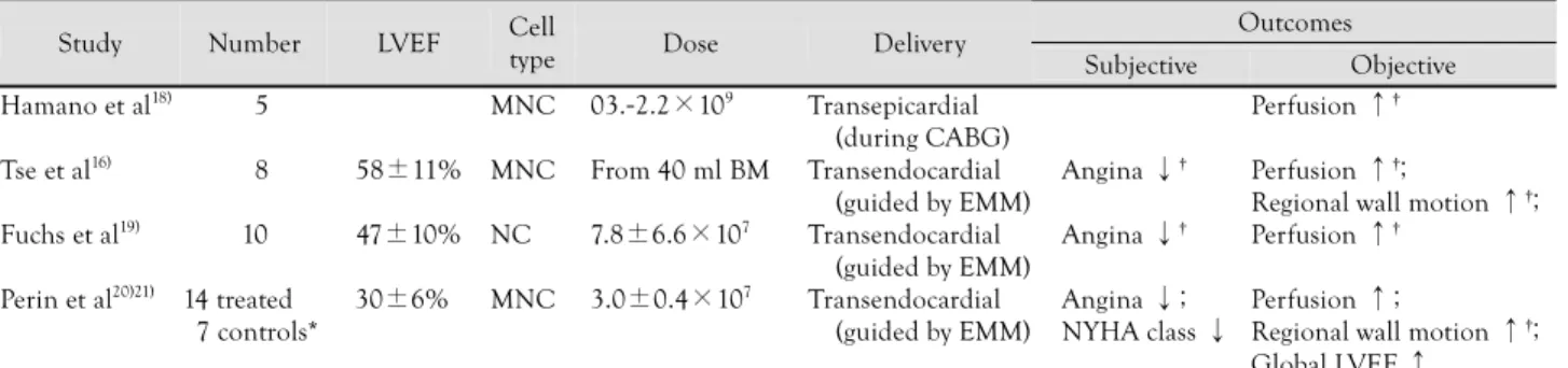
Table 1.
Clinical trials of cell therapy in myocardial ischemia, without a revascularization option
Outcomes
Study Number LVEF Cell
type Dose Delivery
Subjective Objective
Hamano et al
18)05 MNC 03.-2.2×10
9Transepicardial
(during CABG)
Perfusion ↑
†Tse et al
16)08 58±11% MNC From 40 ml BM Transendocardial
(guided by EMM)
Angina ↓
†Perfusion ↑
†;
Regional wall motion ↑
†; Fuchs et al
19)10 47±10% NC 7.8±6.6×10
7Transendocardial
(guided by EMM)
Angina ↓
†Perfusion ↑
†Perin et al
20)21)14 treated
07 controls*
30±6%0 MNC 3.0±0.4×10
7Transendocardial (guided by EMM)
Angina ↓;
NYHA class ↓
Perfusion ↑;
Regional wall motion ↑
†; Global LVEF ↑
LVEF: left ventricular ejection fraction, MNC: bone marrow-derived mononuclear cells, NC: bone marrow-derived nucleated cells, BM: bone mar- row, CABG: coronary artery bypass grafting, EMM: electromechanical mapping, NYHA: New York Heart Association. *: nonrandomized control group, †: effects reported only within cell therapy groups. Values are means±SD
Table 2.
Clinical trials of cell therapy in ischemic cardiomyopathy
Study Number LVEF Cell type Dose Time after MI Delivery Outcomes
‡Menasche et al
22)10 24±4% Myoblasts 8.7±1.9×10
83-228 Months Transepicardial (during CABG)*
Regional wall motion ↑;
Global LVEF ↑ Herreros et al
23)11 36±8% Myoblasts 1.9±1.2×10
83-168 Months Transepicardial
(during CABG)
†Regional wall motion ↑;
Global LVEF ↑;
Viability in Infarct area ↑ Siminiak et al
24)10 25-40% Myoblasts 0.04-5.0×10
74-108 Months Transepicardial
(during CABG)
†Regional wall motion ↑;
Global LVEF ↑ Chachques et al
25)20 28±3% Myoblasts 3.0±0.2×10
8Not reported Transepicardial
(during CABG)*
Regional wall motion ↑;
Global LVEF ↑;
Viability in Infarct area ↑ Smits et al
26)05 36±11% Myoblasts 2.0±1.1×10
824-132 Months Transepicardial
(guided by EMM)
Regional wall motion ↑;
Global LVEF ↑ Stamm et al
27)28)12 36±11% CD 133
+1.0-2.8×10
63-12 weeks Transepicardial
(during CABG)*
Global LVEF ↑;
LVEDV ↓;
Perfusion ↑ Assmus et al
29)51 MNC,
35 CPC
40±11% MNC CPC
1.7±0.8×10
82.3-1.2×10
73-144 Months IC Global LVEF ↑;
(only in MNC group) LVEF: left ventricular ejection fraction, CD 133: bone marrow-derived CD 133
+cells, MNC: bone marrow-derived mononuclear cells, CPC: circu- lating blood-derived progenitor cells, MI: myocardial infarction, CAGB: coronary artery bypass grafting, EMM: electromechanical mapping, IC: intra- coronary, LVEDV: left ventricular end-diastolic volume. *: CABG of noninjected territories only, †: CABG of injected and noninjected territories,
‡: effects only within cell therapy groups. Values are means±SD
with limited blood supply, which may lead to poor cell survival. Direct injection techniques are especially sui- ted for the application of large cells, such as MSCs or myoblasts, which may cause microembolization after intracoronary delivery.
Transendocardial injection
Using an injection needle catheter, advanced across the aortic valve and positioned against the endocardial surface, cells can be directly injected into the left ven- tricular wall.
16)Electromechanical mapping(ex. NOGA*
system) of the endocardial surface can be used to deli- neate viable, ischemic and scarred myocardium before cell injections.
Transepicardial injection
Transepicardial cell injection during open heart sur- gery allows for the direct visualization of the myocar- dium and the targeted application of cells to scarred areas and/or the border zone of an infarct scar. The invasiveness of this approach hampers its use as a stand- alone therapy. Conversely, the efficiency of cell trans- plantation may be difficult to evaluate and ascertain if coronary artery bypass grafting is performed simulta- neously.
Transcoronary vein injection
A catheter system, incorporating an ultrasound tip for guidance and an extendable needle for myocardial access, has been used to deliver BMCs through the coronary veins into normal pig myocardium, and to deliver myo- blasts to areas of nonviable myocardium in a pilot trial in patients with ischemic cardiomyopathy.
17)In contrast to the transendocardial approach, where cells are injec- ted perpendicular to the ventricular wall, the composite catheter system delivers cells parallel to the ventricular wall and deep into the injured myocardium.
Initial Clinical Results of Cell Therapy in Heart Failure
Tables 1 and 2 summarize the results of all clinical trials studying cell-based myocardial repair published to date. It is vital to distinguish between those investigations performed on patients with acute myocardial infarction and those on patients with chronic heart failure due to prior myocardial infarction, not only because of the different cell types and modes of delivery used, but also because fundamentally different pathophysiological pro- cesses are targeted. For example, in patients with acute myocardial infarction, progenitor cell transplantation is predicted to significantly modify postinfarction left ventri- cular(LV) remodeling, through enhanced neovasculari- zation and reduced cardiomyocyte apoptosis, irrespective of long-term engraftment and transdifferentiation. Con-
versely, the former 2 mechanisms acting alone may have little or no benefit in patients with long-established scars, apart from the functional rescue of hibernating myocytes. By comparison, in patients with chronic is- chemic heart disease and old myocardial infarction, the initial attempts at cell-based myocardial repair were more heterogeneous in outcome, most likely due, in part, to the more heterogeneous populations treated(Table 2).
The first such trial used skeletal muscle-derived proge- nitor cells, directly injected into the scarred region of the LV during open heart surgery for coronary artery bypass grafting. Global and regional LV functions were significantly and persistently improved, although con- comitant revascularization complicated the assessment of any benefit.
Indeed, in patients not undergoing simultaneous re- vascularization, transcatheter injection of myoblasts into the scar, resulting from a myocardial infarction 5-6 years earlier, reduced the symptoms of heart failure, but with- out objective evidence of an improved global LV function.
Unfortunately, the enthusiasm for injecting myoblasts into scar tissue for cardiac repair has been dampened by the fact that patients who have received this treat- ment experienced life-threatening arrhythmias. Mecha- nistically, this phenomenon may relate to the lack of electrical coupling of skeletal muscle to neighboring cardiomyocytes or; alternatively, be contingent on coup- ling by the few hybrid cells formed by fusion with adjacent cardiomyocytes, which generate spatially heterogeneous calcium transients. Therefore, skeletal myoblasts trans- plantation currently requires the placement of an im- plantable cardioverter/defibrillator, as a mandatory adjunct to therapy. Importantly, in the one small, non- randomized trial using bone marrow-derived progenitor cells for chronic ischemic heart failure, injection sites were chosen by electromechanical mapping of the LV endocardial surface to find areas of myocardial hiberna- tion: which resulted in significant increases in global LV ejection fraction, with decreased end-systolic volumes and improved exercise capacity. While this functional improvement might be secondary to an improved blood supply to hibernating cardiomyocytes, it is also concei- vable that an area of hibernating myocardium may pro- vide a more favorable microenvironment for the survival and engraftment of injected cells than a cell-depleted scar.
Lessons from Pilot Clinical Trials
Two clinical scenarios following infarction, acute or
chronic, have been subjected to cell transplantation for
cardiac repair. In the case of acute myocardial infarction,
the established safety and suggestive efficacy of intraco-
ronary progenitor cell transplantation provide a cogent
rationale for larger, randomized, double-blind trials and
for the expansion of such studies from Europe to the United States. In the case of chronic ischemic heart fai- lure, an additional question is whether identifying hiber- nating myocardium for direct cell therapy is essential to an effective outcome. Ultimately, it must be proven that cellular therapy aimed at cardiac repair not only improves pump function, but also reduces mortality and morbidity, or both.
Beyond safety and efficacy issues, what else do we need to know clinically? For ischemic disease, the tech- nical armament is in hand for treating patients’ hearts with progenitor cells, but still only at a very early stage, rudimentary experimental knowledge is being applied in the clinical arena; however, a variety of pivotal, but straightforward, utilitarian questions still remain unans- wered(optimal patient selection, usefulness of repeated cell transplantations). Nonischemic heart disease still has not been addressed at all.
The majority of this review focuses on the biological horizons; namely mobilization, homing, neovasculariza- tion and cardiac differentiation.
Cell mobilization
Cytokine-induced mobilization as a way to potentia- lly enhance cardiac repair came as an extrapolation of findings from the results of efforts to increase EPC levels for neovascularization in another context-hind limb ischemia. Indeed, VEGF
30)and GM-CSF
31)were found to augment EPC levels and improve neovascularization, and subsequent studies documented EPC mobilization by numerous other proangiogenic growth factors-stro- mal cell-derived factor-1(SDF-1), angiopoietin-1, pla- cental growth factor and erythropoietin.
32)Based on the report that bone marrow-derived cells can differentiate into cardiomyocytes, hematopoietic stem cell-mobilizing factors, granulocyte-colony stimulating factor(G-CSF) and SCF(Kit ligand) were used to experimentally im- prove cardiac regeneration, which quickly led to the initiation of clinical trials studying the ability of G- CSF to mobilize stem/progenitor cells in patients with coronary artery disease. Adverse vascular events were attributed to G-CSF in patients with intractable angina who were not considered as suitable candidates for re- vascularization and even in patients without cardiac disease. In the future, it may be preferable to use stra- tegies that augment circulating progenitor cells without causing massive inflammation. A second open question, with regard to systemic mobilization, is whether enough progenitor cells will home, where needed, to the sites of cardiac injury. Systemically administered human pro- genitor cells were predominantly trapped by the spleen when given to athymic nude rats, and cardiac regene- ration elicited by treatment with G-CSF plus SCF was documented only for animals lacking a spleen. The use of leukocyte-mobilizing cytokines might be most worth-
while when combined with selective enhancements of progenitor cell homing, or as a prelude to isolating cells for local delivery.
Cell homing
Homing is a multistep cascade, including the initial adhesion to activated endothelium or exposed matrix, transmigration through the endothelium and, finally, migration and invasion of the target tissue. The capa- city to migrate and invade may be pivotal to functional integration, even when cells are injected intramuscula- rly. Particularly in patients who lack the endogenous stimuli incited by acute ischemic injury, the enhance- ment of local homing signals, or the ability of cells to respond, may be of critical importance.
The mechanisms for the homing of progenitor cells to sites of tissue injury are only rudimentarily unders- tood, while homing of hematopoietic progenitor cells to bone marrow has been studied extensively.
33)SDF-1 ap- pears to be one key factor that regulates the trafficking of stem and progenitor cells to ischemic tissue, and local delivery of SDF-1 can enhance EPC recruitment and neovascularization. Cell necrosis causes the release of a chromatin-binding protein, high mobility group box protein 1(HMGB1), whose release acts as an extracel- lular “danger signal”, which may stimulate progenitor cells’ homing. Extracellular HMGB1 attracts mesoan- gioblasts, both in vitro and in vivo, and likely plays a role in muscle regeneration. HMGB1 interacts with the receptor for advanced glycation end products, as well as for Toll-like receptors 2 and 4. The exact mechanisms mediating cell attraction due to HMGB1 are unclear, but may involve additional receptors that remain to be identified.
The adhesion and transmigration of stem and pro- genitor cells are mediated by integrins. Indeed, integrin- dependent adhesion of EPCs is one effect of SDF-1.
34)Many of the chemokines and adhesion molecules in- duced by cardiac ischemic injury are familiar players in other disorders, but different cell types may use diffe- rent mechanisms for homing, and ischemia may differ according to the attractant induced.
Thus, a molecular dissection is essential to define the multiple steps of progenitor cell homing to, and inva- sion of, the myocardium, especially for those cells cur- rently used for clinical cardiac repair and for the other, novel, auspicious cells presently being used in preclinical studies.
Neovascularization
Currently, there is no direct clinical evidence that cellular cardiomyogenesis in fact occurs in the human heart following stem/progenitor cell transplantation.
Angiogenesis, improvements in scar tissue and cytopro-
tection must be considered, along with transdifferentia-
tion, as the most important possible consequences of cell-based therapies for cardiac repair. Of these, proge- nitor cells may most obviously improve neovasculariza- tion, which in turn would augment oxygen supply.
Progenitor cells are expected to be of most benefit to cardiac regeneration or performance when used to treat jeopardized or hibernating cardiomyocytes. Neovascu- larization, in turn, can be mediated by the physical in- corporation of progenitor cells into new capillaries or, in some settings, into perivascular cells. Incorporated progenitor cells of most, if not all, types may release growth factors, which promote angiogenesis by acting on mature endothelial cells. The extent to which pro- genitor cells contribute to vasculogenesis, by becoming physical elements of newly formed vessels angioblasts, versus their action through secreted factors, may depend on the circumstances of the cell type and cardiac in- jury. However, human bone marrow-derived angioblasts exert both types of effect.
35)Cells engineered to overex- press angiogenic factors might enhance both their own survival and that of the recipient myocardium.
Cardiac differentiation
Because most adult cardiac myocytes are terminally differentiated, the regenerative capacity of the infarcted myocardium is limited.
Cardiac myogenesis by noncardiac cells: The possi- bility of cardiomyocyte formation by multipotent pro- genitor/stem cells was first raised by pioneering works, in which ES cells were grafted into mouse myocardium,
36)the one cell type, along with germ cells, for which toti- potency is assured. The formation of functional cardio- myocytes by mouse and human ES cells is proven by adherence to many criteria. Although religious, ethical and political objections to the use of human ES cells have received justifiable attention, human ES cells, being allogeneic, also pose the clinical challenge of immuno- logical barriers, which are obviated in all forms of au- tologous cell therapy. Is the appearance of multiple lineages due merely to a mixed assortment of starting cells? For which cell types, settings, and means of ad- ministration does fusion of donor and host cells create the appearance of transdifferentiation or multiple poten- tials? Is plasticity in propagated cells merely acquired in culture and not a reflection of the native biology? A plausible interpretation is that the MSCs in bone mar- row, not HSCs, chiefly contribute to the creation of new cardiomyocytes following infarction. To date, human trials studying cardiac repair have all employed unfrac- tionated bone marrow cells, presumably including bone marrow MSCs. More recently, several groups have re- ported evidence of extracardiac progenitors in necropsy specimens of the hearts obtained from subjects having undergone sex-mismatched heart transplantation.
37)Cardiac myogenesis by adult cardiac progenitor cells:
The quest for novel heart-forming cells in adult myo- cardium can be traced to several instigating rationales:
the inability of skeletal myocytes to transdifferentiate;
challenges to claims of bone marrow-derived cells’ far- ranging plasticity; and an emerging counter-model of tissue-resident progenitor cells, which share some sig- natures of “stemness”, but are predisposed to differen- tiate into lineages of the organ in which they reside.
Evidence of resident progenitors in the heart has also
recently been reported. Beltrami et al
38)identified clus-
ters of highly proliferating cells in the myocardium,
consisting predominantly of undifferentiated lineage ne-
gative(Lin
-) cells expressing the stem cell marker, c-kit
(c-kit
Pos), and stem cell antigen 1(Sca-1
Pos). These cells
have clonogenic and self-renewing capabilities, and are
capable of differentiating into all myocardial cell types,
including cardiomyocytes, endothelial cells and vascular
smooth muscle cells. These findings suggest that the
resident precursor cells may represent a mechanism for
self-repair of the damaged myocardium. However, the
regenerative capacity of this putative self-repair mecha-
nism has been questioned by several groups, who argued
that the number of cardiac and extracardiac progenitors
that migrate to the heart is insufficient to induce ef-
fective long-term regeneration of myocardium. The me-
chanism of myocardial repair by local or transplanted
cells has not been elucidated. The relative contribution of
transdifferentiation and cell fusion to the regenerative
process remains controversial. Where and how do car-
diac progenitor or stem cells arise in the heart? Various
models can be considered, ranging from persistence as
undifferentiated remnants of heart-forming tissue in the
early embryo, to a hematogenous origin(from bone
marrow or even sites of earlier hematopoiesis, losing
HSC markers in the process), to mechanisms involving
ingrowth of the developing coronary vasculature. As an
example of the first mechanism, postnatal cardioblasts,
numbering just a few hundred per heart, have been
identified on the basis of the persistent expression of a
LIM-homeodomain transcription factor, Isl1, especially
in the atria, right ventricle and outflow tract-regions
where Isl1 is most prevalently expressed during cardiac
organogenesis.
39)By contrast, for cardiac Sca-1
+cells, a
third potential mechanism may be favored, given the
cells’ striking similarities to mesoangioblast, which include
surface labeling, microarray findings and the earliest sites
of marker expression.
40)How many cardiomyocytes, if
any, are generated by these new routes to heart muscle
cell formation in the normal heart after birth? What are
the unassisted contributions of these pathways to car-
diomyocyte formation in disease, as a reserve for the
replacement of dead and dying cells? To answer these
questions, the genetic strategy of ‘fate mapping’ is likely
to be relevant-indelibly tagging cells with an irreversible
marker of their status-using, for example, a Sca-1-driven
gene for Cre recombinase plus a Cre-dependent repor- ter to permanently label the progeny of Sca-1
+cells, even once Sca-1 is no longer expressed.
Cell augmentation for cardiac regeneration
The findings of unexpected persistence of cardiopoie- tic cells in adult hearts and effective cardiac repair by the noncardiac cells in current trials have raised a num- ber of questions. Although cell-based therapy has only taken its first steps toward clinical application, limiting factors have already arisen as targets for future impro- vement. Ultimately, genetic-engineered cells may super- cede naive stem cells.
Firstly, how can one make delivered cells more durable, considering the adverse environment? Most stem cells share the property of stress resistance,
41)but even stem cells die in the absence of blood flow. This concern is especially apt when progenitor cells are directly delive- red into unperfused necrotic myocardium. Targeting in- jections to the margin of injury, where oxygen supply persists, is immediately workable, and biological means to augment the survival of the cells are on the horizon, including the use of angiogenic factors, or modification of MSCs with the survival gene, Akt.
Secondly, how can one restore progenitor cells to nor- mal, where they are deficient in numbers or function?
The functional capacity of bone marrow-derived cells is defective in patients with heart failure,
42)and risk fac- tors for atherosclerosis have been found to correlate in- versely with the numbers and function of circulating EPCs. Stem cell defects also occur as a consequence of aging. Potential remedies include statins, which delay the onset of cellular senescence, telomere “uncapping”
and DNA damage signals in human EPCs. Direct in- terference with telomere-based aging and death signals might also be achieved through the forcible expression of TERT. Antioxidants or growth factors might also be used to promote telomerase activity. TERT and other cell cycle activators may be useful in prolonging donor cell cycling, either ex vivo or, especially if tightly control- led, following engraftment.
Thirdly, what drives the cardiac fate in adult heart- forming cells? While some mechanisms for cardiac specification in injured adult hearts might differ from those in early embryos, better knowledge of the “wiring diagram” for commitment to the cardiac fate is expec- ted to yield useful clues to enhance the differentiation process in susceptible cell types, and even extend the range of donor cells that are well able to form heart muscle.
5)Multiple autocrine or paracrine factors, from host myocardium or donor cells, provide essential ins- tructions to enter the cardiac muscle lineage.
In summary, the existence of cardiac and noncardiac progenitor cells, efficacious in cardiac repair, should stimulate vigorous subsequent inquiry into a means to
activate their migration, survival, growth and differen- tiation. These are questions of great importance, whether one is envisioning the manipulation of cells ex vivo for their subsequent administration, or instead, contempla- ting the activation of latent cells within the injured heart.
Adverse Events
Adverse events in experimental studies using adult stem cell have been relatively rare. However, the deve- lopment of microinfarction has been reported when infusing large MSCs directly into the coronary artery of a dog. In addition, adverse calcification has been shown to be a problem in a rat model, which received either total BM cells, BM-derived multipotent stem cells or saline following coronary ligation.
Serious ventricular arrhythmias developed in patients between 11 to 22 days post-transplantation when ske- letal myoblasts were used for cardiac repair in a phase I clinical trial. This may be explained by the lack of gap junctions between the transplanted myoblasts and the resident cardiomyocytes, or the difference between the action potentials of the two cell types. In contrast, ar- rhythmias were not a problem, per se, with clinical trials using BM-MNCs. However, the administration of any cells via a transendocardial route could induce arrhyth- mias. The major concern of using BM-MNCs may be the development of angiogenic neoplasias, since BM-de- rived EPCs can contribute to tumor neovascularization.
However, no angiogenic neoplasia has yet been repor- ted in clinical trials. One trial, utilizing peripheral blood stem cell, mobilized by G-CSF, for cardiomyo-angioge- nesis, has been stopped. In this study, an intracoronary infusion of stem cells was performed after coronary stenting. The observed increase in restenosis may be partially explained by the study design, but the increase in the leukocyte number to leukemic levels, via plaque growth or destabilization, may be directly responsible.
43)Adverse vascular events and accelerated atherosclerosis have been attributed to G-CSF in patients with intra- ctable angina who were not considered as suitable can- didates for revascularization.
Conclusion and Future Perspectives
Only small, mostly uncontrolled clinical studies, exp- loring the safety and feasibility of stem cell therapy, have so far been conducted. This preliminary clinical evidence suggests that stem cell therapy might be promising.
Upcoming trials will conduct intermediate-size, double-
blind, randomized-controlled clinical trials to establish
the effects of stem cell therapy on surrogate markers,
such as LV systolic function, myocardial perfusion or
exercise capacity. However, there is much caution against
their premature use in clinical settings, with the issues of the optimal cell type, cell number and timing of cell transfer and the appropriate clinical condition, such as acute myocardial infarction versus heart failure, remai- ning to be resolved. Safety of cell protocol remains the key concern. Although studies pertaining to these issues are underway, fundamental questions need to be expe- rimentally addressed. What is the fate of the injected cells after transplantation? How long do they survive?
Do the cells incorporate, or is transient retention suf- ficient to promote functional effects? Cell labeling and molecular imaging techniques need to be developed to track the fate of stem cells in vivo and to correlate the cell retention and engraftment with the functional out- comes. What is the nature and functional relevance of this interaction? Can cardiac stem cells be used for car- diac repair, or is their potential similar to cells obtai- ned from bone marrow? Can the regenerative capacity of transplanted stem cells be enhanced by drugs, cyto- kines or gene therapy approaches? Pharmacological and genetic strategies may help to enhance stem cell reten- tion, engraftment, differentiation and paracrine capa- bility. Additional work will elucidate the mechanisms involved in mobilization, homing, integration and sur- vival of progenitor cells at the sites of implantation. This, in turn, will help define the optimal conditions for their therapeutic application.
REFERENCES
1)
Poss KD, Wilson LG, Keating MT. Heart regeneration in zebra- fish. Science 2002;298:2188-90.
2)
Pasumarthi KB, Nakajima H, Nakajima HO, Soonpaa MH, Field LJ. Targeted expression of cyclin D2 results in cardiomyocyte DNA synthesis and infarct regression in transgenic mice. Circ Res 2005;96:110-8.
3)
Oh H, Wang SC, Prahash A, et al. Telomere attrition and chk2 activation in human heart failure. Proc Natl Acad Sci USA 2003;
100:5378-83.
4)
Losordo DW, Dimmeler S. Therapeutic angiogenesis and vascu- logenesis for ischemic disease: part I. angiogenic cytokines. Cir- culation 2004;109:2487-91.
5)
Olson EN, Schneider MD. Sizing up the heart: development redux in disease. Genes Dev 2003;17:1937-56.
6)
Asahara T, Kawamoto A. Endothelial progenitor cells for postna- tal vasculogenesis. Am J Physiol Cell Physiol 2004;287:C572-9.
7)
Vasa M, Fichtlscherer S, Aicher A, et al. Number and migratory activity of circulating endothelial progenitor cells inversely corre- late with risk factors for coronary artery disease. Circ Res 2001;
89:e1-7.
8)
Rafii S, Lyden D. Therapeutic stem and progenitor cell transp- lantation for organ vascularization and regeneration. Nat Med 2003;9:702-12.
9)
Pittenger MF, Martin BJ. Mesenchymal stem cells and their po- tential as cardiac therapeutics. Circ Res 2004;95:9-20.
10)
Makino S, Fukuda K, Miyoshi S, et al. Cardiomyocytes can be generated from marrow stromal cells in vitro. J Clin Invest 1999;
103:697-705.
11)
Ghostine S, Carrion C, Souza LC, et al. Long-term efficacy of myoblast transplantation on regional structure and function after
myocardial infarction. Circulation 2002;106:I131-6.
12)
Oh H, Bradfute SB, Gallardo TD, et al. Cardiac progenitor cells from adult myocardium: homing, differentiation, and fusion after infarction. Proc Natl Acad Sci U S A 2003;100:12313-8.
13)
Kehat I, Kenyagin-Karsenti D, Snir M, et al. Human embryonic stem cells can differentiate into myocytes with structural and functional properties of cardiomyocytes. J Clin Invest 2001;108:
407-14.
14)
Lee SH, Wolf PL, Escudero R, Deutsch R, Jamieson SW, Thist- lethwaite PA. Early expression of angiogenesis factors in acute myocardial ischemia and infarction. N Engl J Med 2000;342:
626-33.
15)
Kocher AA, Schuster MD, Szabolcs MJ, et al. Neovasculariza- tion of ischemic myocardium by human bone-marrow-derived angioblasts prevents cardiomyocyte apoptosis, reduces remodeling and improves cardiac function. Nat Med 2001;7:430-6.
16)
Tse HF, Kwong YL, Chan JK, Lo G, Ho CL, Lau CP. Angio- genesis in ischaemic myocardium by intramyocardial autologous bone marrow mononuclear cell implantation. Lancet 2003;361:
47-9.
17)
Thompson CA, Nasseri BA, Makower J, et al. Percutaneous trans- venous cellular cardiomyoplasty: a novel nonsurgical approach for myocardial cell transplantation. J Am Coll Cardiol 2003;41:
1964-71.
18)
Hamano K, Nishida M, Hirata K, et al. Local implantation of autologous bone marrow cells for therapeutic angiogenesis in patients with ischemic heart disease: clinical trial and prelimi- nary results. Jpn Circ J 2001;65:845-7.
19)
Fuchs S, Satler LF, Kornowski R, et al. Catheter-based autologous bone marrow myocardial injection in no-option patients with advanced coronary artery disease: a feasibility study. J Am Coll Cardiol 2003;41:1721-4.
20)
Perin EC, Dohmann HF, Borojevic R, et al. Transendocardial, autologous bone marrow cell transplantation for severe, chronic ischemic heart failure. Circulation 2003;107:2294-302.
21)
Perin EC, Dohmann HF, Borojevic R, et al. Improved exercise capacity and ischemia 6 and 12 months after transendocardial injection of autologous bone marrow mononuclear cells for is- chemic cardiomyopathy. Circulation 2004;110 ( Suppl ) :II213-8.
22)
Menasche P, Hagege AA, Vilquin JT, et al. Autologous skeletal myoblast transplantation for severe postinfarction left ventricu- lar dysfunction. J Am Coll Cardiol 2003;41:1078-83.
23)
Herreros J, Prosper F, Perez A, et al. Autologous intramyocardial injection of cultured skeletal muscle-derived stem cells in patients with non-acute myocardial infarction. Eur Heart J 2003;24:
2012-20.
24)
Siminiak T, Kalawski R, Fiszer D, et al. Autologous skeletal myoblast transplantation for the treatment of postinfarction myo- cardial injury: phase I clinical study with 12 months of follow-up.
Am Heart J 2004;148:531-7.
25)
Chachques JC, Herreros J, Trainini J, et al. Autologous human serum for cell culture avoids the implantation of cardioverter- defibrillators in cellular cardiomyoplasty. Int J Cardiol 2004;95 ( Suppl I ) :S29-33.
26)
Smits PC, van Geuns RJ, Poldermans D, et al. Catheter-based intramyocardial injection of autologous skeletal myoblasts as a primary treatment of ischemic heart failure: clinical experience with six-month follow-up. J Am Coll Cardiol 2003;42:2063-9.
27)
Stamm C, Westphal B, Kleine HD, et al. Autologous bone-marrow stem-cell transplantation for myocardial regeneration. Lancet 2003;361:45-6.
28)
Stamm C, Kleine HD, Westphal B, et al. CABG and bone mar-
row stem cell transplantation after myocardial infarction. Thorac
Cardiovasc Surg 2004;52:152-8.
29)
Assmus B, Honold J, Lehmann R, et al. Transcoronary trans- plantation of progenitor cells and recovery of left ventricular function in patients with chronic ischemic heart disease: results of a randomized, controlled trial. Circulation 2004;110 ( Suppl III ) : 238.
30)
Asahara T, Takahashi T, Masuda H, et al. VEGF contributes to postnatal neovascularization by mobilizing bone marrow-derived endothelial progenitor cells. EMBO J 1999;18:3964-72.
31)
Takahashi T, Kalka C, Masuda H, et al. Ischemia- and cytokine- induced mobilization of bone marrow-derived endothelial proge- nitor cells for neovascularization. Nat Med 1999;5:434-8.
32)
Hattori K, Heissig B, Tashiro K, et al. Plasma elevation of stromal cell-derived factor-1 induces mobilization of mature and immature hematopoietic progenitor and stem cells. Blood 2001;
97:3354-60.
33)
Papayannopoulou T. Bone marrow homing: the players, the play- field, and their evolving roles. Curr Opin Hematol 2003;10:214-9.
34)
de Falco E, Porcelli D, Torella AR, et al. Sdf-1 involvement in endothelial phenotype and ischemia-induced recruitment of bone marrow progenitor cells. Blood 2004;104:3472-82.
35)
Kocher AA, Schuster MD, Szabolcs MJ, et al. Neovascularization of ischemic myocardium by human bone-marrow-derived angio- blasts prevents cardiomyocyte apoptosis, reduces remodeling and improves cardiac function. Nat Med 2001;7:430-6.
36)
Klug MG, Soonpaa MH, Koh GY, Field LJ. Genetically selected
cardiomyocytes from differentiating embryonic stem cells form stable intracardiac grafts. J Clin Invest 1996;98:216-24.
37)
Quaini F, Urbanek K, Beltrami AP, et al. Chimerism of the trans- planted heart. N Engl J Med 2002;346:5-15.
38)
Beltrami AP, Barlucchi L, Torella D, et al. Adult cardiac stem cells are multipotent and support myocardial regeneration. Cell 2003;114:763-76.
39)
Laugwitz KL, Moretti A, Lam J, et al. Post-natal isl1+ cardio- blasts enter fully differentiated cardiomyocyte lineages. Nature 2005;433:647-53.
40)
Oh H, Bradfute SB, Gallardo TD, et al. Cardiac progenitor cells from adult myocardium: homing, differentiation, and fusion after infarction. Proc Natl Acad Sci USA 2003;100:12313-8.
41)
Ramalho-Santos M, Yoon S, Matsuzaki Y, Mulligan RC, Melton DA. “Stemness”: transcriptional profiling of embryonic and adult stem cells. Science 2002;298:597-600.
42)
Heeschen C, Lehmann R, Honold J, et al. Profoundly reduced neovascularization capacity of bone marrow mononuclear cells derived from patients with chronic ischemic heart disease. Cir- culation 2004;109:1615-22.
43)