• 검색 결과가 없습니다.

The Impact of Circadian Variation on 12-Month Mortality in Patients With Acute Myocardial Infarction

N/A
N/A
Protected

Academic year: 2022

Share "The Impact of Circadian Variation on 12-Month Mortality in Patients With Acute Myocardial Infarction"

Copied!
9
0
0

로드 중.... (전체 텍스트 보기)

전체 글

(1)

616

Open Access

Introduction

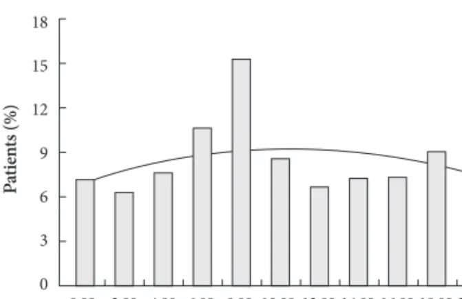
A circadian variation in the frequency of onset of acute myocardial infarction (AMI) has been reported in a number

of studies over the past several decades. A majority of large- scale reports have shown a peak incidence in morning hours, although a secondary peak incidence in the late evening has sometimes been reported.

1-7)

The higher incidence of ST- segment elevation myocardial infarction (STEMI) in early morning has been explained by changes in catecholamine lev- els, fibrinolytic activity, blood pressure, platelet aggregability, coronary tone, and endothelial function.

4)5)8-10)

Differences in the circadian variation of AMI in different regions of the world and in different ethnic groups have also been reported.
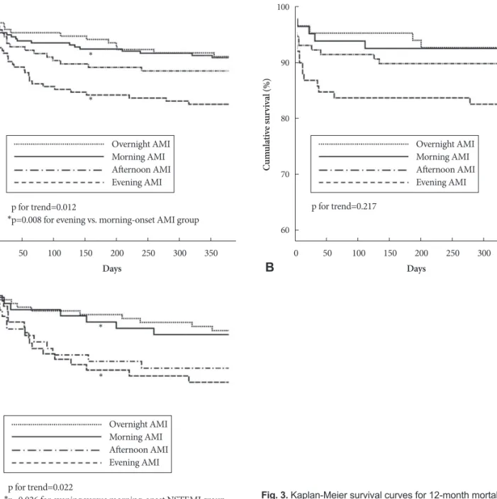
4)5)7)11)

Although associations between the time of onset of AMI and in-hospital or 30-day mortality were suggested in some previous studies,

12)13)

not much is known about the 12- month mortality of AMI, including STEMI and non-STEMI (NSTEMI). The aim of this study was to investigate the impact of circadian variation

Received: February 13, 2010 Revision Received: April 26, 2010 Accepted: May 12, 2010

Correspondence: Yongkeun Cho, MD, Department of Internal Medi- cine, Kyungpook National University Hospital, 50 Samdeok 2-ga, Jung- gu, Daegu 700-721, Korea

Tel: 82-53-420-5528, Fax: 82-53-426-2046 E-mail: choyk@mail.knu.ac.kr

cc

This is an Open Access article distributed under the terms of the Cre- ative Commons Attribution Non-Commercial License (http://creativecom- mons.org/licenses/by-nc/3.0) which permits unrestricted non-commer- cial use, distribution, and reproduction in any medium, provided the origi- nal work is properly cited.

The Impact of Circadian Variation on 12-Month Mortality in Patients With Acute Myocardial Infarction

Myung Hwan Bae, MD

1

, Hyeon Min Ryu, MD

1

, Jang Hoon Lee, MD

1

, Ju Hwan Lee, MD

2

, Yong Seop Kwon, MD

3

, Sang Hyuk Lee, MD

1

, Dong Heon Yang, MD

1

, Hun Sik Park, MD

1

, Yongkeun Cho, MD

1

, Shung Chull Chae, MD

1

, Jae-Eun Jun, MD

1

and Wee-Hyun Park, MD

1

1

Department of Internal Medicine, Kyungpook National University Hospital, Daegu,

2

Department of Internal Medicine, Gumi Cha Hospital, Gumi,

3

Department of Internal Medicine, Busan St. Mary’s Medical Center, Busan, Korea

ABSTRACT

Background and Objectives: Although circadian variation in the onset of acute myocardial infarction (AMI) has been re-

ported in a number of studies, not much is known about the impact of circadian variation on 12-month mortality. The aim

of this study was to investigate the impact of circadian variation on 12-month mortality in patients with AMI. Subjects and

Methods: Eight hundred ninety two patients (mean age 67 ± 12; 66.1% men) with AMI who visited Kyungpook National

University Hospital from November 2005 to December 2007 were included in this study. Patients were divided into groups

based on four 6-hours intervals: overnight (00:00-05:59); morning (06:00-11:59); afternoon (12:00-17:59) and evening

(18:00-23:59). Results: Kaplan-Meier survival curves showed 12-month mortality rates of 9.6%, 9.1%, 12.1%, and 16.7% in

the overnight, morning, afternoon, evening-onset groups, respectively (p=0.012). Compared with the morning-onset AMI

group, the serum creatinine levels (p=0.002), frequency of Killip class ≥3 (p=0.004), and prescription rate of diuretics

(p=0.011) were significantly higher in the evening-onset AMI group, while the left ventricular ejection fraction (p=0.012)

was significantly lower. The proportion of patients who arrived in the emergency room during routine duty hours was signifi-

cantly lower in evening-onset groups irrespective of the presence or absence of ST-segment elevation (p<0.001). According

to univariate analysis, the 12-month mortality rate in the evening group was significantly higher compared to the morning

group (hazard ratio 1.998, 95% confidence interval 1.196 to 3.338, p=0.008). Conclusion: Patients with evening-onset AMI

had poorer baseline clinical characteristics, and this might affect the circadian impact on 12-month mortality. Further stud-

ies are needed to clarify the role of circadian variation on the long-term outcome of AMI. (Korean Circ J 2010;40:616-624)

KEY WORDS: Myocardial infarction; Circadian rhythm.

(2)

on 12-month mortality in patients with AMI.

Subjects and Methods

Study patients

We included 966 consecutive patients with AMI who were admitted to Kyungpook National University Hospital between November 2005 and December 2007. A diagnosis of AMI was made in patients having angina pain lasting more than 15 min- utes that was associated with/without dyspnea or sweating, es- tablished/evolving ST-T wave changes on serial electrocardio- grams, and an increase in levels of cardiac troponin I (cTnI).

cTnI levels were measured on admission and every 6 hours to detect the presence of myocardial injury, using an enzyme im- munoassay based on the sandwich principle (Dimension; Dade Behring, Deerfield, IL, USA); the lower detection limit of this assay was 0.04 ng/mL. Elevated cardiac biomarker levels were defined as having maximal values of cTnI that exceeded the cutoff (99th percentile of the values for a reference control gr- oup) on at least one occasion. The time of onset of AMI was determined by each patient’s report of the chest pain that pro- mpted hospital admission. Seventy-four patients with unclear onset of chest pain, including silent AMI, were excluded from further analysis. In total, 892 patients (mean age 67±12 years;

66.1% men) were included in this study. Informed consent was obtained from each patient.

Data analyses

Each day was divided into 12 equal parts of two hours each, and patients were grouped according to the time of symptom onset. For further analysis, a day was divided again into four equal 6-hour intervals: overnight (00:00 AM-05:59 AM);

morning (06:00 AM-11:59 AM); afternoon (12:00 PM-17:59 PM) and evening (18:00 PM-23:59 PM).

2)14)

Routine duty ho- urs were defined as Monday to Friday between 08:00 AM and 18:00 PM.

13)

Weekends were considered off-duty hours. We an- alyzed the baseline demographic characteristics, initial presen- tation, initial vital signs, results of laboratory tests, and dis- charge medications of the patients. Demographic and clinical characteristics were identified, including age, gender, body mass index (BMI), cardiovascular risk factors (hypertension, diabetes mellitus, hyperlipidemia, and current smoking), and co-morbidities, which indluded previous congestive heart failure and myocardial infarction (MI). Initial vital signs in- cluding the systolic pressure and Killip class were evaluated at admission. Initial blood samples collected at admission, except for those collected to determine peak cTnI, were used for baseline laboratory tests. The left ventricular ejection frac- tion (LVEF) was determined by 2-dimensional echocardiog- raphy during the index hospitalization. The mean symptom- to-door time (STD) and the proportion of patients with STD time ≥6 hours were assessed in patients with typical chest pain.

In patients with STEMI, the median door-to-balloon (DTB) time and the proportion of patients with DTB time ≤90 minutes were also evaluated. Discharge medications assessed includ- ed aspirin, β-blockers, angiotensin converting enzyme in- hibitors (ACE-I)/angiotensin receptor blockers (ARB), and diuretics. Mean follow-up duration was 357±141 days. The primary end-point was 12-month mortality, including both cardiac death and non-cardiac death. Cardiac death was de- fined as death from pump failure, arrhythmia, or mechanical complications, which included ventricular septal rupture and free wall rupture. During the follow-up period, data were ob- tained by reviewing medical records and by telephone inter- view with the patients.

Statistical analyses

Chi-squared goodness-of-fit was performed to test the uniformity of the distribution of patients among the time periods. Data are expressed as mean±SD for continuous vari- ables and percentages for categorical variables. All compari- sons between baseline variables were assessed with the anal-

Fig. 1. The circadian variation of the onset of AMI for the total study population obtained at 2-hour intervals. The distribution is not uniform (p<0.001) and a peak occurring between 8:00 and 9:59 hours was statistically significant (p<0.001). AMI: acute myocardial infarction.

18 15 12 9 6 3 0

Hour of onset of AMI

Pa tie nt s ( % )

0:00- 1:59

2:00- 3:59

4:00- 5:59

6:00- 7:59

8:00- 9:59

10:00- 11:59

12:00- 13:59

14:00- 15:59

16:00- 17:59

18:00- 19:59

20:00- 21:59

22:00- 23:59

Overnight Morning Afternoon Hour of onset of AMI

Evening 50

40 30 20 10 0

50 40 30 20 10

Fr eq ue nc y o f o ns et o f A M I ( % ) 0 M ort alit y r ate (% )

Onset of chest pain 12-month mortality

Fig. 2. The circadian variation of the onset of AMI for the total study population obtained at 6-hour intervals. A morning peak was sta- tistically significant (p<0.001). In contrast, the 12-month mortality rate was significantly higher in the evening-onset AMI group (p=

0.012). AMI: acute myocardial infarction.

(3)

ysis of variance for continuous variables, and with the Pearson’s Chi-square test for categorical variables. A multivariate lo- gistic regression model was used to determine independent predictors for evening-onset AMI. Twelve-month mortality according to the onset time of AMI was evaluated using Ka- plan-Meier survival curve analysis. Univariate analyses were performed to determine the clinical predictors of 12-month mortality. The Cox proportional hazard model was used to calculate the risk of 12-month mortality related to the time of onset of AMI, and adjusted for baseline confounding charac- teristics. For all analyses, a two-sided p<0.05 was considered statistically significant. Statistical analysis was performed using SPSS version 15.0 for Windows (SPSS Inc., Chicago, IL, USA).

Results

Circadian variation in the onset of acute myocardial infarction

The frequency of the onset of AMI as determined by onset of chest pain for all 892 patients is shown in Fig. 1. The distri- bution is not uniform (p<0.001), and a peak was found be- tween 08:00 and 09:59 hours (p<0.001). The distribution of AMI onset was obtained for each 6-hour interval, and a mo- rning peak was found to be statistically significant (p<0.001) (Fig. 2). The numbers of patients grouped according to the AMI onset time were 184 (20.6%), 303 (34.0%), 187 (21.0%), and 218 (24.4%), corresponding to overnight, morning, after- Table 1. Clinical characteristics of patients according to the hour of onset of AMI

Overall (n=892)

Overnight (n=184)

Morning (n=303)

Afternoon (n=187)

Evening

(n=218) p* p

Demographics

Age (years) 066.8±12.0 65.7±13.3 66.8±11.5 67.4±11.0. 67.0±12.5 0.553 0.999

Age ≥65 (%) 59.6 56.0 59.4 62.0 61.0 0.275 0.712

Male (%) 66.1 63.6 65.0 67.9 68.3 0.245 0.427

Body mass index (kg/m

2

) 023.7±3.00 24.0±3.10 23.6±2.90 23.4±3.10. 23.7±2.90 0.421 0.979 Initial presentation

Systolic blood pressure (mmHg) 136.9±29.4 139.1±27.50 136.6±29.30 134.8±30.10. 137.3±30.40 0.562 0.992

Anterior myocardial infarction (%) 53.2 60.1 48.0 53.8 54.2 0.652 0.216

Killip class ≥3 (%) 17.5 16.9 12.6 20.9 22.0 0.027 0.004

Preinfarct angina pectoris (%) 47.0 53.3 45.9 47.6 42.7 0.193 0.467

Mean STD time (minute) 00564±1,157 0618±1,155 0567±1,463 0587±1,076 496±624 0.746 0.923

STD time ≥6 hour (%) 36.3 40.8 36.0 32.1 36.7 0.348 0.865

ER arrival during routine duty-hours (%) 42.0 33.7 70.0 35.3 16.1 <0.001 <0.001 Past history (%)

Diabetes mellitus 26.8 23.4 27.1 24.1 31.8 0.108 0.241

Hypertension 44.8 45.1 43.4 41.7 49.3 0.411 0.181

Hyperlipidemia 23.1 24.2 24.3 21.8 21.5 0.436 0.484

Myocardial infarction 12.4 16.1 10.7 09.6 14.0 0.719 0.427

Current smoker 44.9 43.4 44.9 43.5 47.5 0.479 0.556

LVEF (%) 052.1±10.5 52.4±10.4 53.4±9.70 51.9±10.6 50.1±11.4 0.011 0.012

PCI at index hospitalization (%) 71.6 73.9 71.0 75.4 67.4 0.284 0.440

Laboratory findings

Creatinine (mg/dL) 01.1±1.1 1.0±1.1 1.0±0.8 1.0±0.6 1.3±1.6 0.001 0.002

Cardiac Troponin I (ng/mL) 0055.7±312.5 34.5±52.0 049.5±103.0 41.5±89.4 094.4±612.7 0.197 0.457

Log proBNP (pg/mL) 02.6±0.8 2.6±0.8 2.6±0.8 2.6±0.8 2.7±0.9 0.157 0.532

Discharge medication (%)

Aspirin 94.8 94.8 95.4 94.8 93.9 0.594 0.459

β-blockers 86.3 89.1 85.9 84.4 86.2 0.421 0.911

ACE-I/ARBs 89.2 90.8 89.8 87.8 88.3 0.350 0.607

Diuretics 26.0 25.9 22.3 24.9 29.8 0.073 0.011

* p for all comparisons,

p for the morning vs. evening group. AMI: acute myocardial infarction, STD: symptom-to-door, ER: emergency room,

LVEF: left ventricular ejection fraction, PCI: percutaneous coronary intervention, proBNP: pro-brain natriuretic peptide, ACE-I/ARB: angio-

tensin-converting enzyme inhibitor/angiotensin II receptor blocker

(4)

noon and evening-onset groups, respectively. The incidence of morning-onset AMI was 54% higher than the average of the three other time periods. A morning peak of AMI was found irrespective of the presence or absence of ST-segment elevation (p=0.001 in STEMI and p<0.001 in NSTEMI).

Clinical characteristics of patients according to acute myocardial infarction onset

The clinical characteristics of the patients are shown in Ta-

ble 1. The patients mean age was 67±12 years, and 590 (66.1%) were men. There were no significant differences in age, sex, BMI, systolic blood pressure, preinfarct angina pectoris, lo- cation of AMI, previous history of diabetes mellitus, hyper- tension, hyperlipidemia, MI, use of tobacco, percutaneous cor- onary intervention (PCI) at index hospitalization, peak cTnI, or pro-brain natriuretic peptide (proBNP) levels among the four groups. In the evening-onset AMI group serum creatinine levels (p=0.001) and the frequency of Killip class ≥3 (p=0.027) Table 2. Clinical characteristics of patients according to the hour of onset of STEMI

Overall (n=403)

Overnight (n=86)

Morning (n=134)

Afternoon (n=82)

Evening

(n=101) p* p

Demographics

Age (years) 65.7±11.8 64.8±12.0 65.9±11.5 65.9±10.8 66.0±12.8 0.888 0.973

Age ≥65 (%) 55.8 54.7 57.5 53.7 56.4 0.967 0.875

Male (%) 69.0 65.1 65.7 72.0 74.3 0.104 0.158

BMI (Kg/m

2

) 23.7±3.00 23.8±2.9 23.6±3.20 23.3±2.70 24.2±2.70 0.257 0.130

Initial presentation

Systolic blood pressure (mmHg) 130.3±28.60 134.1±28.00 128.7±27.90 127.7±30.20 131.4±28.70 0.438 0.471

Anterior myocardial infarction (%) 55.0 61.9 46.5 57.5 58.3 0.809 0.079

Killip class ≥3 (%) 15.2 15.3 13.4 15.9 16.8 0.626 0.469

Preinfarct angina pectoris (%) 37.2 44.2 38.1 36.6 30.7 0.298 0.241

Mean STD time (minute) 419±573 455±671 350±484 409±474 487±657 0.294 0.080

STD time ≥6 hour (%) 31.0 36.0 28.4 25.6 34.7 0.889 0.302

Median DTB time, median (range in minute)

80 (23-14,065)

114 (34-12,636)

75 (23-14,065)

81 (27-11,240)

121 (32-13,700)

0.039 0.011

DTB time ≤90 minute (%) 50.3 43.6 58.3 54.7 41.5 0.555 0.020

ER arrival during routine

duty-hours (%) 40.9 26.7 70.1 40.2 14.9 <0.001 <0.001

Past history (%)

Diabetes mellitus 23.4 19.8 21.6 18.3 33.0 0.049 0.051

Hypertension 39.2 38.4 38.1 30.5 48.5 0.245 0.109

Hyperlipidemia 21.3 26.9 22.1 14.1 20.9 0.226 0.518

Myocardial infarction 06.8 07.0 07.5 06.8 05.9 0.791 0.427

Current smoker 49.9 45.3 49.2 57.3 48.5 0.506 0.912

LVEF (%) 51.2±10.4 51.9±10.7 51.8±9.40 51.2±11.0 49.8±11.1 0.527 0.169

PCI at index hospitalization (%) 88.1 91.9 88.1 91.5 82.2 0.079 0.205

Laboratory findings

Creatinine (mg/dL) 0.9±0.6 0.8±0.3 0.9±1.0 0.9±0.2 1.0±0.6 0.354 0.590

Cardiac Troponin I (ng/mL) 102.0±457.5 54.8±59.9 096.0±140.0 082.7±122.5 166.4±893.2 0.383 0.372

Log proBNP (pg/mL) 2.5±0.8 2.4±0.8 2.6±0.8 2.3±0.8 2.6±0.8 0.247 0.970

Discharge medication (%)

Aspirin 97.8 100.0 97.5 96.0 97.7 0.281 0.647

β-blockers 89.0 92.6 86.0 88.0 90.9 0.954 0.275

ACE-I/ARBs 93.4 96.3 93.4 88.0 95.5 0.583 0.526

Diuretics 22.7 21.0 21.5 18.7 29.5 0.234 0.183

* p for all comparison,

p for the morning vs. evening group. STEMI: ST-segment elevation myocardial infarction, BMI: body mass index, STD:

symptom-to-door, DTB: door-to-balloon, ER: emergency room, LVEF: left ventricular ejection fraction, PCI: percutaneous coronary inter-

vention, proBNP: pro-brain natriuretic peptide, ACE-I/ARB: angiotensin-converting enzyme inhibitor/angiotensin II receptor blocker

(5)

were significantly higher, while the LVEF (p=0.011) was sig- nificantly lower. The mean STD time was 564±1,157 minutes, with no significant differences in the mean STD time (p=

0.746) or the proportion of patients with an STD time ≥6 hours (p=0.384) among the four groups. The proportion of pa- tients who arrived in the emergency room (ER) during routine duty hours was significantly higher in the morning-onset AMI group (p<0.001). The discharge prescription rates of aspirin, β-receptor blockers, ACE-I/ARBs, and diuretics were not sig- nificantly different among the four groups.

Compared with the morning-onset AMI group, serum creatinine levels (p=0.002),the frequency of Killip class ≥3 (p=0.004), and the prescription rate of diuretics (p=0.011) were

significantly higher in the evening-onset AMI group, while LVEF (p=0.012) was significantly lower. The proportion of pa- tients who arrived in the ER during routine duty hours was significantly lower in the evening-onset group compared to the morning-onset group (p<0.001).

Subgroup analysis

In the STEMI group (Table 2), the numbers of patients with a previous history of diabetes mellitus was significantly high- er within the evening-onset group (p=0.049). The median DTB time was 80 minutes, and was significantly longer in the over- night-onset and evening-onset groups than in the morning- onset and afternoon-onset groups (p=0.039). The proportion Table 3. Clinical characteristics of patients according to the hour of onset of NSTEMI

Overall (n=489)

Overnight (n=98)

Morning (n=169)

Afternoon (n=105)

Evening

(n=117) p* p

Demographics

Age (years) 67.6±12.2 66.5±14.4 67.6±11.5 68.6±11.1 67.9±12.1 0.659 0.815

Age ≥65 (%) 62.8 57.1 60.9 68.6 65.0 0.142 0.491

Male (%) 63.8 62.2 64.5 64.8 63.2 0.913 0.829

BMI (Kg/m

2

) 23.7±3.00 24.1±3.20 23.7±2.60 23.5±3.30 23.3±2.90 0.294 0.346

Initial presentation

Systolic blood pressure (mmHg) 142.3±28.90 143.6±26.40 142.8±28.90 140.3±28.90 142.4±31.00 0.863 0.907

Anterior myocardial infarction (%) 51.2 58.0 49.6 50.0 48.6 0.332 0.896

Killip class ≥3 (%) 19.5 18.4 11.9 24.8 26.5 0.013 0.002

Preinfarct angina pectoris (%) 55.0 61.2 52.1 56.2 53.0 0.499 0.878

Mean STD time (minute) 0685±1,463 761±1,442 0740±1,896 0726±1,360 504±597 0.500 0.195

STD time ≥6 hour (%) 40.7 44.9 42.0 37.1 38.5 0.258 0.548

ER arrival during routine duty-hours (%) 42.9 39.8 69.8 31.4 17.1 <0.001 <0.001 Past history (%)

Diabetes mellitus 29.7 26.5 31.4 28.6 30.8 0.108 0.915

Hypertension 49.5 51.0 47.6 50.5 50.0 0.951 0.693

Hyperlipidemia 24.6 21.5 26.2 27.7 22.0 0.998 0.451

Myocardial infarction 17.6 24.0 13.3 12.8 22.4 0.953 0.171

Current smoker 40.6 33.3 40.2 44.2 44.0 0.107 0.531

LVEF (%) 52.8±10.5 52.8±10.1 54.6±9.80 52.5±10.2 50.4±11.7 0.017 0.003

PCI at index hospitalization (%) 58.1 58.2 57.4 62.9 54.7 0.798 0.651

Laboratory findings

Creatinine (mg/dL) 1.2±1.3 1.1±1.5 1.0±0.6 1.1±0.7 1.6±2.1 0.001 0.001

Peak cardiac Troponin I (ng/mL) 17.6±53.9 16.7±35.5 12.7±21.9 09.4±15.1 32.8±99.6 0.004 0.034

Log proBNP (pg/mL) 2.7±0.8 2.7±0.7 2.7±0.8 2.7±0.8 2.9±0.9 0.193 0.048

Discharge medication (%)

Aspirin 92.4 90.3 93.8 93.9 90.7 0.971 0.343

β-blockers 84.2 86.0 85.8 81.6 82.4 0.335 0.451

ACE-I/ARBs 85.9 86.0 87.0 87.6 82.4 0.456 0.294

Diuretics 28.6 30.1 22.8 29.6 35.2 0.182 0.027

* p for all comparison,

p for the morning vs. evening group. NSTEMI: non-STEMI, BMI: body mass index, STD: symptom-to-door, ER: emergency room, LVEF: left ventricular ejection fraction, PCI: percutaneous coronary intervention, proBNP: pro-brain natriuretic peptide, ACE-I/ARB:

angiotensin-converting enzyme inhibitor/angiotensin II receptor blocker

(6)

of all patients with a DTB time ≤90 minutes was not signifi- cantly different among the four groups. However, compared with the morning-onset STEMI group, the proportion of pa- tients with a DTB time ≤90 minutes was significantly lower for the evening-onset STEMI group (p=0.039). Serum creatinine levels, frequency of Killip class ≥3, and LVEF were not differ- ent among four STEMI groups.

In the NSTEMI group (Table 3), the frequency of Killip class

≥3 (p=0.013), levels of serum creatinine (p=0.001), and peak cTnI (p=0.004) were significantly higher in the evening-onset NSTEMI group, while the LVEF (p=0.017) was significantly lower. Compared with the morning-onset NSTEMI group, the log proBNP level (p=0.048), prescription rate of diuretics (p=

0.027), frequency of Killip class ≥3, levels of serum creatinine, and peak cTnI were significantly different in the evening-on- set NSTEMI group. The proportion of patients who arrived in

the ER during routine duty hours was significantly lower in the evening-onset group, irrespective of the presence or absence of ST-segment elevation (p<0.001).

Twelve-month mortality

During the study period, 48 (5.4%) patients were lost dur- ing follow-up. The overall mortality rate was 11.7% (n=99), with 10.5% (n=89) from cardiac deaths and 1.2% (n=10) from non- cardiac deaths. No significant difference in the 12-month mor- tality rate was observed between STEMI and NSTEMI groups (11.6% vs. 11.8%, p=NS). Kaplan-Meier survival curves showed 12-month mortality rates of 9.6% (n=17), 9.1% (n= 26), 12.1%

(n=21) and 16.7% (n=35) in the overnight, morning, after- noon and evening-onset groups, respectively (p= 0.012) (Figs.

2 and 3). Although there were no significant differences in in- hospital mortality among the four groups according to univari-

A

C

B

Fig. 3. Kaplan-Meier survival curves for 12-month mortality accord- ing to onset time in patients with all AMI (A), STEMI (B), and NSTE- MI (C). In patients with AMI and NSTEMI, 12-month mortality rates in the evening-onset groups were significantly higher than those in the morning-onset groups. AMI: acute myocardial infarction, STEMI:

ST-segment elevation myocardial infarction, NSTEMI: non-STEMI.

0

0

0 50

50

50 100

100

100 150

150

150 200

200

200 250

250

250 300

300

300 350

350

350 Days

Days

Days p for trend=0.012

*p=0.008 for evening vs. morning-onset AMI group

p for trend=0.022

*p=0.026 for evening versus morning-onset NSTEMI group 100

90

80

70

60

100

90

80

70

60

100

90

80

70

60

Cu m ul at iv e s ur vi va l ( % ) Cu m ul at iv e s ur vi va l ( % ) Cu m ul at iv e s ur vi va l ( % )

Overnight AMI Morning AMI Afternoon AMI Evening AMI

Overnight AMI Morning AMI Afternoon AMI Evening AMI

Overnight AMI Morning AMI Afternoon AMI Evening AMI

*

*

*

*

p for trend=0.217

(7)

ate analysis, 6-month (p=0.006) and 12-month mortality rates (p=0.012) were significantly higher in the evening-onset group than for the three other groups. Compared with the morning onset-AMI group, 6-month {hazard ratio (HR) 2.040, 95% CI 1.181 to 3.523, p=0.011} and 12- month mortali- ty rates [HR 1.998, 95% confidence interval (CI) 1.196 to 3.338, p= 0.008] were significantly higher in the evening onset-AMI group (Table 4). A similar result was obtained for 12-month mortality in patients with NSTEMI. However there were no significant differences between morning- and evening-onset STEMI groups in in-hospital, 6-month, and 12-month mor- tality rates. According to the multivariate Cox proportional- hazards model, evening-onset AMI (HR 0.518, 95% Cl 0.168 to 1.601, p=0.253) was not an independent predictor for 12-month mortality after adjusting for age, gender, ER arrival during off- duty hours, LVEF, Killip class ≥3, prescription of diuretics, lev- els of serum creatinine, peak cTnI, and log proBNP.

Discussion

The main finding in this single center observational study is that 12-month mortality in patients with evening-onset AMI was significantly higher than that in the morning-onset AMI group. This was due to poorer baseline clinical charac- teristics for evening-onset AMI patients. In this study circadi- an variation of 12-month mortality might be secondary to dif- ferences in baseline characteristics at the onset time of AMI.

Killip class and serum creatinine levels have an impact on clinical outcome as surrogates for heart failure and acute renal failure after AMI.

15-18)

In this study, patients with Killip class

≥3, higher levels of serum creatinine, and lower LVEF were found significantly more frequent in evening-onset AMI group compared to the three other onset groups. In a study on the

determinants of MI onset, Mukamal et al.

14)

showed that the risk of heart failure was highest in patients with infarctions that began between 6 PM and midnight and midnight to 6 AM. In another study, the peak between 6 PM and midnight was most pronounced, with patients with a history of conges- tive heart failure experiencing greater onset of MI.

2)

Although the mechanisms underlying these findings were not clear from their study, in this study newly developed heart failure follow- ing AMI, or underlying heart failure might affect the evening peak of 12-month mortality. Poor baseline clinical character- istics including Killip class ≥3, lower LVEF, higher levels of se- rum creatinine, and peak cTnI were also found in the evening- onset NSTEMI group. However, there were no significant differences in baseline clinical characteristics, except for dia- betes mellitus, among the four STEMI groups. An evening peak of 12-month mortality was shown in the NSTEMI group, but was not demonstrated in the STEMI group. We hypothe- sized the differences in the baseline characteristics between the STEMI and NSTEMI groups might explain the difference in 12-month mortality between the two groups.

Outcomes after primary PCI have been reported to be re- lated to the time delay from symptom onset to first-balloon inflation,

19)

and to hospital volume.

20-22)

In this study, the mean STD time was 564 minutes, and one third of patients had STD time ≥6 hours. There were no significant differences in STD time among the four groups. About 70% of patients in the morning-onset AMI group arrived at the ER during routine duty hours. However, only 16% of patients in the evening-on- set AMI group arrived at the ER during routine duty hours. It is well known that patients presenting with AMI during off-du- ty hours have higher in-hospital mortality than patients pre- senting during routine duty hours.

12)13)23)24)

However, quality of care is difficult to measure, and it is unclear whether this affect- Table 4. In-hospital, 6-month, and 12-month mortality rates according to the hour of onset of AMI

Overall (n=844)

Overnight (n=177)

Morning (n=285)

Afternoon (n=173)

Evening

(n=209) p* Evening vs.

morning p

In-hospital mortality (%) OR (95% CI)

All AMI 4.7 3.8 4.6 3.7 6.4 0.281 1.417 (0.661-3.036) 0.370

STEMI 6.7 4.7 7.5 3.7 9.9 0.293 1.363 (0.544-3.410) 0.509

NSTEMI 3.1 3.1 2.4 3.8 3.4 0.692 1.460 (0.358-5.959) 0.598

6-month mortality (%) HR (95% Cl)

All AMI 9.9 7.4 7.6 10.40 14.90 0.006 2.040 (1.181-3.523) 0.011

STEMI 10.70 7.4 10.00 7.4 17.20 0.055 1.762 (0.856-3.629) 0.124

NSTEMI 9.2 8.3 5.6 12.50 12.60 0.086 2.371 (1.026-5.480) 0.043

12-month mortality (%) HR (95% Cl)

All AMI 11.70 9.6 9.1 12.10 16.70 0.012 1.998 (1.196-3.338) 0.008

STEMI 11.60 10.80 10.20 7.6 14.20 0.217 1.732 (0.841-3.567) 0.136

NSTEMI 11.80 8.5 8.2 16.00 16.40 0.022 2.297 (1.106-4.771) 0.026

* p for all comparisons,

p for the morning vs. evening group. AMI: acute myocardial infarction, OR: odds ratio, Cl: confidence interval, STEMI:

ST-segment elevation myocardial infarction, NSTEMI: non-STEMI, HR: hazard ratio

(8)

ed the results of our study. Although the proportion of patients who arrived in the ER during routine duty hours was signifi- cantly higher in the morning-onset group, there were no sig- nificant differences in baseline characteristics between over- night-, morning-, and afternoon-onset AMI groups. Overall, there was no significant difference in the 12-month mortality rate among these three groups.

It is well known that longer DTB time is an independent pre- dictor of short- and long-term outcomes in patients with STE-

MI.

19)25)

Although median DTB time in the STEMI subgroup

was significantly longer among the evening-onset patients, the longer DTB time did not make a difference on 12-month mor- tality in the present study.

One possible explanation for the discrepancy between pre- vious studies and our finding is the different study population.

Previous studies generally included patients who presented acutely to the hospital within 12 or 24 hours of symptom on- set.

19)25)

In this study, we included all consecutive patients with STEMI irrespective of the time from symptom onset to ER ar- rival. We thought the impact of DTB time on short- and long- term outcomes in patients with STEMI would be reduced be- cause of the relatively long STD time.

There are several limitations in this study. First, this was a sin- gle-center observational study with a relatively small sample size. Second, multiple factors could affect the circadian varia- tion and clinical outcome in this population. Finally, because timing of onset of AMI was self-reported by patients, they may have been assigned erroneously to particular groups.

In conclusion, there was a difference between the peak time of onset and 12-month mortality in patients with AMI. The twelve-month mortality rate was higher in the evening-onset AMI group compared to the morning-onset AMI group. Poor- er baseline clinical characteristics included a higher Killip class, higher serum creatinine level, lower LVEF, and ER arrival dur- ing off-duty hours in patients with evening-onset AMI. This might affect the circadian impact on 12-month mortality. We conclude that the time of AMI onset must be considered in the management of patients with AMI. Further studies are need- ed to clarify the role of circadian variation on the long-term outcome of AMI.

Acknowledgments

The authors are deeply indebted to Roberto Patarca, MD, PhD and SungHee Kim, MD, PhD for their useful suggestions for the preparation of this manuscript.

REFERENCES

1) Kinoshita N, Imai K, Kinjo K, Naka M. Longitudinal study of acute myocardial infarction in the southeast Osaka district from 1988 to 2002.

Circ J 2005;69:1170-5.

2) Hjalmarson A, Gilpin EA, Nicod P, et al. Differing circadian patterns of symptom onset in subgroups of patients with acute myocardial in- farction. Circulation 1989;80:267-75.

3) Kim KS, Song YS, Hur SH, et al. Circadian variation in acute myocar-

dial infarction. Korean Circ J 1993;23:173-83.

4) López F, Lee KW, Marín F, et al. Are there ethnic differences in the cir- cadian variation in onset of acute myocardial infarction?: a compari- son of 3 ethnic groups in Birmingham, UK and Alicante, Spain. Int J Car- diol 2005;100:151-4.

5) D’Negri CE, Nicola-Siri L, Vigo DE, Girotti LA, Cardinali DP. Circa- dian analysis of myocardial infarction incidence in an Argentine and Uruguayan population. BMC Cardiovasc Disord 2006;6:1.

6) Rana JS, Mukamal KJ, Morgan JP, Muller JE, Mittleman MA. Circa- dian variation in the onset of myocardial infarction: effect of duration of diabetes. Diabetes 2003;52:1464-8.

7) Kinjo K, Sato H, Sato H, et al. Circadian variation of the onset of acute myocardial infarction in the Osaka area, 1998-1999: characterization of morning and nighttime peaks. Jpn Circ J 2001;65:617-20.

8) Muller JE, Kaufmann PG, Luepker RV, Weisfeldt ML, Deedwania PC, Willerson JT. Mechanisms precipitating acute cardiac events: review and recommendations of an NHLBI workshop. National Heart, Lung, and Blood Institute. Circulation 1997;96:3233-9.

9) Bhalla A, Sachdev A, Lehl SS, Singh R, D’Cruz S. Ageing and circadi- an variation in cardiovascular events. Singapore Med J 2006;47:305-8.

10) Willich SN. Circadian variation and triggering of cardiovascular events.

Vasc Med 1999;4:41-9.

11) Sari I, Davutoglu V, Erer B, et al. Analysis of circadian variation of acute myocardial infarction: afternoon predominance in Turkish popu- lation. Int J Clin Pract 2009;63:82-6.

12) Henriques JP, Haasdijk AP, Zijlstra F. Outcome of primary angioplas- ty for acute myocardial infarction during routine duty hours versus dur- ing off-hours. J Am Coll Cardiol 2003;41:2138-42.

13) Magid DJ, Wang Y, Herrin J, et al. Relationship between time of day, day of week, timeliness of reperfusion, and in-hospital mortality for pa- tients with acute ST-segment elevation myocardial infarction. JAMA 2005;

294:803-12.

14) Mukamal KJ, Muller JE, Maclure M, Sherwood JB, Mittleman MA.

Increased risk of congestive heart failure among infarctions with night- time onset. Am Heart J 2000;140:438-42.

15) Park SR, Kang YR, Seo MK, et al. Clinical predictors of incomplete ST- segment resolution in the patients with acute ST segment elevation myo- cardial infarction. Korean Circ J 2009;39:310-6.

16) Lee CH, Joo SJ, Park DS, et al. Remodeling and changes of systolic and diastolic functions of left ventricle after acute myocardial infarction: com- parison according to killip class at admission. Korean Circ J 1998;28:

1727-39.

17) Al Suwaidi J, Reddan DN, Williams K, et al. Prognostic implications of abnormalities in renal function in patients with acute coronary syndromes.

Circulation 2002;106:974-80.

18) Naidu SS, Selzer F, Jacobs A, et al. Renal insufficiency is an indepen- dent predictor of mortality after percutaneous coronary intervention. Am J Cardiol 2003;92:1160-4.

19) Cannon CP, Gibson CM, Lambrew CT, et al. Relationship of symptom- onset-to-balloon time and door-to-balloon time with mortality in patients undergoing angioplasty for acute myocardial infarction. JAMA 2000;

283:2941-7.

20) Magid DJ, Calonge BN, Rumsfeld JS, et al. Relation between hospital primary angioplasty volume and mortality for patients with acute MI treat- ed with primary angioplasty vs thrombolytic therapy. JAMA 2000;284:

3131-8.

21) Canto JG, Every NR, Magid DJ, et al. The volume of primary angio- plasty procedures and survival after acute myocardial infarction. Na- tional Registry of Myocardial Infarction 2 Investigators. N Engl J Med 2000;342:1573-80.

22) Vakili BA, Kaplan R, Brown DL. Volume-outcome relation for physi- cians and hospitals performing angioplasty for acute myocardial in- farction in New York state. Circulation 2001;104:2171-6.

23) Assali AR, Brosh D, Vaknin-Assa H, et al. The impact of circadian

variation on outcomes in emergency acute anterior myocardial infarc-

tion percutaneous coronary intervention. Catheter Cardiovasc Interv

2006;67:221-6.

(9)

24) Dominguez-Rodriguez A, Garcia-Gonzalez M, Abreu-Gonzalez P. Out- come of primary angioplasty for ST-segment elevation myocardial in- farction during routine duty hours versus during off-hours: results of a single-center in Spain. Int J Cardiol 2007;119:227-9.

25) Berger PB, Ellis SG, Holmes DR Jr, et al. Relationship between delay

in performing direct coronary angioplasty and early clinical outcome

in patients with acute myocardial infarction: results from the Global Use

of Strategies to Open Occluded Arteries in Acute Coronary Syndromes

(GUSTO-IIb) Trial. Circulation 1999;100:14-20.

수치

Fig. 1. The circadian variation of the onset of AMI for the total  study population obtained at 2-hour intervals
Fig. 3. Kaplan-Meier survival curves for 12-month mortality accord- accord-ing to onset time in patients with all AMI (A), STEMI (B), and  NSTE-MI (C)

참조

관련 문서

Conclusion: The mean CGRP level in overweight and obese children with headache was not significantly higher than in overweight and obese children without headache. There

Second, analyzing students' level of interest in the unit of Clothes and Life Style revealed that there was significant difference between genders and students in the two

As a result of comparing with the temperature change, the coldest month mean temperature (January, February, December) showed positive value in the whole

Through a review of electronic medical records, we analyzed the applied BCSs, perioperative hematologic changes, and morbidity and mortality in JW patients

Comparing pharmacological variables, 12-month follow-up group showed higher rate of prescription of antianxiety drug at discharge(p=0.020). Conclusion The none

(Background) Gallbladder wall thickening(GWT) and gallbladder contraction are often observed in patients with acute hepatitis.. The incidence of acute hepatitis A

According to the month and the date, the forward speed and direction, the present position and the position at 12 hours before of the existing typhoon,

Conclusion: No evidence of a substantial increase in all-cause mortality was found during the 2020 pandemic period in South Korea, as a result of a large decrease in deaths