aAssistant Professor, Karadeniz Technical University, Department of Orthodontics, Trabzon, Turkey.
bAssociate Professor and Chair, Department of Orthodontics, Faculty of Dentistry, Erciyes University, Kayseri Turkey, and Visiting Professor, King Saud University, Riyadh, Saudi Arabia.
cAssociate Professor, Karadeniz Technical University, Department of Operative Dentistry, Trabzon, Turkey.
Corresponding author: Metin Nur.
Karadeniz Technical University, Department of Orthodontics, Trabzon, Black Sea 61080, Turkey.
+90 462 3774747; e-mail, [email protected].
Received January 12, 2010; Last Revision March 26, 2010;
Accepted May 31, 2010.
DOI:10.4041/kjod.2010.40.4.267
Effects of conventional and self-etching adhesive systems on bond strength of orthodontic attachments
bonded to erupted and unerupted teeth
Metin Nur, DDS, PhD,a Tancan Uysal, DDS, PhD,b Cemal Yesilyurt, DDS, PhD,c Mehmet Bayram, DDS, PhDa
Objective: The aim of this study was to evaluate and compare the shear bond strength (SBS) and failure-mode of orthodontic buttons bonded to erupted and unerupted teeth with conventional and self-etching adhesive systems. Methods: Eighty-four erupted and 84 unerupted, human third-molar teeth were used. For both groups, the buccal surfaces of each tooth were assigned one of the following type of adhesive systems (n = 12). A, Conventional systems: 1, Transbond XT (3M Unitek, Monrovia, CA, USA); 2, Prime & Bond NT (Dentsply/Caulk, Milford, USA); 3, Single Bond (3M ESPE, Minnesota, USA); and B, Self-etching adhesives; 4, Clearfil SE Bond (Kuraray, Okayama, Japan); 5, Transbond Plus (3M Unitek, Monrovia, CA, USA); 6, Clearfil S3 (Kuraray, Tokyo, Japan); 7, G Bond (GC, Tokyo, Japan). The SBSs of the attachments and the adhesive remnant index (ARI) scores were recorded. Data were analyzed with analysis of variance (ANOVA), independent-sample t-test and chi-square tests. Results: When the SBSs of erupted and unerupted teeth were compared, only the Clearfil-SE Bond and G-Bond were significantly different. Bond strengths of all adhesive systems were higher in unerupted teeth than erupted teeth, except the Single-Bond system. Conclusions: When using conventional adhesives, bond- ing to erupted and unerupted teeth may not be significantly different. However, clinicians need to take into consid- eration the types of self-etching systems before usage. (Korean J Orthod 2010;40(4):267-275)
Key words: Bonding, Bracket, Resin, Adhesive
INTRODUCTION
Manufacturers have continuously introduced new ad- hesives in dentistry that are more reliable, i.e. stronger, adhere better, less prone to leakage at margins and/or easier to handle.1 As new materials and techniques are introduced, orthodontists adopt some of these innova- tions and add them to their armamentarium,1 including the use of self-etching primers, resin modified glass ionomer cement (RMGIC), chlorhexidine varnishes and different adhesives etc.
Odontogenesis, a series of events taking place from bud formation stage until the completion of calcifica- tion and maturation of the tooth, is a complex pro- cess.2 Upon eruption, the outermost layer of enamel is
immature and not fully calcified.2 This outer layer then begins to calcify due to the effects of salivary mi- nerals. Although not well understood, it is known that changes in both mineral and organic components of the enamel are involved during post-eruptive maturation.3 It has been shown that the hydroxyl (OH−) group of hydroxyl-apatite crystals is absent in less mature enam- el, but it can be found in mature enamel by using Fourier-transform-infrared-spectrometry.4 Ooya5 carried out a scanning electron microscope (SEM) study and showed that lingual and buccal surfaces of mature teeth have a prism-less enamel structure. However, the same areas of newly erupted teeth are prismatic.5 Compositional analyses of successive layers of enamel suggest that mineralization can take place to a depth of 0.5 mm for some time after eruption.6 Since there are structural differences between mature and newly erupt- ed teeth, it is logical to expect differences in bond strengths of orthodontic attachments bonded to mature teeth versus newly erupted or unerupted teeth.2 The acid-etch bonding technique is commonly used in orthodontic clinics for attaching brackets. For bond- ing application, phosphoric acid etching is recom- mended for composite resin adhesives and poly-acrylic acid etching for resin-modified glass-ionomer cements (RMGIC),7 however both of these etching techniques require rinsing and air-drying. To simplify orthodontic bonding, self-etching primer (SEP) systems, which combines the steps of acid etching, rinsing and pri- ming8 reduces the clinical steps and saves clinical op- eration time, because the procedure requires simply air-drying after application. According to White9 SEPs are easily manipulated and used, resulting in comfort for the patients and decreasing the chair time by 65%.
Because newly erupted or unerupted teeth have compositional and structural differences in their enamel minerals,6,10 less etching time might be needed to cre- ate the surface irregularities required for bonding, or more time might be needed for etching mature teeth.11 Tüfekçi et al.11 investigated the differences in shear bond strength (SBS) between newly erupted (taken from 13 - 14 year old patients) and mature (taken from
>23 year old patients) premolar teeth when using both conventional and self-etching techniques for bonding orthodontic appliances and concluded that bond
strength does not appear to be affected by the post- eruptive enamel maturation process. Jacobs et al.12 in- vestigated the acid etching times and bonding charac- teristics of erupted and impacted teeth from young (12 to 24 years of age) and older (over 50 years of age) persons. They found that, differences in composition and surface structure of enamel between unerupted teeth and those that had been exposed to the oral envi- ronment do not appear to be large enough to cause a statistically significant difference in bond strength.
Oliver13 evaluated the SBS of orthodontic attachments to enamel by using conventional adhesive systems, from unerupted and erupted young permanent teeth and their results gave no significant difference in bond strength between the two groups.
No research has been published in the literature that has compared the SBS values and failure-modes of or- thodontic attachments bonded to unerupted teeth with conventional and self-etching adhesive systems.
Thus, the aim of this study was to evaluate and compare the SBS and adhesive remnant index (ARI) scores of orthodontic buttons bonded with conventional and self-etching adhesives to erupted and unerupted teeth. For the purposes of this study, the null hypoth- esis assumed that there were no statistically significant differences between the SBS values and the site of bond-failure of orthodontic buttons bonded to erupted and unerupted teeth that prepared by conventional and self-etching methods.
MATERIAL AND METHODS
One hundred sixty-eight extracted, sound, human third-molar teeth were used in the study. The criteria for tooth selection included: intact enamel not sub- jected to any pretreatment chemical agents (e.g. hydro- gen peroxide), no cracks and gross-irregularities and no caries. Two groups of specimens were equally prepared according to the developmental stage of the teeth:
erupted and unerupted. Teeth were collected from pa- tients between ages of 18 and 30 years. The “erupted teeth” were completely erupted into the oral cavity with no surfaces covered by gingival soft tissue. The
“unerupted (impacted) teeth” were those teeth that had no exposure to the oral cavity; they included both soft-
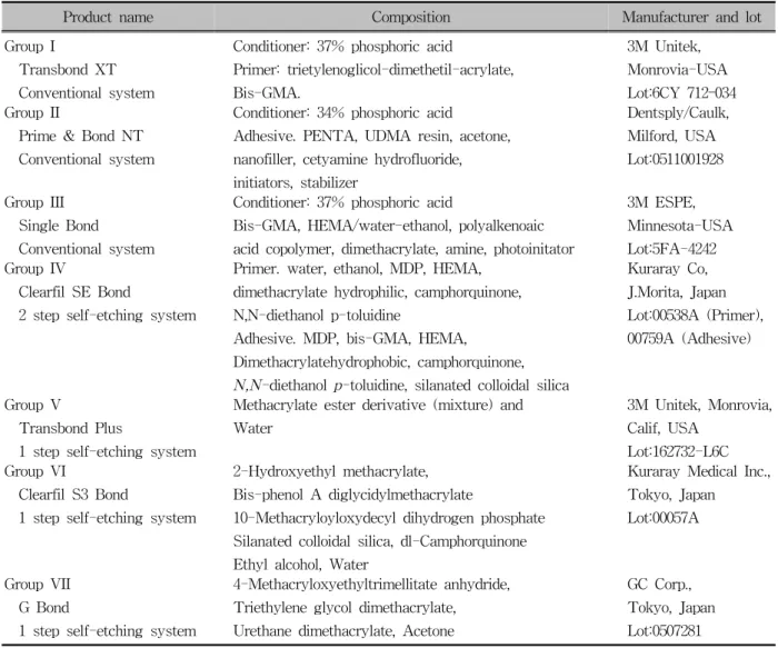
Table 1. Composition of the adhesive systems used in this study
Product name Composition Manufacturer and lot
Group I
Transbond XT Conventional system
Conditioner: 37% phosphoric acid
Primer: trietylenoglicol-dimethetil-acrylate, Bis-GMA.
3M Unitek, Monrovia-USA Lot:6CY 712–034 Group II
Prime & Bond NT Conventional system
Conditioner: 34% phosphoric acid
Adhesive. PENTA, UDMA resin, acetone, nanofiller, cetyamine hydrofluoride, initiators, stabilizer
Dentsply/Caulk, Milford, USA Lot:0511001928
Group III Single Bond
Conventional system
Conditioner: 37% phosphoric acid
Bis-GMA, HEMA/water-ethanol, polyalkenoaic acid copolymer, dimethacrylate, amine, photoinitator
3M ESPE, Minnesota-USA Lot:5FA-4242 Group IV
Clearfil SE Bond
2 step self-etching system
Primer. water, ethanol, MDP, HEMA, dimethacrylate hydrophilic, camphorquinone, N,N-diethanol p-toluidine
Adhesive. MDP, bis-GMA, HEMA,
Dimethacrylatehydrophobic, camphorquinone, N,N-diethanol p-toluidine, silanated colloidal silica
Kuraray Co, J.Morita, Japan Lot:00538A (Primer), 00759A (Adhesive)
Group V
Transbond Plus
1 step self-etching system
Methacrylate ester derivative (mixture) and Water
3M Unitek, Monrovia, Calif, USA
Lot:162732-L6C Group VI
Clearfil S3 Bond
1 step self-etching system
2-Hydroxyethyl methacrylate, Bis-phenol A diglycidylmethacrylate
10-Methacryloyloxydecyl dihydrogen phosphate Silanated colloidal silica, dl-Camphorquinone Ethyl alcohol, Water
Kuraray Medical Inc., Tokyo, Japan Lot:00057A
Group VII G Bond
1 step self-etching system
4-Methacryloxyethyltrimellitate anhydride, Triethylene glycol dimethacrylate, Urethane dimethacrylate, Acetone
GC Corp., Tokyo, Japan Lot:0507281 and hard-tissue impactions. Following extraction, the
teeth were immediately placed in distilled water at room-temperature and stored until the bonding procedure. The root of each tooth was embedded into an acrylic (Imicryl, Konya, Turkey) cylindrical block.
Metallic buttons (G&H Wire, Greenwood, USA) were used in the study. The average button base sur- face area was determined to be 9.43 mm2 from the manufacturer’s instructions.
Table 1 shows the primer and adhesive systems that were used in the current study. The buttons were bonded to the mounted teeth following one of theseven adhesive protocols according to the manufacturers’
instructions. Each group contained 12 specimens. Con- ventional etching and adhesive systems were used in
Groups I - III; and self-etching systems in groups IV - VII.
A 37% phosphoric acid gel (Ventura Gel Acondicio- nador, Madespa, Spain) was applied to the enamel for 15-seconds and the teeth were then rinsed with water spray for 30-seconds and air dried for 20 seconds.
After surface preparation, liquid primer was applied to the etched surface in the conventional groups.
Activation procedures for the self-etching primer were performed according to the manufacturer’s in- structions. Self-etching primer (Table 1) was gently rubbed onto the enamel surface for approximately three seconds with the disposable applicator supplied with the system. Then, a moisture-free air source was used to deliver a gentle burst of air to the enamel.
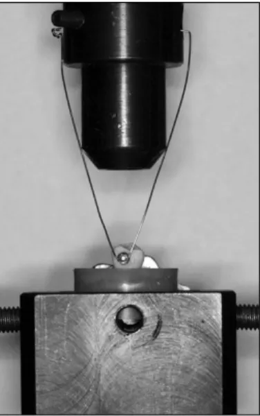
Fig 1. Shear application using the testing machine.
After etching/priming, bonding agent was photo- polymerized in all groups for 10-seconds. To exclude possible differences in bond strength caused by the or- thodontic composite used, the same material (Trans- bond XT, 3M Unitek, Monrovia, CA, USA) was ap- plied under all buttons. Standard edgewise premolar stainless-steel brackets (3M Unitek, Monrovia, CA, USA) were positioned in the center of the crown and firm pressure was applied. Any excess composite was removed. Before light-curing, the buttons were slightly pressed with bracket holder and excess adhesive was removed with a scaler. A light-emitting diode (LED) (SmartLite, Dentsply, Milford, USA) was used for cur- ing the composite, 20 seconds from both the mesial and distal sides. The same clinician carried out all bonding procedures in all groups (C.Y.). The teeth were then placed in distilled water at 37oC for 24 hours before testing.
Debonding procedure
Each toothwas oriented with a guiding device, so that its tooth surfaces were parallel to the shear-force dur- ing the test. A gingivo-occlusal load was applied to the button, producing a shear force from the button (Fig 1). A computer, electronically connected to the Lloyd testing machine (Lloyd instruments, Foreham, Hamp-
shire, UK), recorded the results of each test. The SBSs were measured at a crosshead speed of 1 mm/min. The force required to remove the buttons was measured in Newtons (N), and the SBS (1 megapascal-MPa = 1 N/mm2) was then calculated by dividing the force val- ues by the button base area (9.43 mm2).
Evaluation of the residual adhesive
After debonding, all the teeth and buttons were eval- uated under a stereomicroscope (Nikon, SMZ-1B, Osaka, Japan) by another operator (M.B.) who was blinded to the group allocation, under 10× magnifica- tion for the adhesive remnant index (ARI)14 scores: 0, no adhesive remaining on tooth; 1, less than half of the enamel bonding site covered with adhesive; 2, more than half of the enamel bonding site covered with adhesive; 3, the enamel bonding site covered en- tirely with adhesive.
Scanning electron microscope evaluation
For SEM investigations erupted and unerupted tooth specimens were used to evaluate the enamel surfaces.
Tooth were transferred to 70% ethanol and dehydrated in increasing concentrations of ethanol. Specimens were gradually dehydrated through a graded series of ethanol, air-dried and mounted on SEM stubs so that the relevant area of interest could be seen, sputter coat- ed with 10 nm of platinum in a Polaron E5100 SEM coating unit (Polaron Equipment Ltd, Hertfordshire, England), and examined in a Hitachi S 2500 SEM (Hitachi Ltd, Tokyo, Japan) at an operating voltage of 15 kV. The SEM photomicrographs were taken at 500× and 1500× magnification for visual inspection.
Statistical analysis
The Shapiro-Wilks normality test and the Levene variance homogeneity test were applied to the SBS data. The data showed normal distribution, and there was homogeneity of variances between the groups.
Thus, the statistical evaluation of SBS values between test groups was performed using parametric tests.
Descriptive statistics, including mean, standard devi-
Table 4. One-way analysis of variance (ANOVA) results of shear bond strength test for unerupted teeth groups
Sum of squares df Mean square F Significance
Between groups 13.364 6 2.227 0.821 0.557
Within groups 208.850 77 2.712
Total 222.214 83
Table 3. One-way analysis of variance (ANOVA) results of shear bond strength test for erupted teeth groups
Sum of squares df Mean square F Significance
Between groups 14.860 6 2.477 1.058 0.395
Within groups 180.283 77 2.341
Total 195.144 83
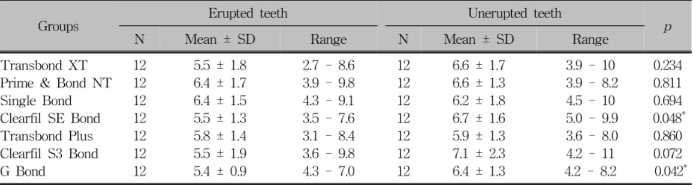
Table 2. Descriptive statistics of the shear bond strength values (MPa) of adhesive groups for erupted and unerupted teeth and results of the independent sample t-test
Groups Erupted teeth Unerupted teeth
N Mean ± SD Range N Mean ± SD Range p
Transbond XT 12 5.5 ± 1.8 2.7 - 8.6 12 6.6 ± 1.7 3.9 - 10 0.234
Prime & Bond NT 12 6.4 ± 1.7 3.9 - 9.8 12 6.6 ± 1.3 3.9 - 8.2 0.811
Single Bond 12 6.4 ± 1.5 4.3 - 9.1 12 6.2 ± 1.8 4.5 - 10 0.694
Clearfil SE Bond 12 5.5 ± 1.3 3.5 - 7.6 12 6.7 ± 1.6 5.0 - 9.9 0.048*
Transbond Plus 12 5.8 ± 1.4 3.1 - 8.4 12 5.9 ± 1.3 3.6 - 8.0 0.860
Clearfil S3 Bond 12 5.5 ± 1.9 3.6 - 9.8 12 7.1 ± 2.3 4.2 - 11 0.072
G Bond 12 5.4 ± 0.9 4.3 - 7.0 12 6.4 ± 1.3 4.2 - 8.2 0.042*
SD, standard deviation. *p < 0.05.
ation, minimum and maximum values were calculated for all groups of the erupted and unerupted teeth. An independent sample t-test was undertaken to compare the SBS values of the same adhesive system between the erupted and unerupted teeth groups. The SBS val- ues were analyzed by one-way analysis of variance (ANOVA) to determine significance of differences among 7 adhesive systems for each tooth type (erupted and unerupted), separately. To analyze the failure sites, contingency tables were designed and subjected to the chi-square test. The statistical significance level was established at p < 0.05.
Scoring of the ARI scores were repeated 4 months after the first measurement. Paired sample t-test was applied to the first and second data. It was found that the differences between the first and second measure- ments of the ARI scores were insignificant. The in-
tra-observer intraclass correlation coefficient was 0.90 for erupted teeth and 0.94 for unerupted teeth.
RESULTS
The descriptive statistics and the results of in- dependent sample t-test are presented in Table 2.
When the SBS values of erupted and unerupted teeth were compared, significant differences were found in two self-etching adhesives (Clearfil SE Bond and G Bond). Bond strengths of all adhesive systems were higher in unerupted teeth than erupted teeth, except for the Single-Bond system. Thus, the SBS part of the null hypothesis of this study was rejected.
The ANOVA comparisons of the 7 bonding systems for erupted and unerupted teeth groups are shown in Table 3 and Table 4, respectively. No statistically sig-
Erupted teeth Unerupted teeth
Test groups ‐‐‐‐‐‐‐‐‐‐‐‐‐‐‐‐‐‐‐‐‐‐‐‐‐‐‐‐‐‐‐‐‐‐‐‐‐‐‐‐‐‐‐‐‐‐‐‐‐‐‐‐‐‐‐‐‐‐‐‐‐‐‐‐‐‐‐‐‐‐‐‐‐‐‐‐ ‐‐‐‐‐‐‐‐‐‐‐‐‐‐‐‐‐‐‐‐‐‐‐‐‐‐‐‐‐‐‐‐‐‐‐‐‐‐‐‐‐‐‐‐‐‐‐‐‐‐‐‐‐‐‐‐‐‐‐‐‐‐‐‐‐‐‐‐‐‐‐‐‐‐‐‐
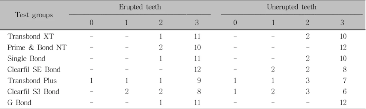
0 1 2 3 0 1 2 3
Transbond XT - - 1 11 - - 2 10
Prime & Bond NT - - 2 10 - - - 12
Single Bond - - 1 11 - - 2 10
Clearfil SE Bond - - - 12 - 2 2 8
Transbond Plus 1 1 1 9 1 1 3 7
Clearfil S3 Bond - 2 2 8 1 2 3 6
G Bond - - 1 11 - - - 12
ARI scores: 0, No adhesive left on the toothsurface; 1, less than 50% of adhesive left on the tooth surface; 2, more than 50% of adhesive left on the tooth surface; 3, all adhesive left on the tooth surface along with the impression of the button base.
Table 5. Frequency distribution of Adhesive Remnant Index (ARI) scores of 7 groups evaluated for erupted and un- erupted teeth
nificant differences were found among 7 adhesive groups for both erupted and unerupted teeth (p > 0.05).
The ARI scores for the adhesive systems are listed in Table 5. The data distributions indicated that bond failure occurred more frequently at the button-adhesive interface. For both teeth groups and adhesive systems the distribution of the ARI scores was similar and showed no significant differences. Thus, the failure- mode part of the present null hypothesis was not rejected.
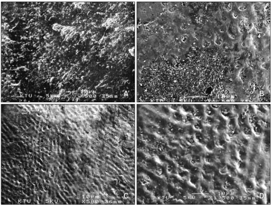
Photomicrographs for SEM observations of erupted and unerupted enamel are provided in Fig 2. Remark- able differences in the buccal enamel surfaces of erupt- ed and unerupted teeth were observed. The surface of erupted tooth has an unclear and irregular structure.
However, the unerupted tooth has a prismatic enamel structure.
DISCUSSION
Sheen et al.15 reported that bond strength inolder permanent teeth was greater than in younger teeth, re- gardless of etching time. Bhaskar16 found that the en- amel surfaces of unerupted and recently erupted teeth are completely covered with pronounced perikymata and rod-ends. With age, the perikymata and rod-ends may wear away. As a result of time changes in the or-
ganic portion of enamel, presumably near the surface, teeth may become harder and thereby reinforce the bond strength.16 Oliver13 investigated the bond strength of orthodontic attachments to enamel from two groups of teeth (erupted premolars and unerupted canines) and concluded that the bond strength of the enamel/adhe- sive interface is, in fact, different for erupted and un- erupted enamel. Tüfekçi et al.11 reported that there were no differences in bond strengths between teeth with mature and newly erupted enamel etched with ei- ther self-etching adhesive or conventional etching tech- niques. Almy2 indicated that the post-eruptive enamel maturation process may have little effect on bond strength values when etched either conventionally or with 3M Unitek self-etching primer. Using a subjective measurement of etching patterns when viewed under SEM, Nordenvall et al.17 reported that more deep re- tentive surfaces were obtained when conventionally etching newly erupted teeth for 15-seconds and mature teeth for 60-seconds. In the present study, due to pos- sible effects of the post-eruptive enamel maturation process, the erupted and unerupted teeth groups were analyzed for bond strength differences and significant differences were found in two self-etching groups (Clearfil SE Bond and G Bond) between the erupted and unerupted teeth. Conventional groups’ SBS values were not found statistically different and agree with previous work which also found no differences in bond
Fig 2. Scanning electron micrographs of erupted (A, × 500; B, × 1,500) and unerupted (C, × 500; D, × 1,500) molar teeth.
Fig 3. Attachment base design used in this study.
strengths between erupted and unerupted teeth.13 Many orthodontic attachment base designs are in clinical use today. Sharma-Sayal et al.18 found that at- tachment base designssignificantly affected mean shear bond strength. Brackets with foil-mesh bases have also been shown to have higher bond strengths than those with integral milled bases.18 Additionally, a reduction in bond strength was found associated with the reduc- tion of base surface area.19 In the present study, shear bond strength of the attachments were lower than pre- vious studies. We thought that the lower results of this study can be explained by the attachment base design (Fig 3) and base surface area. We did not use foil- mesh bases in the present study; and base surface area was lower than the conventional brackets.
Present findings of the comparisons of different ad- hesive systems do not agree with previous studies that have reported lower bond strength values with self- etching adhesives. No statistically significant differ- ences were determined among 7 adhesive groups in both erupted and unerupted teeth. A self-etching adhe- sive, Clearfil S3 Bond showed the highest mean SBS
value in unerupted teethand conventional etching/bond- ing systems, Prime & Bond NT and Single Bond showed the highest mean value in erupted teeth. This is some- what in accordance with the results of Buyukyilmaz et al.20 that reported higher bond strengths with the 3M self-etching primer. The findings from the current study do agree with Dorminey et al.21 who found no difference between conventional etching and the 3M self-etching primer when used according to the manu- facturer’s instructions.
The ARI scores in the current study indicated that the differences in the amount of adhesive remaining on
the enamel surfaces after debonding were not sig- nificant when the erupted and unerupted teeth groups were compared. The mode of bond failure of erupted and unerupted teeth was usually at the adhesive inter- face (at the button-resin interface-Score 3). Tüfekçi et al.11 reported that, there were significant differences in ARI scores between mature and newly erupted teeth.
Newly erupted teeth had more adhesive bond failures, whereas mature teeth had more cohesive bond failures, suggesting some differences between the 2 groups in the quality of the bond formed.
The mode of bond failure in this study for both con- ventional and self-etching systems was usually at the adhesive interface (at the button-resin interface-Score 3) and showed no statistically significant differences.
These results agree with some previous works. Bishara et al.22 found that self-etching primers left more adhe- sive on the teeth. Other studies claimed that less adhe- sive was left on the teeth in the self-etching primer group compared with the conventional group.20,23,24 These conflicting results can be attributed to the highly subjective nature of ARI scores and the fluoride con- tent in the enamel of the teeth tested.10
According to SEM evaluations, buccal enamel surfa- ces of erupted and unerupted teeth revealed aspects which varied from each other. The SEM photographs confirmed that the prismatic view of the surface of an erupted tooth is lost via calcium and other mineral pre- cipitation during post-eruptive maturation. This situa- tion might be an advantage for strong bonding of self-etch adhesives to unerupted enamel surfaces. Self- etching adhesives do not require a separate acid-etch step. They are composed of aqueous mixtures of acidic functional monomers, generally phosphoric acid esters, with a pH relatively higher than that of phosphoric acid-etching gels.25 Thus, self-etching adhesives do not etch enamel to the level obtained with phosphoric acid26 on erupted tooth surfaces. However, because un- erupted teeth lack post-eruptive maturation, self-etching systems may optimize the etching of unerupted enamel to the level obtained with phosphoric acid.
CONCLUSION
After our encouraging laboratory findings and hav-
ing in mind all the shortcomings of an in vitro setting we concluded that:
1. The SBS values between erupted and unerupted teeth were not significantly different between each other, except for two self-etching adhesives (Clearfil SE (Bond and G Bond).
2. Among investigated adhesive systems, there were no differences in SBSs between teeth that were pre- pared for bonding with conventional and self-etch- ing systems.
3. For both tooth types (erupted and unerupted) and adhesive systems (conventional and self-etching) the distribution of the ARI scores indicated that bond failures were more frequently at the button-adhesive interface.
- 국문초록 -
치아 맹출 유무에 대한 자가부식 접착제에 의한 교정용 부착장치의 접착강도
Metin Nur, Tancan Uysal, Cemal Yesilyurt, Mehmet Bayram
이번 연구의 목적은 맹출 또는 미맹출된 치아에 교정용 버 튼을 부착 후 자가부식 접착제(self-etching adhesive)의 사 용 유무에 대한 전단결합강도(shear bond strength)와 탈락 모드를 비교하고자 함이다. 각각 84개의 맹출 또는 미맹출된 제3대구치를 사용하였다. 각각 치아의 협측면을 다음의 부착 시스템 그룹으로 할당하였다. A, 기존방식: 1, Transbond XT (3M Unitek); 2, Prime & Bond NT (Dentsply/Caulk); 3, Single Bond (3M ESPE`); B, 자가부식 접착제; 4, Clearfil SE Bond (Kuraray); 5, Transbond Plus (3M Unitek); 6, Clearfil S3 (Kuraray); 7, G Bond (GC). 부착물의 전단강도 와 접착제 잔류지수를 측정하였으며 결과값은 ANOVA와 independent t-test 및 chi-square 검증을 통해 분석되었다.
맹출 또는 미맹출된 치아의 전단결합강도를 비교하였을 때 Clearfil SE와 G Bond에서 유의한 차이가 관찰되었다.
Single Bond를 제외한 모든 접착시스템에서 맹출보다는 미 맹출 치아면에서 높은 접착강도가 관찰되었다. 기존 접착제 를 사용하는 경우 맹출 또는 미맹출 치아에 대한 접착강도에 는 차이가 없을 수 있다. 그러나 임상의사는 사용 전에 self-etching system의 종류를 고려할 필요가 있다.
주요 단어: 접착제, 브라켓, 레진, 미맹출 치아
REFERENCES
1. Bishara SE, Ajlouni R, Soliman MM, Oonsombat C, Laffoon JF, Warren J. Evaluation of a new nano-filled restorative mate- rial for bonding orthodontic brackets. World J Orthod 2007;8:
8-12.
2. Almy D. Bonding properties of newly erupted and mature hu- man premolars. Master of Science Thesis, Virginia Common- wealth University 2004.
3. Crabb HS. The porous outer enamel of unerupted human premolars. Caries Res 1976;10:1-7.
4. Bonar LC, Shimizu M, Roberts JE, Griffin RG, Glimcher MJ.
Structural and composition studies on the mineral of newly formed dental enamel: a chemical, x-ray diffraction, and 31P and proton nuclear magnetic resonance study. J Bone Miner Res 1991;11:1167-76.
5. Ooya K. A scanning electron microscopic study on the differ- ences between newly erupted teeth and old teeth with refer- ence to fissure enamel surfaces and contents. Bull Tokyo Med Dent Univ 1977;24:89-102.
6. Brudevold F, Aasenden R, Bakhos Y. A preliminary study of posteruptive maturation of teeth in situ. Caries Res 1982;16:
243-8.
7. Sfondrini MF, Cacciafesta V, Pistorio A, Sfondrini G. Effects of conventional and high-intensity light-curing on enamel shear bond strength of composite resin and resin-modified glass- ionomer. Am J Orthod Dentofacial Orthop 2001;119:30-5.
8. Romano FL, Tavares SW, Nouer DF, Consani S, Borges de Araujo Magnani MB. Shear bond strength of metallic ortho- dontic brackets bonded to enamel prepared with Self-Etching Primer. Angle Orthod 2005;75:849-53.
9. White LW. An expedited indirect bonding technique. J Clin Orthod 2001;35:36-41.
10. Kotsanos N, Darling AI. Influence of posteruptive age of en- amel on its susceptibility to artificial caries. Caries Res 1991;25:241-50.
11. Tüfekçi E, Almy DM, Carter JM, Moon PC, Lindauer SJ.
Bonding properties of newly erupted and mature premolars.
Am J Orthod Dentofacial Orthop 2007;131:753-8.
12. Jacobs G, Kuftinec MM, Showfety KJ, von Fraunhofer JA.
Bonding characteristics of impacted versus erupted permanent teeth. Am J Orthod 1986;89:242-5.
13. Oliver RG. Bond strength of orthodontic attachments to enam- el from unerupted and erupted young permanent teeth. Eur J Orthod 1986;8:123-6.
14. Artun J, Bergland S. Clinical trials with crystal growth con- ditioning as an alternative to acid-etch enamel pretreatment.
Am J Orthod 1984;85:333-40.
15. Sheen DH, Wang WN, Tarng TH. Bond strength of younger and older permanent teeth with various etching times. Angle Orthod 1993;63:225-30.
16. Bhaskar SN. Orban’s oral histology and embryology. 9th ed.
St. Louis, Toronto, London: The C.V. Mosby; 1980. p. 46-106.
17. Nordenvall KJ, Brannstrom M, Malmgren O. Etching of decid- uous teeth and young and old permanent teeth: a comparison between 15 and 60 seconds of etching. Am J Orthod Dentofa- cial Orthop 1980;78:99-108.
18. Sharma-Sayal SK, Rossouw PE, Kulkarni GV, Titley KC. The influence of orthodontic bracket base design on shear bond strength. Am J Orthod Dentofacial Orthop 2003;124:74-82.
19. MacColl GA, Rossouw PE, Titley KC, Yamin C. The relation- ship between bond strength and orthodontic bracket base sur- face area with conventional and microetched foil-mesh bases.
Am J Orthod Dentofacial Orthop 1998;113:276-81.
20. Buyukyilmaz T, Usumez S, Karaman AI. Effect of self-etching primers on bond strength - are they reliable? Angle Orthod 2003;73:64-70.
21. Dorminey JC, Dunn WJ, Taloumis LJ. Shear bond strength of orthodontic brackets bonded with a modified 1-step etchant and primer technique. Am J Orthod Dentofacial Orthop 2003;
124:410-3.
22. Bishara SE, VonWald L, Laffoon JF, Warren JJ. Effect of a self-etch primer/adhesive on the shear bond strength of ortho- dontic brackets. Am J Orthod Dentofacial Orthop 2001;119:
621-4.
23. Larmour CJ, Stirrups DR. An ex vivo assessment of a bonding technique using a self-etching primer. J Orthod 2003;30:225-8.
24. Davari AR, Yassaei S, Daneshkazemi AR, Yosefi MH. Effect of different types of enamel conditioners on the bond strength of orthodontic brackets. J Contemp Dent Pract 20071;8:36-43.
25. Tay FR, Sano H, Carvalho R, Pashley EL, Pashley DH. An ultra-structural study of the influence of acidity of self-etching primers and smear layer thickness on bonding to intact dentin.
J Adhes Dent 2000;2:83-98.
26. Miller MB. Self-etching adhesives: solving the sensitivity conundrum. Pract Proced Aesthet Dent 2002;14:406.