Proliferation of vascular smooth muscle cells is 2+ response accompanied by changes in ryanodine receptors (RyRs)
6
0
0
전체 글
(2) 432. EJ Kim, et al. CICR. A second goal of the present study was to elucidate alterations of RyRs that occurred in rat aortic smooth muscle cells (RASMCs) during culture. To accomplish these goals, we directly examined Ca2+ release via activation of 2+ concentration of internal RyRs by measuring the Ca stores in permeabilized cells and comparing RyR expression levels in freshly dissociated and cultured RASMCs. In this study, we found that RyRs are present and mediate CICR in freshly dissociated RASMCs but disappear during cell culture.. METHODS Preparation of RASMCs RASMCs were dissociated from the thoracic aorta of 8to 9-wk-old Sprague-Dawley rats. Dissected aortas were cut and cleaned of fat and connective tissue in ice-cold phosphate buffered saline (PBS) containing 1.06 mM KH2PO4, 155.17 mM NaCl, and 2.97 mM Na2HPO4 (pH 7.4). The tissues were first digested by incubating in PBS containing 0.5% papain (Sigma, St Louis, Mo, USA), 0.37% DL-dithiothreitol, and 0.44% bovine serum albumin (BSA, Sigma) for 10 min with shaking at 37oC, and then digested by incubating in PBS containing 1% collagenase (Wako, Tokyo, Japan) o for 15∼16 hours with shaking at 4 C. After digestion, the tissues were washed five times with PBS at 4oC and gently triturated with a fire-polished Pasteur pipette in PBS to obtain a single-cell suspension. Cell culture Dissociated cells were resuspended and plated on culture dishes. RASMC cultures were maintained in Dulbecco’s Modified Eagle Medium (DMEM) supplemented with 10% fetal bovine serum (FBS) and 1% antibiotic-antimycotic uno der a humidified atmosphere of 5% CO2-95% O2 at 37 C. Experiments on cultured RASMCs were performed on passage 11∼18 cells. The purity of RASMCs was verified by staining for smooth muscle-specific α-actin (Santa Cruz Biotechnology, Santa Cruz, CA, USA). All cells were α-actin positive. 2+. Cytosolic Ca. measurement in RASMCs 2+. For measurement of [Ca ]i, RASMCs were loaded with 2μM Fura-2/AM, 7.5% BSA, and 0.03% F127 for 20 min at 37oC. Fura-2/AM-loaded cells were transferred to a glass coverslip at the bottom of a perfusion chamber for mounting. Cells were continuously perfused at room temperature with HEPES-buffered physiological saline solution (HEPESPSS) containing 140 mM NaCl, 4 mM KCl, 5 mM HEPES, 1.28 mM CaCl2, 1 mM MgCl2, and 11 mM glucose (pH 7.4) using an electronic-controlled perfusion system (Warner Instrument, Hamden, CT, USA). RyRs were stimulated by 2+ perfusing with 20 mM caffeine. Cytosolic Ca was meas2+ ured in Ca -free HEPES-PSS to eliminate any possibility 2+ 2+ of caffeine-induced Ca entry. Cytosolic Ca imaging was carried out using an inverted Olympus IX71 microscope equipped with a 40X fluorescence objective. [Ca2+]i was determined from the ratio of Fura-2/AM fluorescence at excitation wavelengths of 380 nm and 340 nm using a polychrome V monochromator (Til Photonics, Pleasanton, CA, USA). Images were obtained at an emission wavelength of. 510 nm using a SNAP HQ2 camera (Photometrics, Tuscon, AZ, USA). Measurement of SR Ca2+ release in permeabilized cells RASMCs, adhered to a poly-L-lysine-coated coverslip at the bottom of the perfusion chamber, were incubated with 10μM mag-fura-2/AM, 7.5% BSA, and 0.03% F127 for 30 min at 37oC, and then permeabilized for 80∼100 s with 20μM β-escin in intracellular medium (ICM; 125 mM KCl, 19 mM NaCl, 10 mM HEPES, and 1 mM EGTA, pH 7.3). Permeabilized RASMCs were washed with ICM for 5 min to remove cytosolic dye and then were superfused for 60∼ 80 s with loading buffer (125 mM KCl, 19 mM NaCl, 10 mM HEPES, 1 mM EGTA, 0.650 mM CaCl2 [free Ca2+, 200 nM], 1.4 mM MgCl2, and 3 mM Na2ATP, pH 7.3) to activate 2+ 2+ SERCA and load Ca stores. After Ca loading, RASMCs were superfused with release buffer (125 mM KCl, 19 mM NaCl, 10 mM HEPES, 1 mM EGTA, and 3 mM Na2ATP, 2+ pH 7.3) to inactivate SERCA. Ryanodine-sensitive Ca release channels were activated by adding 10 mM caffeine, 2+ 10μM ryanodine, or 200 nM Ca to the release buffer. The emission of mag-fura-2/AM above 505 nm following excitation at 340 nm and 380 nm was recorded using the TILL Photonics imaging system. Immunocytochemistry RASMCs were allowed to attach to coverslips for 3 h in PBS at room temperature, followed by fixation with 4% paraformaldehyde for 15 min. Fixed RASMCs were rinsed with PBS for 5 min, permeabilized with 0.2% Triton-X 100 (Sigma) for 5 min, and blocked with 10% normal rabbit serum (Jackson Immunoresearch Laboratories, PA, USA) for 2 h. Immunocytochemistry was carried out using goat polyclonal anti-IP3R (Santa Cruz Biotechnology) and goat polyclonal anti-RyR (Santa Cruz Biotechnology) primary antibodies, and a Cy3-conjugated rabbit anti-goat secondary antibody (Jackson Immunoresearch Laboratories). RASMCs o were incubated overnight at 4 C with freshly antibodies diluted 1:50 in a 10% normal rabbit serum solution. RASMCs were rinsed with PBS and then incubated with secondary antibody in a dark chamber. The coverslip was then mounted and dried for at least 2 h. RyR immunofluorescence images were collected using a confocal microscope (Carl Zeiss, Germany) and then processed using Photoshop 7.0 software (Adobe, Mountain View, CA, USA). Drugs Inositol 1,4,5-trisphosphate (IP3) was purchased from Biomol Research Laboratories (Plymouth, PA, USA), and ryanodine was purchased from Tocris Bioscience (Ballwin, MO, USA). Fura-2/AM and mag-fura-2/AM were purchased from Invitrogen (Carlsbad, CA, USA). PBS was purchased from GIBCO BRL (Grand Island, NY, USA). Caffeine, βescin, and other chemicals used in the preparation of buffers were purchased from Sigma Aldrich Chemical Co..
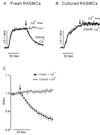
(3) 433. Ryanodine-receptors Lose during Culture. RESULTS Effects of caffeine in RASMCs. feine in cultured RASMCs (Fig. 1C). Taken together, these 2+ results suggest that caffeine directly releases Ca from the 2+ channel. SR via a caffeine-sensitive Ca 2+. 2+. The effects of caffeine, a RyR activator [16,18], were investigated in freshly dissociated RASMCs and cultured RASMCs. The cytosolic Ca2+ response to caffeine was meas2+ ured in the absence of extracellular Ca to rule out the 2+ possibility of extracellular Ca influx. After the addition of 20 mM caffeine to dissociated RASMCs, [Ca2+]i abruptly increased and slowly declined to basal levels (Fig. 1A). This 2+ from intraresult means that caffeine mobilized Ca cellular Ca2+ stores in freshly dissociated RASMCs. In con2+ trast, no cytosolic Ca response to caffeine was observed in cultured RASMCs (Fig. 1B). To confirm that caffeine directly induced Ca2+ release from the SR, we permeabilized cells using 20μM β-escin 2+ by activating SERCA. and loaded the SR with Ca Addition of 10 mM caffeine reduced the concentration of 2+ Ca in SR in permeabilized, dissociated RASMCs. However, little or no change was observed after perfusion of caf-. Induction of Ca release from intracellular Ca 2+ stores by altering [Ca ]i concentration. It is well known that increased [Ca ]i activates RyR to 2+ release Ca from SR via a CICR mechanism [16-18]. To demonstrate CICR, we measured the Ca2+ concentration of 2+ intracellular Ca stores in permeabilized RASMCs after 2+ 2+ altering [Ca ]i. Ca -free release buffer did not induce a change in intracellular Ca2+ stores in permeabilized, dis2+ sociated RASMCs (Fig. 2A, bright line), indicating that Ca 2+ leak channel. is not released from the SR via a Ca Perfusing permeabilized, dissociated RASMCs with release 2+ buffer containing 200 nM Ca decreased the concentration 2+ of Ca in intracellular stores (Fig. 2A, C). In contrast, cultured RASMCs treated in the same manner did not respond 2+ to 200 nM Ca (Fig. 2B, C). These results indicate that 2+ 2+ 2+ cytosolic Ca induces Ca release from intracellular Ca. Fig. 1. Effects of caffeine on RASMCs. Representative traces of caffeine-induced cytosolic Ca2+ responses in freshly dissociated RASMCs (A) and cultured RASMCs (B). (C) The application of 20 mM caffeine in Ca2+-free HEPES-PSS buffer induced a rapid and large cytosolic Ca2+ increase only in freshly dissociated RASMCs. 2+ 2+ Effects of caffeine on Ca release from intracellular Ca stores in permeabilized freshly dissociated and cultured RASMCs. The data were obtained from four experiments. The arrows indicate the 2+ starting points of drug perfusion. Caffeine (10 mM) induced Ca release from the SR in freshly dissociated RASMCs (■), but not in cultured RASMCs (□).. Fig. 2. CICR in permeabilized RASMCs. Representative traces of CICR in freshly dissociated RASMCs (A) and cultured RASMCs 2+ 2+ (B). The fine line indicates Ca -free solution. Ca (200 nM) induced Ca2+ release only in freshly dissociated RASMCs. (C) Effects of Ca2+ on Ca2+ release in permeabilized freshly dissociated and cultured RASMCs. The data, normalized to the initial 10s period prior to 200 nM Ca2+ perfusion, were obtained from four experiments. The arrows indicate the starting points of 200 nM 2+ 2+ 2+ Ca perfusion. Ca (200 nM) triggered Ca release from the SR in freshly dissociated RASMCs (■), but had no effect in cultured RASMCs (□).. 2+.
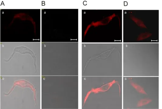
(4) 434. EJ Kim, et al. stores via RyRs in dissociated RASMCs but not in cultured RASMCs. Induction of Ca2+ release from intracellular Ca2+ stores by ryanodine The plant alkaloid ryanodine has dual effects on RyR activity. At low concentrations, ryanodine activates RyRs 2+ and induces Ca release from the SR [19]. However, at high concentrations, ryanodine inhibits RyRs. To determine 2+ 2+ if Ca release channel in intracellular Ca stores was activated by ryanodine, we treated cells with 10μM ryanodine, a concentration that activates RyRs. Application of 10μM ryanodine to permeabilized dissociated RASMCs de2+ in intracellular stores creased the concentration of Ca (Fig. 3A, 3C), whereas ryanodine failed to induce SR Ca2+ release in cultured RASMCs (Fig. 3B, 3C). Expression of RyRs and IP3Rs in RASMCs Finally, we examined the expression of RyRs to determine if the loss in sensitivity to the SR Ca2+-mobilizing 2+ agents, caffeine, Ca and ryanodine, was associated with an alteration in the expression of RyRs during cell culture. In freshly dissociated RASMCs, RyR immunofluorescence was clearly detected, exhibiting a primarily cytoplasmic distribution with nuclear exclusion (Fig. 4A). However RyRs were not expressed in cultured RASMCs (Fig. 4B). The disappearance of RyR expression in cultured RASMCs 2+ is consistent with the Ca imaging results (see Figs. 1∼3). The expression of IP3Rs, the other major type of SR Ca2+release channel, was also investigated by immunofluorescence in dissociated RASMCs and cultured RASMCs. IP3Rs were expressed in both freshly dissociated and cultured RASMCs, exhibiting a primarily cytoplasmic pattern in both types of RASMCs (Fig. 4C, D). Although these results do not exclude the possibility that IP3R subtypes are altered during culture, they indicate that IP3R expression. 2+. Fig. 3. Ryanodine-induced Ca release in permeabilized RASMCs. Representative traces of ryanodine-induced Ca2+ release in freshly dissociated RASMCs (A) and cultured RASMCs (B). (C) Effects of 2+ ryanodine on Ca release in permeabilized freshly dissociated RASMCs and cultured RASMCs. The data, normalized to the initial 10s period prior to ryanodine application, were obtained from four experiments. The arrow indicates the starting points of ryanodine perfusion. Ryanodine (10μM) induced Ca2+ release from the SR in freshly dissociated RASMCs (■), but not in cultured RASMCs (□).. Fig. 4. Expression of RyRs and IP3Rs in RASMCs. Expression of RyRs in freshly dissociated RASMCs (A) and cultured RASMCs (B). Expression of IP3Rs in freshly dissociated RASMCs (C) and cultured RASMCs (D). (a) Immunocytochemistry; (b) interference contrast micrographs; (c) merged images. Immunocytochemistry was performed using primary anti-RyRs and anti-IP3Rs antibodies, as described in Methods. The data show that RyRs are only present in freshly dissociated RASMCs (n=5), whereas IP3Rs are present in both freshly dissociated RASMCs and cultured RASMCs (n=5). Scale bars, 5μm (A, C) and 10μm (B, D)..
(5) 435. Ryanodine-receptors Lose during Culture. is retained.. DISCUSSION 2+. RyRs are Ca channels that are located in the SR membrane [10,11]. RyRs are physiologically activated by in2+ creased levels of cytosolic Ca resulting from extracellular 2+ 2+ Ca entry or Ca release from intracellular stores [16-18]. Because it is experimentally difficult to measure Ca2+ release from the SR, many researchers have used caffeine as 2+ a RyR activator, and measured [Ca ]i as an index of RyR function [12,13]. Caffeine is known to have off-target ef2+ fects, including blocking IP3Rs and store-operated Ca entry [14,15]. Thus, results obtained using caffeine are an imprecise indicator of RyR function. In the present study, we permeabilized the plasma membrane of RASMCs and then 2+ 2+ directly monitored the Ca concentration of internal Ca stores to provide direct evidence of operational RyRs. We observed that perfusion of permeabilized dissociated 2+ reRASMCs with a high concentration of cytosolic Ca 2+ 2+ sulted in Ca release from internal Ca stores. We also 2+ showed that caffeine or ryanodine stimulated Ca release 2+ stores in permeabilized dissociated from internal Ca RASMCs, and confirmed expression of RyRs in these cells by immunocytochemistry. Collectively, these results provide direct evidence that RyRs are expressed and operational in freshly dissociated RASMCs. Several studies have reported that the caffeine-sensitive VSMC population is gradually decreased or abolished as a function of days in culture [20,21]. These studies have suggested that a decline in caffeine sensitivity may reflect a reduction in RyR expression [20] or alteration of RyR subtype [21]. Although each RyR subtype has a different sensitivity to caffeine [22-24], all three types of RyR are activated by 10 mM caffeine [22,25]. Thus, if the RyR subtype is switched in cultured RASMCs, these cells should still retain the ability to respond to caffeine at concentrations ≥10 mM . However, in the current study, we found that intact (unpermeabilized) cultured RASMCs showed no response 2+ to 20 mM caffeine. Moreover, ryanodine and Ca failed 2+ to trigger Ca release in permeabilized cultured RASMCs. Using a polyclonal anti-RyR antibody, we also confirmed that RyRs are not expressed in cultured RASMCs. These data argue against a switch in RyR subtype during cell culture and instead suggest that RyRs disappear. 2+ The Ca spike characteristic of the transient increase 2+ in [Ca ]i is known to be related to the function of RyRs [26,27]. For example, this Ca2+ spike contributes to the regulation of tone in vasoconstrictor-contracted smooth muscle [26], and promotes activation of the cAMP response element-binding protein, which leads to cell-cycle arrest in G1 phase [28,29]. Consistent with this latter observation, it has been reported that blocking RyRs with high concentrations of ryanodine enhances cell proliferation of VSMCs during culture [30]. A study showed that RyR1 is decreased at the mRNA level in thymomas [31], suggesting an association between disruption of the Ca2+ spike and enhanced cell proliferation in cancer, implying that RyR expression and function may inhibit cell proliferation. In contrast to the potential inhibitory role of RyRs in cell proliferation, IP3Rs, the 2+ other major type of SR Ca -release channel, are known 2+ to regulate [Ca ]i, which promotes cell proliferation [32,33]. In the current study, we confirmed that IP3Rs were 2+ expressed in cultured RASMCs; we also observed Ca re-. lease following activation of IP3Rs (data not shown), indicating that these channels are functional. The above results are consistent with the idea that cell proliferation is enhanced by activation of IP3Rs in the absence of RyRs. In vivo, VSMCs generally exhibit a contractile phenotype and do not proliferate. However, VSMCs recover their proliferative function in certain contexts, such as vessel-injury repair and vascular pathologies, including atherosclerosis, hypertension, and vein-graft failure [1,3,4]. Because studies on RyRs under conditions of vascular injury and atherosclerosis are lacking, the role of RyRs in the development of vascular disease remains unclear. Thus, further investigation of RyR expression levels in vivo in relevant pathological settings is needed.. ACKNOWLEDGEMENTS This study was supported by the Myung-Gok Research Fund of Konyang University (2007), and Basic Research Program through the National Research Foundation (NRF) of Korea funded by the Ministry of Education, Science and Technology (2010-0012568).. REFERENCES 1. Chamley-Campbell J, Campbell GR, Ross R. The smooth muscle cell in culture. Physiol Rev. 1979;59:1-61. 2. Schwartz SM, Campbell GR, Campbell JH. Replication of smooth muscle cells in vascular disease. Circ Res. 1986;58:427444. 3. Halayko AJ, Salari H, MA X, Stephens NL. Markers of airway smooth muscle cell phenotype. Am J Physiol. 1996;270:L10401051. 4. Thyberg J. Differentiated properties and proliferation of arterial smooth muscle cells in culture. Int Rev Cytol. 1996;169: 183-265. 5. Berridge MJ. Inositol trisphosphate and calcium signalling. Nature. 1993;361:315-325. 6. Ghosh TK, Bian JH, Short AD, Rybak SL, Gill DL. Persistent intracellular calcium pool depletion by thapsigargin and its influence on cell growth. J Biol Chem. 1991;266:24690-24697. 7. Short AD, Bian J, Ghosh TK, Waldron RT, Rybak SL, Gill DL. Intracellular Ca2+ pool content is linked to control of cell growth. Proc Natl Acad Sci USA. 1993;90:4986-4990. 8. Waldron RT, Short AD, Meadows JJ, Ghosh TK, Gill DL. Endoplasmic reticulum calcium pump expression and control of cell growth. J Biol Chem. 1994;269:11927-11933. 9. Wang Y, Chen J, Wang Y, Taylor CW, Hirata Y, Hagiwara H, Mikoshiba K, Toyo-oka T, Omata M, Sakaki Y. Crucial role of type 1, but not type 3, inositol 1,4,5-trisphosphate (IP3) receptors in IP3-induced Ca2+ release, capacitative Ca2+ entry, and proliferation of A7r5 vascular smooth muscle cells. Circ Res. 2001;88:202-209. 10. Savineau JP. Is the translocon a crucial player of the calcium homeostasis in vascular smooth muscle cell? Am J Physiol Heart Circ Physiol. 2009;296:H906-9067. 11. Hirota S, Helli P, Janssen LJ. Ionic mechanisms and Ca2+ handling in airway smooth muscle. Eur Respir J. 2007;30: 114-133. 12. Côrtes SF, Lemos VS, Stoclet JC. Alterations in calcium stores in aortic myocytes from spontaneously hypertensive rats. Hypertension. 1997;29:1322-1328. 13. Côrtes SF, Lemos VS, Corriu C, Stoclet JC. Changes in angiotensin II receptor density and calcium handling during proliferation in SHR aortic myocytes. Am J Physiol. 1996;271: H2330-2338. 14. Choi KJ, Kim KS, Kim SH, Kim DK, Park HS. Caffeine and.
(6) 436. 15. 16. 17. 18.. 19. 20.. 21.. 22. 23.. 24.. EJ Kim, et al. 2-aminoethoxydiphenyl borate (2-APB) have different ability to inhibit intracellular calcium mobilization in pancreatic acinar cell. Korean J Physiol Pharmacol. 2010;14:105-111. Hume JR, McAllister CE, Wilson SM. Caffeine inhibits InsP3 responses and capacitative calcium entry in canine pulmonary arterial smooth muscle cells. Vascul Pharmacol. 2009;50:89-97. Islam MS. The ryanodine receptor calcium channel of betacells: molecular regulation and physiological significance. Diabetes. 2002;51:1299-1309. Kamishima T, Quayle JM. Ca2+-induced Ca2+ release in cardiac and smooth muscle cells. Biochem Soc Trans. 2003;31: 943-946. Choi KJ, Cho DS, Kim JY, Kim BJ, Lee KM, Kim SH, Kim DK, Kim SH, Park HS. Ca2+-induced Ca2+ release from internal stores in INS-1 rat insulinoma cells. Korean J Physiol Pharmacol. 2011;15:53-59. Masumiya H, Li P, Zhang L, Chen SR. Ryanodine sensitizes the Ca2+ release channel (ryanodine receptor) to CaCa2+ activation. J Biol Chem. 2001;276:39727-39735. Vallot O, Combettes L, Jourdon P, Inamo J, Marty I, Claret M, Lompré AM. Intracellular Ca2+ handling in vascular smooth muscle cells is affected by proliferation. Arterioscler Thromb Vasc Biol. 2000;20:1225-1235. Berra-Romani R, Mazzocco-Spezzia A, Pulina MV, Golovina VA. Ca2+ handling is altered when arterial myocytes progress from a contractile to a proliferative phenotype in culture. Am J Physiol Cell Physiol. 2008;295:C779-790. Perez CF, Voss A, Pessah IN, Allen PD. RyR1/RyR3 chimeras reveal that multiple domains of RyR1 are involved in skeletaltype E-C coupling. Biophys J. 2003;84:2655-2663. Copello JA, Barg S, Sonnleitner A, Porta M, Diaz-Sylvester P, Fill M, Schindler H, Fleischer S. Differential activation by Ca2+, ATP and caffeine of cardiac and skeletal muscle ryanodine receptors after block by Mg2+. J Membr Biol. 2002;187: 51-64. Fessenden JD, Wang Y, Moore RA, Chen SR, Allen PD, Pessah. 25.. 26. 27. 28.. 29.. 30.. 31. 32. 33.. IN. Divergent functional properties of ryanodine receptor types 1 and 3 expressed in a myogenic cell line. Biophys J. 2000;79: 2509-2525. Nakai J, Ogura T, Protasi F, Franzini-Armstrong C, Allen PD, Beam KG. Functional nonequality of the cardiac and skeletal ryanodine receptors. Proc Natl Acad Sci USA. 1997;94:10191022. Nelson MT, Cheng H, Rubart M, Santana LF, Bonev AD, Knot HJ, Lederer WJ. Relaxation of arterial smooth muscle by calcium sparks. Science. 1995;270:633-637. Dreja K, Nordström I, Hellstrand P. Rat arterial smooth muscle devoid of ryanodine receptor function: effects on cellular Ca2+ handling. Br J Pharmacol. 2001;132:1957-1966. Arnould T, Vankoningsloo S, Renard P, Houbion A, Ninane N, Demazy C, Remacle J, Raes M. CREB activation induced by mitochondrial dysfunction is a new signaling pathway that impairs cell proliferation. EMBO J. 2002;21:53-63. Giebler HA, Lemasson I, Nyborg JK. p53 recruitment of CREB binding protein mediated through phosphorylated CREB: a novel pathway of tumor suppressor regulation. Mol Cell Biol. 2000;20:4849-4858. Wilkerson MK, Heppner TJ, Bonev AD, Nelson MT. Inositol trisphosphate receptor calcium release is required for cerebral artery smooth muscle cell proliferation. Am J Physiol Heart Circ Physiol. 2006;290:H240-247. Kusner LL, Mygland A, Kaminski HJ. Ryanodine receptor gene expression thymomas. Muscle Nerve. 1998;21:1299-1303. Jaggar JH, Nelson MT. Differential regulation of Ca2+ sparks and Ca2+ waves by UTP in rat cerebral artery smooth muscle cells. Am J Physiol Cell Physiol. 2000;279:C1528-1539. Gomez MF, Stevenson AS, Bonev AD, Hill-Eubanks DC, Nelson MT. Opposing actions of inositol 1,4,5-trisphosphate and ryanodine receptors on nuclear factor of activated T-cells regulation in smooth muscle. J Biol Chem. 2002;277:37756-37764..
(7)
수치
관련 문서
Activation of Autophagy by Mangiferin Protects Auditory Hair Cells from Oxidative stress Induced Ototoxicity.. Gyeongmin
Taken together, these results suggested that latex containing the ficin inhibited the cell growth and induced apoptosis by caspase and Bcl-2 family signaling pathway in
Conclusion: In conclusion, the relaxant effects of neuromuscular blockers on the uterine smooth muscle may be transmitted via nicotinic acetylcholine receptors
Activated Foxo3a can inhibit the activation of p53 by DNA damage [46] and Resulted in a significant down-regulation of both p21 and p53 expression in Foxo3a- siRNA U2OS cells
The biological effect of ionizing radiation is a consequence of the energy transfer, by ionization and excitation, to cells in the body. Factors of radiation effects
The photovoltaic(PV) power generation system transforms unlimited and pollution-free solar energy directly into electronic energy by using solar cells
• Generation of different specialized kinds of cells from zygote (fertilized egg) or other precursor cells.. – Generate blood cells, muscle
Glutamate excitotoxicity induced by excessive activation of NMDA receptor causes various damage to cells, which leads to cell death.. In previous studies, increased ROS
