- 80 -
KISEP Case Reports J Rhinol 7((((1)))), 2000
Three Cases of Hemangioma in Nasal Septum
Seon Tae Kim, M.D., Seung Hwan Kim, M.D., Gi Young Gu, M.D. and Heung Eog Cha, M.D.
ABSTRACT
Nasal septal hemangioma is a disease entity which is found most commonly at the anterior part of the nasal septum. The etiology of the nasal septal hemangioma remains controversial. But its development appears to be the result of post-traumatic proliferation of local blood vessels. It is generally an isolated finding, not part of a systemic disorder. In general, the best method of treatment is a wide resection of the tumor with a cuff of underlying mucosa and perichondrium. We present three cases of septal hemangioma which were presented as unilateral epistaxis. They were completely removed with laser photocoagulation under the endoscopic approach. The postoperative course has been gone well without recurrence.
KEY WORDS:Hemangioma·Nasal septum·Laser
INTRODUCTION
The hemangioma is a frequently found disease in the head and neck area and rarely found in the nasal cavity or sinus. Clinically, its main complaints include nasal ob- struction and nasal bleeding.1)2)
The hemangioma in the nasal cavity is often confused with bleeding polyposis or angiofibromatous polyp, and commonly occurs in the anterior nasal septum in Little’s area.2)3) We experienced three cases of hemangioma oc- curring in the nasal septum and report the cases together with clinical discussion.
CASE Case 1:
A 31-year-old male patient visited our hospital on November 7, 1997 with a main complaint of frequent nasal bleeding. Since about a month before he visited
the hospital, he had suffered from occasional nasal bl- eeding but did not seek any medical treatment, which made the symptom worsen and develop nasal obstru- ction.
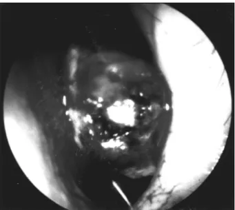
We found a dark-red polypoid mass in the left nasal cavity and also found that his nasal septem was deviated to the left side (Fig. 1). Through sinus CT, the sinus was observed to be normal while the nasal septum was dev- iated to the left side and a soft tissue density of 1×2 cm size was observed within the left nasal cavity (Fig.
2). Through endoscopic surgery under local anesthesia, the tumor was removed as en bloc, without breaking it into pieces, by using a laser cautery at the basal portion.
There was not much bleeding during the operation and the basal portion was located in the anterior nasal se- ptum in Little’s area. A microscopic observation found irregular, wide vascular lumen and extended cavernous blood vessels, some of which showed cavernous hema- ngioma where the blood vessels are filled with thrombus (Fig. 3).
Case 2:
A 48-year-old female patient visited the hospital on January 13, 1998 complaining of occasional nasal ble- eding over the previous two months and gradual nasal obstruction. There was nothing special found in the fa- mily history or past medical history while the general status was good. A red-brown tumor occurring from the Department of Otorhinolaryngology-Head and Neck Surgery,
Gil Hospital,Gachon Medical School, Inchon, Korea
Address correspondence and reprint requests to Seon-Tae Kim, M.D., Department of Otolaryngology-Head and Neck surgery Gil Medical Center Gachon Medical School Guwoldong 1198 Namdonggu, Inchon, 405-760, Korea
Tel:82-32-460-3323, Fax:82-32-467-9044 E-mail:[email protected]
Accepted for publication on February 23, 2000
Kim et al:Hemangioma in Nasal Septum / 81
nasal septum was found in the left-side nasal cavity. Th- rough an endoscopic surgery under local anesthesia the tumor was removed in a mass by using a laser cautery through the left-side nostril. There was not much blee- ding during the operation.
Through a pathohistological examination, it was found to be a cystic type tumor, 1×1×2 cm in size, which sh- owed partial bleeding. It had a form of mixed hemangi- oma which was a mixture of capillary vessels which are consisting of singlelayer, well differentiated endothelial cells, and large and irregular vascular lumen (Fig. 4).
Case 3:
A 24-year-old female patient visited the hospital on January 10, 1999 with main complaints of gradual nasal obstruction which had developed over the past two mo- nths and frequent nasal bleeding. There was nothing sp- ecial found in her family history or past history and her general status was relatively good. A dark-red tumor from the anterior nasal septum was found in the left-side na- sal cavity. The sinus CT showed that the sinus was in normal status and revealed a tumor of 1×1 cm size de- nsity in the left-side nasal cavity. The surgery was con- ducted under local anesthesia by using an endoscope and laser. The tumor was removed en bloc and there was not much bleeding during the operation.
A pathohistological examination observed a 1×1×
Fig. 1. Endoscopic intranasal finding of case 1. This photograph shows a hemorrhagic polypoid mass at the left Little’s area wh- ich has smooth surface with focal congestion.
Fig. 2. Preoperative PNS axial CT finding of case 1. Homoge- neous mass with smooth margin is attached at the left side of nasal septum.
Fig. 3. Microscopic finding of cavernous type hemangioma.
There are cavernous, blood filled structures, lined by a single layer of endothelial cells (H & E, ×100).
Fig. 4. Microscopic finding of mixed type hemangioma. Large irregular vascular lumina with thin endothelial cell layer are dispersed among the small vascular spaces and some conne- ctive tissues (H & E, ×100)
82 / J Rhinol 7(1), 2000
1 cm dark-red tumor which showed partial bleeding.
A mixed hemangioma was also observed which was an irregular mixture of capillary vessels consisting of well differentiated endothelium cells and cavernous ti- ssues.
DISCUSSION
The hemangioma in nasal septum is a rare disease which is most commonly found in white females older than 40 years of age.2) While the exact etiology has not been found yet, Ash, et al.,4) maintained that the hema- ngioma is a kind of tumor because it is destructive and has blood vessels showing aspects similar to tumors. Wi- llis, et al.,5) reported that every benign blood vessel proliferation is not a true tumor, but a harmartoma or congenital anomaly. It may be caused by opening of bl- ood vessels previously closed by birth. Besides, prolif- eration of local blood vessels and an increase of regional hydrostatic pressure caused by repeated local stimulation are known to influence the occurrence of hemangioma.6) It commonly occurs in the anterior nasal septum in Li- ttle’s area because the area has a rich blood vessel di- stribution and is largely exposed to repeated digital tra- uma.7)
Symptoms may include unilateral nasal bleeding and gradual nasal obstruction over a period of six months.
Pyogenic granuloma, hemangiopericytoma and angiof- ibroma are often distinguished disease. Pyogenic gran- uloma, most similar to hemangioma, features nasal ble- eding and a fastgrowing unilateral bleeding tumor, and is commonly reported during a pregnancy.8) It is different from the hemangioma in its lobular arrangement of bl- ood vessels.8)9) The size of the hemangioma which is limited to the inside nasal cavity ranges from a few mm to more than 2cm, and sometimes it is spreading out to the surrounding area of the nasal cavity to invade nei- ghboring structures.7) By conducting a CT, the size of the tumor and the degree of its invasion into neighbo- ring structures can be obtained.10) The angiography, in particular, is helpful in distinguishing the hemangioma from the nasopharynx angiofibroma in cases of severe invasion into neighboring areas.
Histological features include the rich blood vessels consisting of singlelayer endothelial cells which extend irregularly between a small volume of connective tissue.
Pathohistological classification categorizes it in three ty- pes;capillary blood vessel, cavernous and mixture in accordance with the size of prominent vessel.6)11) In this study, all three cases occurred in the anterior nasal sep- tum, and the age ranged from the 20s to the 40s with two female patients. Through histological examination, two cases were classified as mixture type and one case belonged to cavernoustype hemangioma.
Though most of congenital hemangioma is disappea- red naturally, the nasal septal hemangioma requires fu- ndamental treatments. Radiation therapy, use of a hard- ening agent, and fundamental or supporting surgery can be applied but the surgical method is most effective in localized cases. When the hemangioma is removed, a wide-range excision of the surrounding nasal mucosa with the perichondrium is necessary in order to prevent recurrence. Because blood vessels can form in the cart- ilage or bone tissue, the recurrence rate is high when the perichondrium is not removed.2)7) In the past, the lateral nasal excision approach was used to have a clear view of the anterior nasal septum, but today, an endoscope approach can be used to remove the tumor in a mass through cautery of the basal portion without breaking it into pieces, which reduces bleeding significantly.10) In this study, we used a diode laser (Diomed, U.K.) in all the three cases to remove the basal portion of heman- gioma without much bleeding. The postoperative course has been gone well without recurrence.
REFERENCES
1) Jung DH, Jang HS, Kang JW. 2 cases of hemangioma in the nasal septum. Korean J Otolarygol 1982;25:205-80.
2) Wenig BL, Sciubba JJ, Cohen A, Abramson AL. Nasal septal he- mangioma. Otolaryngol Head Neck Surg 1985;93:436-41.
3) Dettelbach MA, Weissman JL, Singh J, Eibling DE.“Bleeding po- lyp”of the osseous nasal septum: A rarely seen lesion. Am J Ot- olaryngol 1995;16:341-6.
4) Ash JE, Old JW. Hemangiomas of the nasal septum. Trans Am Acad Opthalmol Otolaryngol 1950;54:350-6.
5) Willis RA. Pathology of tumors. London, Butterworth & Co;1948.
p.700-2.
6) Batsakis JG: Tumors of the Head and Neck, Clinical and patholo- gical Considerations, 2nd ed. Baltimore, Williams and Wilkins Co;
1979. p.291-6.
7) Myer CM, Gluckman JL. Hemangioma of the nasal septum. Ear, Nose and Throat 1983;62:536-7.
8) Ochi JW, Kearns DB, Seid AB, Pransky SM, Krous HF. Do ang- iomas of the nasal septum exist? Int J of Ped Otorhinolaryngol 1990;90:169-73.
Kim et al:Hemangioma in Nasal Septum / 83
9) Yousry ES, Awad AS. Lobular capillary haemangioma (pyogenic granuloma) of the nose. The J of laryngol and Otology 1997;11:
941-5.
10) Dillon WP, Som PM, Rosenau W. Hemangioma of the nasal vault:
MR and CT features. Radiology 1991;180:761-5.
11) Arnold WJ, laissue JA, Friedmann I, Naumann HH.Disease of the Head and Neck. An atlas of Histopathology. New York: Thieme Medical Pub;1987. p.1.24-5.