materials used for bone resorption
around implants after crestal approach sinus lifting: a retrospective study
Young-Kyun Kim1,3, Junho Lee2,*, Ji-Young Yun1, Pil-Young Yun1, In-Woong Um4
1Department of Oral & Maxillofacial Surgery, Section of Dentistry, Seoul National University Bundang Hospital, Seongnam, Korea
2Department of Periodontology, Seoul National University School of Dentistry, Seoul, Korea
3Department of Oral & Maxillofacial Surgery, Seoul National University School of Dentistry, Seoul, Korea
4R&D Institute, Korea Tooth Bank, Seoul, Korea
Research Article
J Periodontal Implant Sci 2014;44:216-221 http://dx.doi.org/10.5051/jpis.2014.44.5.216
Purpose: This retrospective study compares the amount of bone resorption around implants between an autogenous tooth bone graft (AutoBT) and a synthetic bone graft after a bone- added crestally approached sinus lift with simultaneous implant placements.
Methods: In all, 37 patients participated in this study. Seventeen patients were grouped as group I and underwent an AutoBT-added sinus lift using the crestal approach. The remaining 20 patients were grouped as group II and underwent synthetic bone grafting. Both groups received the implant placements simultaneously. Of the 37 participating patients, only 22 patients were included in the final results: Eleven patients of group I and 11 patients of group II. Before the surgery, the distance from the alveolar crest to the sinus floor was mea- sured using panoramic radiography. After the surgery, the distance was measured again from the neck of the implant thread to the most superior border of the added graft materials.
Then, the amount of sinus lift was calculated by comparing the two panoramic radiographs.
After a year, a panoramic radiograph was taken to calculate the resorption of the bone graft material from the radiograph that was taken after the surgery. The significance of the re- sorption amount between the two types of graft materials was statistically analyzed.
Results: The bone height was increased to an average of 4.89 mm in group I and 6.22 mm in group II. The analysis of panoramic radiographs 1 year after the surgery showed an average bone resorption of 0.76 mm and 0.53 mm, respectively. However, the degree of lifting (P=0.460) and the amount of bone-grafted material resorption (P=0.570) showed no sta- tistically significant difference.
Conclusions: Based on this limited study, AutoBT can be considered a good alternative bone graft to a synthetic bone graft in a bone-added sinus lift, when extraction is necessary prior to the surgery.
Keywords: Bone substitutes, Demineralized dentin matrix, Dental implants, Osseointegration, Sinus floor augmentation.
Received: Jul. 10, 2014 Accepted: Aug. 20, 2014
*Correspondence:
Junho Lee
Department of Periodontology, Seoul National University School of Dentistry, 103 Daehak-ro, Jongno-gu, Seoul 110-799, Korea
E-mail: [email protected] Tel: +82-2-564-2875
Fax: +82-2-561-2855
INTRODUCTION
Often, the present bone level is insufficient for implant placements in the posterior maxil- lary area due to postextraction pneumatization. In these cases, sinus lifting with a bone graft has been recommended. This technique prevents surgeons from placing short implants to obtain a decrease in the implant-to-crown ratio as well as high failure rates of up to 44% [1].
Extensive studies have been conducted with respect to sinus floor lifting using various techniques and graft materials [2,3]. Further, among the existing techniques, the crestal and lateral approaches are the most widely performed ones [4], and choosing between the
This is an Open Access article distributed under the terms of the Creative Commons Attribution Non-Commercial License (http://creativecommons.org/licenses/by-nc/3.0/).
two depends on the degree of invasiveness and clinical findings such as the residual ridge height and evidence-based indications.
When the crestal approach was decided, Summers proposed the use of an osteotome to lift the Schneiderian membrane and condense the bone graft materials [5]. A clinician can expect a maxillary sinus floor lift of 4–5 mm if the present bone height is at least 6 mm with a possible immediate implant placement [5,6].
Once the lifting technique has been decided, choosing the right grafting material can contribute to favorable outcomes. An autoge- nous bone graft can be considered the gold standard of bone grafts due to its favorable osteogenic ability [7]. An autograft promotes osteogenesis from its own growth factors that are capable of differ- entiating cells into osteoblasts [8]. However, because of the compli- cations and morbidities caused by an autogenous graft, clinicians prefer to use the commercially available allograft, xenograft, or synthetic bone graft materials.
In search of a near-gold standard graft with low technique sensi- tivity, yet possessing the ideal characteristics of osteoconductivity, osteoinductivity, and osteogenicity, Kim et al. [9] have focused on human teeth, particularly dentin as an intraoral autogenous graft material based on the studies conducted by Bessho et al. [10], Urist et al. [11,12], Yeomans and Urist [13]. Bessho et al. [10] successfully extracted bone morphogenic proteins (BMP) from the dentin ma- trix of rabbit teeth confirming that BMP induced the formation of new bone. Moreover, noncollagenous proteins such as osteocalcin, osteonectin, phosphoprotein, and sialoprotein in the dentin are known to be involved in bone calcification [14].
Finally, Kim et al. [15] conducted basic studies on the component analysis of autogenous tooth bone graft (AutoBT) material. AutoBT was made from freshly extracted wisdom teeth, deciduous teeth, or premolars for orthodontic treatment. It consists of 55% inorganic materials and 45% organic materials. AutoBT includes four types of calcium phosphate: hydroxyapatite (HA), tricalcium phosphate (TCP), octacalcium phosphate, and amorphous calcium phosphate [15].
The organic components of AutoBT are mainly type I collagen and noncollagenous proteins [7]. Collagen fibers were observed in the vicinity of dentinal tubules [9].
On the other hand, Osteon (Genoss, Suwon, Korea) is a 100% syn- thetic bone graft material. It consists of 30% β-TCP and 70% HA.
This osteoconductive synthetic bone graft material was chosen to be compared with AutoBT because of its popularity and variety of uses in reconstructive dentistry in Korea.
The present study evaluates the use of AutoBT in comparison to Osteon in bone resorption around implants after a crestally ap- proached sinus lift during the 1-year follow-up.
MATERIALS AND METHODS
The study was conducted with the authorization of Seoul Na- tional University Bundang Hospital Institutional Review Board (No.:
B-1007-105-105).
Patient selection
This study included medically controlled patients (American Soci- ety of Anesthesiologists physicial status 1 and 2 patients) who un- derwent a bone-added crestally approached sinus lift with simulta- neous implant placements from January 2008 to December 2010 at the Seoul National University Bundang Hospital Dental Department.
Of all types of bone grafting materials used during the procedure, AutoBT and Osteon were compared in this study in terms of the re- sorption levels around the implants.
A total of 37 patients met the inclusion criteria. Of these 37 pa- tients, 17 patients received AutoBT (group I) and 20 patients re- ceived Osteon (group II) when the sinus lift procedures were per- formed.
In group I, six patients were excluded from the study because they missed the 1-year follow-up and the follow-up radiographs.
As a result, only 11 patients with a total of 18 implants were in- cluded in the statistical analysis. Of the 11 patients, 8 were male and 3 were female. The mean age was 57.5 years.
In group II, a total of 20 patients underwent the Osteon-added crestal approach sinus lift for implant placements. A total of 26 implants were placed in patients belonging to group II. However, of the 20 patients, 9 patients were excluded from the final analysis, because no follow-up panoramic radiograph was available at the 1-year follow-up. Hence, a total of 16 implants placed in 11 pa- tients were finally considered. Group II consisted of five males and six females, and the average age of the patients in this group was 63.9 years.
AutoBT preparation
Patients in group I needed teeth extraction for the AutoBT fabri- cation. Teeth were extracted in the prospective implant placement area for eight patients due to nonrestorability of the existing teeth.
For the remaining three patients, the teeth were already missing at the implant sites. Thus, the third molars and/or teeth that needed extraction due to pathology involvement from areas other than the implant placement site were used. The extracted teeth were then stored in 75% ethyl alcohol until they were sent to the Korea Tooth Bank along with the patients’ signed consent for AutoBT fabrication.
Surgical procedure
For the surgical procedures, the patients were prepared and draped in a sterile environment. Preoperative rinse with 2% chlorhexidine was provided to each patient before the surgery.
In both groups, a sinus lift with the crestal approach was per- formed using an Sinus Crestal Approach kit (Neobiotech, Seoul, Ko- rea). Initial drilling was done up to a depth of 1 mm less than the residual bone height as measured using a panoramic radiograph.
Then, the maxillary sinus inferior wall was drilled using the S-ream- er with a stopper 1 mm longer than the initial drilling length. The sinus membrane was lifted while inserting the bone graft material (AutoBT or Osteon) using a bone carrier and a bone condenser. Fol- lowing the sinus lift, the implants were placed simultaneously.
Types of implants and placement areas
Four types of implant fixtures (SuperLine [Dentium, Seoul, Ko- rea], Sinus Quick [Neobiotech, Seoul, Korea], Ostem GSIII [Ostem, Seoul, Korea], and Zimmer [Zimmer Dental, Warsaw, IN, USA]) were used for patients in group I. The diameters and lengths of the fix- tures ranged from 4 mm to 6 mm and from 8 mm to 11.5 mm, re- spectively. The same types of fixtures were used in patients of group II. The implant fixture diameter ranged from 3.5 mm to 6 mm with the length ranging from 8 mm to 12 mm (Table 1).
In group I, 18 implants were placed at the bicuspid and/or molar areas; two implants were placed in the bicuspid areas and 16 im- plants, in the molar areas. In group II, 16 implants were placed: one implant in the bicuspid and 15 implants in the molar areas (Table 2).
Radiograph analysis
All panoramic radiographs were taken by the same machine (Or- thoceph OC100CR, Instrumentarium Imaging, Tuusula, Finland) with the same settings and the same program (Infinitt PACS, Infinitt Healthcare Co., Seoul, Korea). For ensuring minimum discrepancy, all measurements in this study were recorded by two clinicians who
were not involved in any of the operations considered in this study.
None of the patient’s medical or social history was exposed to them, either. The measurements recorded by the two clinicians were aver- aged and statistically compared by using a Mann-Whitney analysis with PASW Statistics ver. 18.0 (SPSS Inc., Chicago, IL, USA). Further, these two measurements were found to be not statistically signifi- cant with a P-value of 0.561.
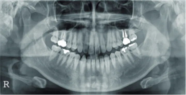
Height of residual bone prior to surgery: The vertical distance from the alveolar ridge to the most inferior sinus floor at the pro- jected implant placement site was measured using the panoramic radiograph prior to the surgery (Fig. 1).
Bone height after surgery: The distance from the neck of the implant fixture to the uppermost bone level above the implant fixture was measured. The enlargement ratio was calculated using the length of the implant placed, from every radiograph. By sub- tracting the bone height pre- and postsurgery, we calculated the grafted bone height (Fig. 2).
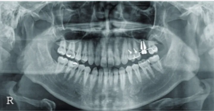
Bone height one year after surgery: The measurement was car- ried out from the neck of the implant fixture to the apex of the added bone graft material above the implant fixture in the pan- oramic radiograph taken during the 1-year follow up (±2 months) (Fig. 3).
Statistical analysis
An independent variable t-test was conducted using PASW Sta- tistics ver. 18.0 (SPSS Inc., Chicago, IL, USA), and data were analyzed to determine whether there were significant differences in variables Table 1. Length and diameter of implants used in groups I and II.
No. of implants
Group I (n=18) Group II (n=16) Implant length (mm)
8–8.5 10–11.5 12
4 14 0
3 12 1 Diameter (mm)
3.4–3.8 4–4.3 4.5–4.8 5–5.3 6
0 3 3 9 3
2 0 2 8 4
Group I: A group of patients who received autogenous tooth bone graft grafting during crestally approached sinus lifting surgery, group II: A group of patients who received synthetic bone (Osteon, Genoss, Suwon, Korea) grafting during crestally approached sinus lifting surgery.
Table 2. Implant placement area in groups I and II.
Area Group I (n=18) Group II (n=16)
Bicuspid 2 1
Molar 16 15
Group I: A group of patients who received autogenous tooth bone graft grafting during crestally approached sinus lifting surgery, group II: A group of patients who received synthetic bone (Osteon, Genoss, Suwon, Korea) grafting during crestally approached sinus lifting surgery.
Figure 2. The distance from the neck of the implant fixture to the upper most bone level above the implant fixture was measured to be 14.84 mm in this specific panoramic radiograph immediately after the implant surgery (black line).
Figure 1. The vertical distance from the alveolar ridge to the most inferior si- nus floor at the projected implant placement site was 7.45 mm in this specif- ic panoramic radiograph before the implant surgery (black line).
such as the degree of resorption between AutoBT or Osteon a year after surgery. A P-value of less than 0.05 was deemed statistically significant.
RESULTS
In group I, the average healing period between the first and the second operations was 3.61 months with an average prosthetics loading period of 18 months. Overall, the average follow-up period after surgery was 23.8 months. Before the operation, the distance between the alveolar crest and the sinus floor ranged from a mini- mum of 6.32 mm to a maximum of 12.10 mm, with a mean of 9.64 mm. AutoBT-added sinus floor lifting increased the bone height with a minimum of 2.62 mm to a maximum of 8.15 mm. The aver- age increased bone height was 4.89 mm. The analysis of the pan- oramic radiograph taken 1 year after surgery showed bone graft resorption ranging from 0.15 mm to 2.51 mm, with an average of 0.76 mm (Table 3). The periapical radiograph showed crest bone resorption of 0.07 mm 1 year after the prosthetics became func- tional.
In group II, the average healing period was 4.88 months with overall observation follow-up periods of 29.1 months after the sur- gery. The presurgery distance between the crest and the sinus floor ranged from a minimum of 6.04 mm to a maximum of 11.33 mm, with a mean of 9.22 mm. Sinus floor lifting resulted in minimum lifting of 3.19 mm, maximum lifting of 9.32 mm, and average lift- ing of 6.22 mm. The analysis of panoramic radiography conducted 1 year after surgery showed lifting reduction ranging from a mini- mum of 0.10 mm to a maximum of 1.50 mm, with an average of 0.53 mm (Table 3). The periapical radiograph showed crest bone resorption of 0.04 mm 1 year after the prosthetics became func- tional. In group II, one of the 16 implants failed due to mobility from the lack of osseointegration. The implant was removed and replaced with a new implant with a larger diameter and longer length. The subsequent osseointegration was successful.
The statistical analysis showed no statistically significant differ- ence between groups I and II in terms of the initial distance be-
tween the crest and the sinus floor (P=0.973) and the degree of lifting (P=0.460). In particular, the statistical analysis of the amount of bone-grafted material resorption between AutoBT and Osteon showed no statistically significant differences at the 1-year follow up (P=0.570).
DISCUSSION
Different types of sinus lifting techniques were applied depend- ing on the existing residual bone height. When the panoramic ra- diograph showed a residual bone height of less than 4 mm during the initial diagnosis, two-phase implant placement through sinus lifting via the lateral approach was recommended. If the residual bone height was between 4 mm and 6 mm, one-phase sinus lifting via the lateral approach was recommended. However, sinus lifting using the crestal approach was suggested, if the residual bone height was greater than 6 mm [5].
Once the Schneiderian membrane is lifted, the bone graft mate- rial is added to increase the bone height. Demineralized freeze-dried bone allograft has been actively used due to its osteoconductive and osteoinductive abilities. Additionally, xenografts such as Bio- Oss (Geistlich-Pharma AG, Wolhusen, Switzerland), which are osteo- conductive bovine-derived bones, are also widely used.
The synthetic bone graft material, Osteon is also one of the choic- es of grafting material. It consists of 30% β-TCP and 70% HA. It has a porous structure with 300- to 500-μm pores, which is similar to the human cancellous bone. Furthermore, it provides an environ- ment wherein an osteoblast can migrate into it. Studies have been conducted regarding Osteon’s utility as a bone graft material in si- nus floor lifting resulting in favorable outcomes [16,17].
The newly developed AutoBT is also gaining popularity in hospi- tal dental clinics and private practices when extraction is neces- sary. In an actual clinical setting, the clinician may decide on the appropriate form and size of the particles to be used. AutoBT can be processed as either powdered or block bone graft material.
Powdered bone graft particles can be made in sizes of 0.5–1 mm and 1–2 mm. The block bone graft form can be used in the hori- zontal and/or vertical augmentation of an alveolar ridge and ex- traction wound reconstruction [18]. The bone graft materials used Figure 3. The measurement was made from the neck of the implant fixture
to the apex of the added bone graft material above the implant fixture 1 year after surgery (±2 months). The vertical distance was measured as 13.25 mm in this specific panoramic radiograph (black line).
Table 3. Changes resulting from using either AutoBT or Osteon in crestally approached sinus lift.
Mean initial bone
height (mm) Mean increase in
bone height (mm) Mean resorption of bone height (mm) Tooth bone graft
material (AutoBT) 9.64 4.89 0.76
Synthetic bone graft
material (Osteon) 9.22 6.22 0.53
P-value* 0.973 0.460 0.570
Autogenous tooth bone graft (AutoBT) and Osteon (Genoss, Suwon, Korea).
*Statistically significantly different from bone resoprtion between two types of bone graft material (P<0.05).
in sinus floor lifting should show low levels of resorption to main- tain the stability of the implant over an extended period of time.
AutoBT made from crown mainly consists of highly crystalline calcium phosphate, resulting in slow resorption. Materials with a high crystalline content are not easily decomposed by osteoclasts, resulting in poor osteoconductive properties [19]. Meanwhile, Au- toBT made from the root has a low-crystalline structure. Low-crys- talline calcium phosphate is known to have osteoinductive and os- teoconductive healing tendencies [20]. Moreover, good bony re- modeling by osteoconduction can be expected because the main minerals of bone tissue are low-crystalline apatite as well [15].
Usually, an autogenous bone graft shows high bone resorption requiring more harvest volume at the donor site and a second op- eration [21]. Although AutoBT possesses osteoconductive and os- teoinductive potential in that the healing process is very similar to that of free autogenous bone grafts, it showed successful results in maintaining the graft volume until a year after surgery in the pres- ent study. The highly crystalline structure of the enamel portion in the AutoBT powder probably caused a slow resorption of the graft- ed material with a relatively slow remodeling process.
A previous study on sinus lifting using a crestal approach with an autogenous, allogenous, xenogenous, or synthetic bone graft material showed an average reduction of 0.62 mm in transplants during 6-month follow-up periods [22]. Another study on sinus lifting using the lateral approach with xenografts (Bio-Oss) result- ed in an average bone resorption of 1.8 mm a year after the sur- gery [23]. Although that study performed the lateral approach in- stead of the crestal approach used in the present study, AutoBT showed comparably less mean bone resorption.
However, the evaluation of bone resorption through panoramic radiography is a 2-dimensional evaluation and shows significant image magnification and/or distortion. Furthermore, accurately evaluating the degree of mineralization is difficult. Nevertheless, radiographs are still useful since they enable a general overview in the amount of bone and an evaluation of the form of the sinus [4].
Hence, the magnification of the digitalized panoramic radiograph was calculated using the actual length of the implant placed. This calculated magnified ratio was applied in measuring the initial bone height as well.
The small collected sample size and an evaluation of the bone resorption without a computed tomography (CT) scan are the limi- tations of the present study. Since it is a retrospective study, a CT scan evaluation could not be performed during the initial periods.
However, since this is the first study comparing AutoBT to the syn- thetic bone graft material in a crestally approached sinus lift, the present report is noteworthy.
Indeed, when AutoBT was used in crestally approached sinus floor lift procedures, similar levels of bone resorption were observed a year after surgery to the synthetic bone graft materials, Osteon. No serious complications or implant failures were noted with the use of AutoBT. Within the limitations of the current knowledge, AutoBT, with comparable clinical results to Osteon when used for crestally
approached sinus lift procedures with simultaneous implant place- ments, may replace the xenogenous, allogenous, and synthetic bone graft materials that are currently widely in use. Furthermore, with further investigations and improvements of the AutoBT graft material, we may expect to overcome clinical limitations such as bone resorption associated with the use of an autogenous bone graft alone in certain procedures.
CONFLICT OF INTEREST
No potential conflict of interest relevant to this article was re- ported.
ACKNOWLEDGEMENTS
This study was supported by a grant of the Korea Healthcare Technology R&D Project, Ministry of Health and Welfare, Republic of Korea (A102065).
ORCID
Young-Kyun Kim http://orcid.org/0000-0002-7268-3870 Junho Lee http://orcid.org/0000-0002-0009-441X Ji-Young Yun http://orcid.org/0000-0002-5634-5054 Pil-Young Yun http://orcid.org/0000-0001-6097-1229 In-Woong Um http://orcid.org/0000-0002-4628-3662
REFERENCES
1. Jaffin RA, Berman CL. The excessive loss of Branemark fixtures in type IV bone: a 5-year analysis. J Periodontol 1991;62:2-4.
2. Buchmann R, Khoury F, Faust C, Lange DE. Peri-implant conditions in periodontally compromised patients following maxillary sinus augmentation: a long-term post-therapy trial. Clin Oral Implants Res 1999;10:103-10.
3. Raghoebar GM, Timmenga NM, Reintsema H, Stegenga B, Vissink A. Maxillary bone grafting for insertion of endosseous implants:
results after 12-124 months. Clin Oral Implants Res 2001;12: 279- 86.
4. Kim SM, Park JW, Suh JY, Sohn DS, Lee JM. Bone-added osteo- tome technique versus lateral approach for sinus floor elevation:
a comparative radiographic study. Implant Dent 2011;20:465-70.
5. Summers RB. A new concept in maxillary implant surgery: the os- teotome technique. Compendium 1994;15:152, 154-6, 158 passim.
6. Zitzmann NU, Scharer P. Sinus elevation procedures in the re- sorbed posterior maxilla. Comparison of the crestal and lateral approaches. Oral Surg Oral Med Oral Pathol Oral Radiol Endod 1998;85:8-17.
7. Kim YK, Kim SG, Byeon JH, Lee HJ, Um IU, Lim SC, et al. Develop- ment of a novel bone grafting material using autogenous teeth.
Oral Surg Oral Med Oral Pathol Oral Radiol Endod 2010;109:
496-503.
8. Block MS, Kent JN. Sinus augmentation for dental implants: the use of autogenous bone. J Oral Maxillofac Surg 1997;55:1281-6.
9. Kim YK, Kim SG, Yun PY, Yeo IS, Jin SC, Oh JS, et al. Autogenous teeth used for bone grafting: a comparison with traditional grafting materials. Oral Surg Oral Med Oral Pathol Oral Radiol 2014;117:e39-45.
10. Bessho K, Tagawa T, Murata M. Purification of rabbit bone mor- phogenetic protein derived from bone, dentin, and wound tissue after tooth extraction. J Oral Maxillofac Surg 1990;48:162-9.
11. Urist MR, Nakata N, Felser JM, Nogami H, Hanamura H, Miki T, et al. An osteosarcoma cell and matrix retained morphogen for nor- mal bone formation. Clin Orthop Relat Res 1977;(124):251-66.
12. Urist MR, Mikulski A, Boyd SD. A chemosterilized antigen-extract- ed autodigested alloimplant for bone banks. Arch Surg 1975;110:
416-28.
13. Yeomans JD, Urist MR. Bone induction by decalcified dentine im- planted into oral, osseous and muscle tissues. Arch Oral Biol 1967;
12:999-1008.
14. Ritchie HH, Ritchie DG, Wang LH. Six decades of dentinogenesis research. Historical and prospective views on phosphophoryn and dentin sialoprotein. Eur J Oral Sci 1998;106 Suppl 1:211-20.
15. Kim YK, Kim SG, Oh JS, Jin SC, Son JS, Kim SY, et al. Analysis of the inorganic component of autogenous tooth bone graft mate- rial. J Nanosci Nanotechnol 2011;11:7442-5.
16. Kim YK, Yun PY, Lim SC, Kim SG, Lee HJ, Ong JL. Clinical evalua- tions of OSTEON as a new alloplastic material in sinus bone graft-
ing and its effect on bone healing. J Biomed Mater Res B Appl Biomater 2008;86:270-7.
17. Bae JH, Kim YK, Kim SG, Yun PY, Kim JS. Sinus bone graft using new alloplastic bone graft material (Osteon)-II: clinical evalua- tion. Oral Surg Oral Med Oral Pathol Oral Radiol Endod 2010;109:
e14-20.
18. Kim YK. Development of autogenous teeth bone graft material and clinical evaluation. J Korean Dent Assoc 2011;49:159-69.
19. Kim GW, Yeo IS, Kim SG, Um IW, Kim YK. Analysis of crystalline structure of autogenous tooth bone graft material: X-Ray dif- fraction analysis. J Korean Assoc Oral Maxillofac Surg 2011;37:
225-8.
20. Kim YK, Lee HJ, Kim KW, Kim SG, Um IW. Guide bone regenera- tion using autogenous teeth: case reports. J Korean Assoc Oral Maxillofac Surg 2011;37:142-7.
21. Zins JE, Whitaker LA. Membranous versus endochondral bone:
implications for craniofacial reconstruction. Plast Reconstr Surg 1983;72:778-85.
22. Lee JY, Kim YK, Bae JH, Kim SG. Clinical study of sinus membrane elevation using minimally invasive crestal approach. J Korean As- soc Oral Maxillofac Implantol 2008;12:4-16.
23. Kim YK, Yun PY, Kim SG, Kim BS, Ong JL. Evaluation of sinus bone resorption and marginal bone loss after sinus bone grafting and implant placement. Oral Surg Oral Med Oral Pathol Oral Radiol Endod 2009;107:e21-8.