http://dx.doi.org/10.12671/jkfs.2014.27.1.36
36
Copyright ⓒ 2014 The Korean Fracture Society. All rights reserved.
This is an Open Access article distributed under the terms of the Creative Commons Attribution Non-Commercial License (http://creativecommons.org/licenses/
by-nc/3.0) which permits unrestricted non-commercial use, distribution, and reproduction in any medium, provided the original work is properly cited.
Received November 12, 2012
Revised (1st) January 28, 2013, (2nd) May 12, 2013, (3rd) September 4, 2013
Accepted October 26, 2013
Address reprint requests to: Sang-Ho Lee, M.D.
Department of Orthopedic Surgery, Gwangmyeong Sungae Hospital, 36 Digital-ro, Gwangmyeong 423-711, Korea
Tel: 82-2-2680-7528ㆍFax: 82-2-2680-7755 E-mail: [email protected]
ITST 골수강내 정을 이용한 불안정 대퇴골 전자간 골절 치료에서 대전자부 분쇄 유무에 따른 치료 결과 비교
송경섭⋅이상호 ⋅정성훈⋅이수건⋅홍성하
광명성애병원 정형외과
Unstable Intertrochanteric Fracture Treated with ITST: A Comparative Study between Groups with and without Comminution of Greater Trochanter
Kyung-Sub Song, M.D., Sang-Ho Lee, M.D. , Seong-Hun Jeong, M.D., Su-Keon Lee, M.D., Sung-Ha Hong, M.D.
Department of Orthopedic Surgery, Gwangmyeong Sungae Hospital, Gwangmyeong, Korea
Purpose: To evaluate whether the radiological and clinical results of treatment with intertrochanteric/subtrochanteric (ITST) nail on unstable intertrochanteric fractures are combined with comminution of the greater trochanter or not.
Materials and Methods: We reviewed the results on 210 cases of unstable intertrochanteric fractures (grouped 88 patients with comminution of greater trochanter [GT] and 122 patients without comminution of GT) treated with ITST nail from January 2007 to October 2011, which was to be followed-up for more than 12 months.
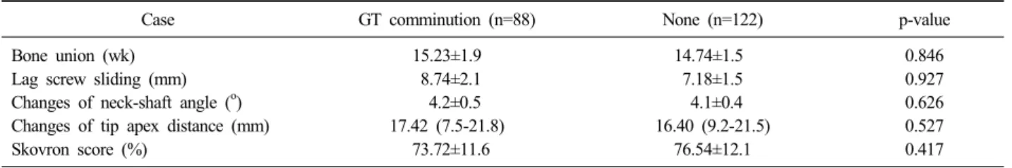
Results: The mean union time was 15.2 weeks in the study group (combined with comminution of GT). The mean union time was 14.7 weeks in control group (no comminution of GT). The lag screw sliding was 8.7 mm in the study group and 7.2 mm in the control group. Changes of neck-shaft angle was 4.2o in study group and 4.1o in control group. Tip-apex distance was 17.4 mm in study group and 16.4 mm in control group. The complications were 4 cases in each study group and control group. The clinical results checked by Skovron recovery scores decreased similarly in both groups, 73.7% in study group and 76.5% in control group. There were no significant differences in both groups according to radiological and clinical results.
Conclusion: The comminution of great trochanter does not affect on the radiological and clinical results when using the ITST nail of unstable intertrochanteric fractures.
Key Words: Femur, Unstable intertrochanteric fracture, ITST
서 론
골다공증을 가진 노령인구의 증가에 따라 대퇴골 전자간 골절의 빈도는 지속적으로 증가하고 있으며 분쇄가 심하여 불안정 골절이 많다8,10,13). 그동안 대퇴골 전자간 골절의 치 료 시에 압박 고나사, 골수정이 주로 사용되어 왔다. 골수 정을 통한 치료는 근위골편의 활강을 효과적으로 조절할 수 있고 골수 내로 삽입함으로써 생역학적 및 구조적으로 활강 고나사에 비해 안정적인 것으로 알려져 있으며1,6,11,14,15,19)
골절양상과 무관하게 수술 직후 적극적인 재활 및 조절적 인 체중 부하가 가능한 이점이 있어 유용하게 사용되고 있 다. 골수정의 이런 이점에도 불구하고 대퇴골 근위 간부 골절 및 지연 나사의 골두 천공 등의 고정 실패는 지속적 으로 보고되고 있고2,4,12,18,20,21,25)
골유합에 영향을 주는 인 자로 불안정 골절 양상, 심한 골다공증, 부적절한 정복 및 지연 나사의 위치 등이 있으며 이런 인자의 존재 시 고정 실패는 20% 이상으로 증가한다3,7,9,17,22,23,26)
. 이외에도 고전 자부 골절, 골수정 삽입구인 대전자부의 분쇄상, 내측 및 전방 피질골의 불완전한 정복 등이 고정 실패의 예에서 발 견되었다5). 최근의 대다수의 대퇴부 골수정의 치료는 앙와 위 상에서 비만 환자에게 유리하고 피부절개가 이상와 삽 입점보다 적은 대전자 삽입점을 이용하고 있다. 대전자부 의 분쇄가 심할 경우 삽입점을 정확히 촉지하기 어렵고 삽 입 시 분쇄가 더 심해져 골절 정복이 유지되지 않고 정확 한 골수정 삽입이 힘들다. 저자들은 금속정으로 불안정 대 퇴골 전자간 골절을 치료 시 대전자부 분쇄 유무에 따른 치료 결과 비교에 관한 보고가 거의 없어 이 두 군에 대한 임상적 및 방사선적 결과를 비교하고 차이점을 분석하고자 하였다.
대상 및 방법
1. 연구 대상
2007년 1월부터 2011년 10월까지 불안정 대퇴골 전자간 부 골절로 진단하여 intertrochanteric/subtrochanteric (ITST) 골수강내 정을 이용하여 치료받은 210예 중 대전자부 분쇄 가 있는 환자군 88예와 분쇄가 없는 환자군 122예를 대상 으로 하였다. 평균 연령은 74.2세(59-91세), 남녀의 성비는 5:7이었다. 추시 기간은 평균 14.9개월(12-19개월)이었다.
골절의 분류는 Evans 분류를 이용하여 안정성 골절과 불안 정성 골절로 나누었고 본 연구에 선택된 환자는 모두 불안 정성 골절이었다.
2. 수술 방법
한 명의 집도의에 의해 이루어졌고 ITST 골수강내 정을 사용하였다. 수술 방법은 모든 환자에서 골절 테이블을 이 용하여 앙와위 위치에서 반대측 다리는 쇄석위(lithotomy position)로 하였고 환측의 다리는 방사선 투시기가 투과할 수 있도록 하였다. 전후면 방사선상 골절부위 내측 피질골 의 중첩을, 측면 방사선상 전방 피질골의 중첩을 얻기 위 해 노력하였다. 지연 나사의 골두 내 위치 및 길이는 중앙 보다는 하방 및 후하방에 위치하도록 하였고 첨단-정점 거
리는 20 mm를 넘지 않도록 하였다. 모든 환자에서 비관혈 적 정복을 시행하였고 180 mm인 표준 ITST 골수강내 정 을 사용하였다. 재활치료는 술 후 골절부위 안정성 및 환 자의 전신상태에 따른 차이가 있었으나 원칙적으로 1-2주 간 체중 부하를 제한하고 이후 부분 체중 부하를 시행하였다.
3. 평가 방법
방사선적 추시상 골절선을 지나는 가골의 음영이 보이며 골소주의 재형성 및 피질골의 연속성을 기준으로 골유합을 판정하였다. 대퇴골 전자부의 함몰 정도는 수술 직후 및 최종 추시 사진의 전후면 방사선 사진상에서 지연나사의 활강거리를 측정하였고 지연 나사의 골두 내 위치 및 대퇴 골 경간각의 변화를 측정하였다. 방사선적 고정 실패는 10 도 이상의 경간각의 변화로 내반 변형을 보이거나 지연 나 사의 골두 천공 및 15 mm 이상의 과도한 활강이 있는 경 우로 정의하였다27). 임상적 결과로는 Skovron score를 Shon 등28)이 변형시킨 평가표에 따라 분석하였고 수상 전 과 술 후 12개월이 지난 후 기능상태를 백분율로 표시하였다.
통계 프로그램은 SPSS ver. 16.0.2 (SPSS Inc., Chicago, IL, USA)를 이용하였고, 결과에 대한 통계적 유의성은 in- dependent t-test을 이용하여 검정하였다.
결 과
총 210예의 불안정 대퇴골 전자간부 골절 중 대전자부 분쇄가 동반된 88예 중 4예(4.5%)에서 내반 변형과 지연나 사의 과도한 활강 등 불유합 소견이 관찰되었고 이를 제외 하고 평균 15.2주에 골유합을 보였다(Fig. 1). 분쇄가 없는 군에서는 4예(3%)에서 불유합 소견이 관찰되었고 평균 14.7주에 골유합을 관찰할 수 있었으며(Fig. 2) 통계적으로 유의한 차이는 보이지 않았다(p>0.05). 지연나사의 활강 정도는 분쇄가 있는 군에서 평균 8.7 mm, 없는 군에서 7.2 mm로 통계적 유의성은 없었으며(p>0.05) 대퇴경간각 변화는 각각 평균 4.2o, 4.1o로 통계적 유의성은 없었다 (p>0.05). 첨단-정점 거리 또한 각각 평균 17.4 mm, 16.4 mm로 측정되어 통계적으로 의의가 없었다(p>0.05). 수술 후 기능적 회복 지수 평가는 Shon 등28)이 변형시킨 평가 표에 따라 수술 후 12개월째 외래 추시 중 문진이나 전화 통화를 통해 평가했으며 수상 전 91%에서 술 후 12개월째 각각 평균 76.5%, 73.7%로 두 군 사이에 유사한 하락을 보였다(Table 1). 합병증은 추시 중 방사선 결과로 평가했 으며 대전자부 분쇄골절이 동반된 군에서는 총 4예 중 내 반 변형이 1예, 지연 나사의 과도한 활강이 3예였고, 분쇄 가 없는 군에서는 총 4예 중 내반 변형이 2예, 지연 나사
Fig. 1. (A) Preoperative radiographs of a 74-year-old female show an unstable intertrochanteric fracture with comminution of the greater trochanter. (B) Preoperative computed tomography shows comminution on the greater trochanter. (C) Postoperative 4-months radiographs show the complete union without deformity.
Fig. 2. (A) Preoperative radiographs of a 61-year-old female show an unstable intertrochanteric fracture without comminution of the greater trochanter. (B) Preoperative computed tomography shows no comminution on the greater trochanter. (C) Postoperative 2-months radiographs show the complete union without deformity.
Table 1. Comparison between Presence of GT Comminution or without in Unstable Intertrochanteric Fracture
Case GT comminution (n=88) None (n=122) p-value
Bone union (wk) Lag screw sliding (mm) Changes of neck-shaft angle (o) Changes of tip apex distance (mm) Skovron score (%)
15.23±1.9 8.74±2.1 4.2±0.5 17.42 (7.5-21.8)
73.72±11.6
14.74±1.5 7.18±1.5 4.1±0.4 16.40 (9.2-21.5)
76.54±12.1
0.846 0.927 0.626 0.527 0.417 Values are presented as mean±standard deviation or median (range). GT: Greater trochanter.
의 과도한 활강이 2예였다. 내반 변형이 있던 3예에서 전 신 상태가 양호하지 못한 2예를 제외하고 1예에서 이극성 인공관절 반치환술로 재치환술을 시도하였다. 과도한 활강 으로 인한 하지 단축 및 대퇴 전자 외측부 동통을 호소한 5예 중 3예에서 골유합 시점 이후 내고정술 제거술을 시행 하였고 2예에서 이극성 인공 관절 반치환술을 시행하였다.
고 찰
대퇴골 전자간부 골절의 치료로서 골수정은 다른 치료에 비해 수술시간, 출혈량, 하지 길이 단축 및 재원기간을 줄 여준다는 장점과 더불어 구조적 및 생역학적으로 안정적이 라고 알려져 유용하게 사용되고 있다. 이러한 이점에도 불 구하고 대퇴골 근위간부 골절 및 지연 나사의 골두 천공과 같은 고정실패는 지속적으로 보고되고 있다2,4,12,18,20,21,25)
. 지 연 나사의 골두 천공은 본 연구에서는 보고되지는 않았지만 0%-11.8%까지 보고되고 있으며, 주로 지연 나사의 대퇴골 두 내 부적절한 위치 및 불량한 정복에 기인하는 것으로16,24) 고정실패를 초래하는 가장 중요한 합병증으로 알려져 있고, 이외에 고전자부 골절, 대전자부의 분쇄상, 내측 및 전방 피질골의 불완전한 해부적 정복 등이 추가적인 위험인자가 될 수 있음이 보고되었다5).
대전자부의 분쇄상이 동반된 경우 상부에 근위골편의 활 강에 따른 지지대가 단단한 원통형의 골수정 외에는 없기 때문에 고정 실패의 가능성이 높아지고 고전자부 골절이 동반된 경우에는 골수정 삽입 시 대퇴경부 기저부 골절 형 태로 전환되어 지연 나사의 축을 따라 근위 골편의 회전에 의한 고정 실패의 위험도가 증가할 것으로 보고된 바가 있 다6). 또한 골수정 삽입 전 유도핀 삽입 시 기준점을 잡기 가 힘들고 술자가 원하는 삽입 방향으로 확공 시에 분쇄가 더 심해져 정복이 유지가 안되는 어려움이 발생한다. 본 연구에서 사용한 ITST 골수강내 정의 경우 회전 방지핀이 나 근위 마개를 사용하여 골두 경부 골편의 회전과 붕괴를 방지할 수 있고 근위 골편의 활강을 조절할 수 있다는 점16) 에서 대전자부의 분쇄가 동반 시 고정실패의 위험을 줄일
수 있었던 것으로 생각한다.
저자들의 연구에서는 대상 환자군의 크기가 충분히 크지 않고 짧은 추시 기간 및 고정 실패의 기전 분류에 있어 다 른 기전의 동반 및 혼재 여부를 확인하지 못한 것 등에서 한계가 있으며 앞으로 더 많은 환자에 대한 장기 추시 및 추가적인 연구가 필요할 것으로 생각한다.
결 론
대퇴골 전자간부 골절 환자에서 골수정 삽입술 시 고정 실패의 위험인자를 술 전 정확히 파악하는 것이 중요하다.
ITST 골수강내 정을 이용한 대퇴골 전자간 골절에서 대전 자부의 분쇄 유무는 임상적, 방사선적 결과에 영향을 미치 지 않았다. 그러므로 대전부의 분쇄가 동반된 대퇴골 전자 간 골절에서 ITST 골수강내 정은 유용한 치료 방법이라고 생각한다.
References
1) Boriani S, Bettelli G, Zmerly H, et al: Results of the multicentric Italian experience on the Gamma nail: a re- port on 648 cases. Orthopedics, 14: 1307-1314, 1991.
2) Bridle SH, Patel AD, Bircher M, Calvert PT: Fixation of intertrochanteric fractures of the femur. A randomised prospective comparison of the gamma nail and the dynam- ic hip screw. J Bone Joint Surg Br, 73: 330-334, 1991.
3) Brink PR, Bolhuis RJ, Runne WC, De Vries AC: Low nail-plate fixation and early weight-bearing ambulation for stable trochanteric fractures. J Trauma, 27: 491-495, 1987.
4) Butt MS, Krikler SJ, Nafie S, Ali MS: Comparison of dynamic hip screw and gamma nail: a prospective, randomized, controlled trial. Injury, 26: 615-618, 1995.
5) Chang JD, Kim TY, Hwang JH, Min SK, Yoo JH:
Analysis of the fixation failure in intertrochanteric hip fractures treated with hip nailing. J Korean Fract Soc, 25:
169-176, 2012.
6) Davis J, Harris MB, Duval M, D'Ambrosia R: Pertro- chanteric fractures treated with the Gamma nail: technique and report of early results. Orthopedics, 14: 939-942, 1991.
7) Davis TR, Sher JL, Horsman A, Simpson M, Porter BB, Checketts RG: Intertrochanteric femoral fractures.
Mechanical failure after internal fixation. J Bone Joint Surg Br, 72: 26-31, 1990.
8) Dimon JH, Hughston JC: Unstable intertrochanteric frac- tures of the hip. J Bone Joint Surg Am, 49: 440-450, 1967.
9) Doherty JH Jr, Lyden JP: Intertrochanteric fractures of the hip treated with the hip compression screw: analysis of problems. Clin Orthop Relat Res, (141): 184-187, 1979.
10) Evans EM: The treatment of trochanteric fractures of the femur. J Bone Joint Surg Br, 31: 190-203, 1949.
11) Geller JA, Saifi C, Morrison TA, Macaulay W: Tip- apex distance of intramedullary devices as a predictor of cut-out failure in the treatment of peritrochanteric elderly hip fractures. Int Orthop, 34: 719-722, 2010.
12) Goldhagen PR, O'Connor DR, Schwarze D, Schwartz E: A prospective comparative study of the compression hip screw and the gamma nail. J Orthop Trauma, 8:
367-372, 1994.
13) Guyton JL: Intertrochanteric fractures of the hip. In:
Canale ST, Campbell WC eds. Campbell's operative orthopaedics. 9th ed. St. Louis, Mosby: 2188-2190, 1998.
14) Halder SC: The Gamma nail for peritrochanteric fractures.
J Bone Joint Surg Br, 74: 340-344, 1992.
15) Jeon HS, Park BM, Song KS, Kim HG, Yun JJ: The comparison between ITST (TM) (intertrochanteric/subtro- chanteric) & DHS (dynamic hip screw) in unstable femur intertrochanteric fracture. J Korean Fract Soc, 22: 131- 137, 2009.
16) Kawaguchi S, Sawada K, Nabeta Y: Cutting-out of the lag screw after internal fixation with the Asiatic gamma nail. Injury, 29: 47-53, 1998.
17) Larsson S, Friberg S, Hansson LI: Trochanteric fractures.
Influence of reduction and implant position on impaction and complications. Clin Orthop Relat Res, (259): 130-139, 1990.
18) Leung KS, So WS, Shen WY, Hui PW: Gamma nails and dynamic hip screws for peritrochanteric fractures. A randomised prospective study in elderly patients. J Bone Joint Surg Br, 74: 345-351, 1992.
19) Lindsey RW, Teal P, Probe RA, Rhoads D, Davenport S, Schauder K: Early experience with the gamma inter- locking nail for peritrochanteric fractures of the proximal femur. J Trauma, 31: 1649-1658, 1991.
20) Liu M, Yang Z, Pei F, Huang F, Chen S, Xiang Z: A meta-analysis of the Gamma nail and dynamic hip screw in treating peritrochanteric fractures. Int Orthop, 34: 323- 328, 2010.
21) Mahomed N, Harrington I, Kellam J, Maistrelli G, Hearn T, Vroemen J: Biomechanical analysis of the Gamma nail and sliding hip screw. Clin Orthop Relat Res, (304): 280-288, 1994.
22) Mainds CC, Newman RJ: Implant failures in patients with proximal fractures of the femur treated with a sliding screw device. Injury, 20: 98-100, 1989.
23) Mulholland RC, Gunn DR: Sliding screw plate fixation of intertrochanteric femoral fractures. J Trauma, 12: 581- 591, 1972.
24) Nishiura T, Nozawa M, Morio H: The new technique of precise insertion of lag screw in an operative treatment of trochanteric femoral fractures with a short intramedullary nail. Injury, 40: 1077-1083, 2009.
25) Radford PJ, Needoff M, Webb JK: A prospective rand- omised comparison of the dynamic hip screw and the gamma locking nail. J Bone Joint Surg Br, 75: 789-793, 1993.
26) Sernbo I, Johnell O, Gentz CF, Nilsson JA: Unstable intertrochanteric fractures of the hip. Treatment with Ender pins compared with a compression hip-screw. J Bone Joint Surg Am, 70: 1297-1303, 1988.
27) Shin DK, Kwun KW, Kim SK, Lee SW, Choi CH, Kim KM: Proximal Femoral Nail (PFN) for Femur Intertro- chanteric Fracture. J Korean Fract Soc, 15: 328-335, 2002.
28) Shon WY, Park JH, Kil KH, Jeon SJ, Suh SW:
Functional recovery after operative treatment of hip frac- tures in the elderly. J Korean Orthop Assoc, 33: 968-973, 1998.
Copyright ⓒ 2014 The Korean Fracture Society. All rights reserved.
This is an Open Access article distributed under the terms of the Creative Commons Attribution Non-Commercial License (http://creativecommons.org/licenses/
by-nc/3.0) which permits unrestricted non-commercial use, distribution, and reproduction in any medium, provided the original work is properly cited.
http://dx.doi.org/10.12671/jkfs.2014.27.1.36
ITST 골수강내 정을 이용한 불안정 대퇴골 전자간 골절 치료에서 대전자부 분쇄 유무에 따른 치료 결과 비교
송경섭⋅이상호 ⋅정성훈⋅이수건⋅홍성하
광명성애병원 정형외과
목 적: 골수강내 정을 이용한 불안정 대퇴골 전자간 골절 치료 시 대전자부 분쇄 유무에 따른 치료 결과를 비교, 분석하고자 하였다.
대상 및 방법: 2007년 1월부터 2011년 10월까지 ITST 골수강내 정으로 불안정 대퇴골 전자간 골절로 치료받고 12개월 이상 추시 관찰이 가능했던 210예 중 일반 방사선 사진에서 대전자부 분쇄(골절선이 두 개 이상)가 있는 군(88예), 없는 군(122예)으 로 나누어 이 두 군의 치료 결과를 임상적 그리고 방사선적으로 비교, 분석하였다.
결 과: 평균 골유합 시기는 분쇄가 있는 경우와 없는 경우 각각 15.2주, 14.7주가 걸렸고 지연 나사의 활강은 각각 8.8 mm, 7.2 mm였다. 대퇴골 경간각 변화는 각각 4.2o, 4.1o로 측정되었고 첨단-정점 거리는 각각 17.4 mm, 16.4 mm였다. 합병증은 두 군 모두 4예였다. 임상적 결과로 Skovron 기능적 회복 지수 평가표에서 두 군 간에 73.7%, 76.5%로 유사한 하락을 보였다.
방사선 및 임상적 결과에서 두 군 간의 유의한 차이는 없었다.
결 론: 대전자부 분쇄 유무는 ITST 골수강내 정을 이용한 불안정 대퇴골 전자간 골절 치료에서 임상적, 방사선적 결과에서 영향을 미치지 않았다.
색인 단어: 대퇴골, 불안정 전자간 골절, ITST 골수강내 정
접수일 2012. 11. 12 수정일 1차 2013. 1. 28, 2차 2013. 5. 12, 3차 2013. 9. 4 게재확정 2013. 10. 26 교신저자 이 상 호
광명시 디지털로 36, 광명성애병원 정형외과
Tel 02-2680-7528, Fax 02-2680-7755, E-mail [email protected]
41