19
<접수일:2010년 6월 21일, 수정일:2010년 9월 3일, 심사통과일:
2010년 10월 23일>
통신저자:김 완 욱
경기도 수원시 팔달구 지동 93번지 성빈센트병원 류마티스내과 E-mail:[email protected]
한국인 전신홍반루푸스 환자에서 골밀도 감소의 위험인자
박윤정ㆍ박보형ㆍ민도준ㆍ김완욱 가톨릭대학교 의과대학 내과학교실
Risk Factors for Low Bone Mineral Density in Korean Patients with Systemic Lupus Erythematosus
Yun-Jung Park, Bo-Hyoung Park, Do-June Min, Wan-Uk Kim
Division of Rheumatology, Department of Internal Medicine, The Catholic University of Korea, Suwon, Korea
Objective. To determine the degree and risk factors for de- creased bone mineral density (BMD) in patients with sys- temic lupus erythematosus (SLE).
Methods. One hundred and one patients with SLE and 57 age- and gender- matched healthy controls were enrolled in this study. The BMD was measured by dual energy X-ray absorptiometry (DXA). The laboratory findings and clinical variables evaluated in the SLE patients consisted of disease duration, SLE disease activity index (SLEDAI), and medications, including mean and cumulative dose of glucocorticoid. At the time of the clinical and laboratory assessment, the levels of serum osteocalcin, serum FSH/
LH, urine deoxypyridinoline (DPD), and serum cytokines, such as IL-6 and soluble receptor activator of NF-kB li- gand (RANKL), were determined in SLE patients using a enzyme-linked immunosorbent assay.
Results. The BMD T score decreased in patients with SLE compared to the healthy controls (−1.11 versus −0.41, p=0.001 at lumbar spine, −0.84 versus −0.01, p<0.001
at femur neck, −1.20 versus −0.45, p<0.001 at total hip, respectively). Osteoporosis and osteopenia was present in 16.8% and 46.5% of patients, respectively. Multiple re- gression analysis revealed a low BMD in the lumbar spine to be associated with increased FSH, low BMI and cumu- lative glucocorticoid dose. A low BMD in the hip and fe- mur neck was associated with increased FSH, low BMI, and duration of glucocorticoid. On the other hand, the lev- els of osteocalcin, deoxypyridinoline (DPD), IL-6, and solu- ble RANKL were similar in patients with a low BMD and those with normal BMD.
Conclusion. Osteoporosis and osteopenia are more com- mon in young Korean SLE patients than in control sub- jects. Elevated FSH, low BMI, and the use of glucocorti- coid are independent risk factors linked to a decreased BMD in Korean patients with SLE.
Key Words. Systemic lupus erythematosus, Bone mineral density, FSH, BMI, Glucocorticoi
서 론
전신홍반루푸스(이하 루푸스) 는 지난 10년간 진단과 치 료법에서 많은 발전이 있었고 그 결과 사망률을 현저히 줄 어들어 과거에는 5년 생존율이 51%정도였으나 최근에는 10년 생존율이 90%정도로 향상되었다 (1-3). 루푸스 환자 의 생존율이 증가됨에 따라, 동맥경화증, 암 등과 같은 장 기 합병증이 증가하는 추세이며, 골다공증 역시 골절 및 사망의 위험도가 높은 장기 합병증의 하나로서 이에 대한
관심이 점점 더 커지고 있다 (4-6).
골다공증은 대사성 골질환 중의 하나로 골강도의 손상으 로 인한 골절의 위험이 증가되는 골격계 질환이다. 골강도 는 골밀도와 골의 질로 결정이 되며, 골질의 감소와 관련 하여 조절 불가능한 위험 인자들로는 나이, 성별, 골절의 가족력 등의 유전적 및 환경적 요소들이 있다 (7,8). 이외 에도 에스트로겐의 감소, 칼슘섭취나 비타민 D의 부족, 육 체적 활동이나 체중의 감소, 흡연 및 만성적 당질코르티코 이드 사용 등과 같은 조절 가능한 위험인자들도 골다공증 발생에서 중요한 역할을 한다 (9-14). 류마티스관절염에서 의 만성 염증 상황도 골밀도 감소에서 중요한 역할을 한다 (15-18). 만성 염증성 질환의 하나인 루푸스 역시 골다공증 의 위험이 증가된다고 알려져 있다 (19- 22). 루푸스에서 골다공증은 당질코르티코이드 사용, 염증성 사이토카인에 지속적인 노출, 조기 폐경, 광과민성으로 인한 햇빛 노출 의 감소 등과 관련되어 있을 것으로 추정된다. 여러 가지 요인들이 복합적으로 작용할 것으로 생각되지만 아직까지 그 정확한 원인 및 기전에 대해서는 불분명하다 (22-25).
루푸스나 골다공증은 모두 그 발병 양상에 있어서 인구 학적, 유전학적인 차이를 보이는 질환이다. 그럼에도 불구 하고 루푸스 환자들을 대상으로 하는 골다공증 및 골밀도 감소에 관한 연구는 주로 해외에 국한되어 있으며, 우리나 라 환자들을 대상으로 한 연구는 없는 실정이다. 이에 저자 들은 한국인 루푸스 환자에서 골밀도 감소의 정도를 결정 하고 골밀도 감소와 관련된 위험인자들에 관해 알아보고 자 하였다.
대상 및 방법 대상 환자 및 대조군
2007년 1월부터 2010년 4월까지 내원한 환자 중 1982년 미국 류마티스학회에서 정한 진단기준을 만족하는 루푸스 환자들 중 101명을 대상으로 하였고 (26), 연령과 성별이 부합된 건강한 정상인을 57명을 대조군으로 비교하였다.
흡연력이 있거나, 알코올중독, 만성 염증성장질환, 종양, 급 만성 감염으로 치료 중이거나, 쿠싱증후군, 비스포스포 네이트나 여성호르몬 제제를 복용 중인 환자들은 연구에 서 제외하였다. 정상 생리가 끝나고 12개월이 지난 경우를 폐경이라고 정의하였고, 우리나라 평균 폐경 나이가 50.2
±3.7세임을 고려하여 (27), 46.5세 이전에 폐경 된 경우를 조기 폐경이라고 정의하였다. 24시간 소변채집에서 0.5 g 이상의 지속적 단백뇨 존재가 있거나, 세포성 원주가 존재 하거나, 신조직 검사로 확인된 경우 루푸스 신염으로 정의 하였다. 골밀도 검사 시 대상 환자들의 나이, 키, 체중, 신 체활동 여부, 당뇨병 유무, 갑상선 기능저하증 유무 등이 조사되었고, 당뇨와 갑상선 기능 이상이 있는 경우 연구에
서 제외하였다. 말초혈액 검사, 혈청 생화학 검사, 혈청 보 체 검사, 항핵항체 검사, 항 dsDNA 항체 검사, 소변 검사, 루푸스의 이환 기간, SLE disease activity index (SLEDAI) (28), 약물 복용력도 함께 조사하였다. 당질코르티코이드 는 사용 여부 및 용량, 총 누적 사용기간 및 용량을 모두 조사하였고, 면역억제제, 칼슘보충제, 항응고제 복용 여부 도 함께 조사되었다. 본 연구는 병원 윤리심의위원회의 승 인을 받았다.
골밀도 검사(Bone densitometry: BMD)
척추(요추1번에서 4번까지)와 대퇴골(대퇴경부(femoral neck), 대전자(trochanter)와 전자간(intertrochanter))에서 골 밀도를 측정하였다. 골밀도의 측정은 이중에너지 방사선 흡수법(dual energy X-ray absorptiometry; Delphi-W, Hologic) 으로 숙련된 검사자가 시행하였다. 척추 골밀도는 요추 1 번부터 4번까지 측정하여 평균치를 요추 골밀도로 분석하 였고, 대퇴골에서는 경부, 대전자, 전자간에서 측정하였다.
T-점수는(환자 BMD-성별 부합 대조군의 최대 BMD)/성별 부합 대조군의 최대BMD의 표준편차로 정의하였다. 세계 보건기구 (WHO)의 정의에 따라 −1에서 −2.5 이내의 T- 점수를 보이는 경우 골감소증으로, −2.5 이하의 T-점수를 보이는 경우 골다공증으로 하였다 (29).
생화학적 골표지자 검사 및 호르몬 검사
환자들은 골밀도 검사와 동시에 골흡수의 생화학적 표지 자로서 deoxypyridinoline (DPD)과 골형성의 생화학적 표지 자로서 osteocalcin이 각각 소변과 혈청에서 8시간 금식 후 아침에 측정되었다. 또한 골형성과 흡수의 조절인자로서 난포자극 호르몬(follicle stimulating hormone: FSH)과 황체 형성호르몬(luteinizing hormone: LH), 사이토카인으로 os- teoprotegerin (OPG), soluble receptor activator of NF-kB li- gand (sRANKL). interleukin-6 (IL-6) 등이 효소면역검사법 (enzyme-linked immunosorbent assay, ELISA)방법으로 혈청 내에서 8시간 금식 후 아침에 측정되었다.
통계분석
결과가 정규분포를 따르는 경우는 평균±표준편차로 표 시하였고, 정규분포를 따르지 않는 경우는 중앙값[범위]로 표시하였다. 평균값의 비교는 따라 Independent t-test 또는 Mann-Whitney U test, ANOVA 또는 Kruskal Wallis test를 시행하였고, 교차분석은 카이제곱 검정(chi-square test) 또 는 Fisher’s exact test를 시행하였다. 상관분석은 Pearson 상 관분석을 하였으며, p<0.05일 때 통계적으로 유의 하다고 판정하였다. 다중회귀분석은 단계별 선택방법(stepwise conditional modeling)을 사용하였고, 모델에 포함하는 변수
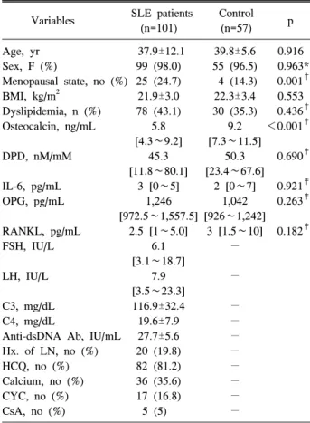
Table 1. Characteristics of the SLE patients and age-and gender-matched control subjects
SLE patients Control
Variables p
(n=101) (n=57)
Age, yr 37.9±12.1 39.8±5.6 0.916
Sex, F (%) 99 (98.0) 55 (96.5) 0.963*
Menopausal state, no (%) 25 (24.7) 4 (14.3) 0.001†
BMI, kg/m2 21.9±3.0 22.3±3.4 0.553
Dyslipidemia, n (%) 78 (43.1) 30 (35.3) 0.436†
Osteocalcin, ng/mL 5.8 9.2 <0.001‡
[4.3∼9.2] [7.3∼11.5]
DPD, nM/mM 45.3 50.3 0.690‡
[11.8∼80.1] [23.4∼67.6]
IL-6, pg/mL 3 [0∼5] 2 [0∼7] 0.921‡
OPG, pg/mL 1,246 1,042 0.263‡
[972.5∼1,557.5] [926∼1,242]
RANKL, pg/mL 2.5 [1∼5.0] 3 [1.5∼10] 0.182‡
FSH, IU/L 6.1 −
[3.1∼18.7]
LH, IU/L 7.9 −
[3.5∼23.3]
C3, mg/dL 116.9±32.4 −
C4, mg/dL 19.6±7.9 −
Anti-dsDNA Ab, IU/mL 27.7±5.6 −
Hx. of LN, no (%) 20 (19.8) −
HCQ, no (%) 82 (81.2) −
Calcium, no (%) 36 (35.6) −
CYC, no (%) 17 (16.8) −
CsA, no (%) 5 (5) −
The data is reported as mean±SD or median [interquartile range:
IQR]. BMI: body mass index, DPD: deoxypyridinoline, IL-6:
interleukin-6, OPG: osteoprotegerin, RANKL: receptor activator NF-kB ligand, FSH: follicule stimulating hormone, LH:
luteinizing hormone, LN: lupus nephritis, HCQ: hydroxychlo- roquine, CYC: cyclophosphamide, CsA: cyclosporine A. Analy- sis was done by Independent t-test, *Fisher’s exact test, †Pearson Chi-Square, ‡Mann-Whitney U test
Figure 1. Bone mineral density at different sites in the patients with SLE and age-and gender-matched healthy controls. The BMD T-score was significantly lower in the SLE patients than in the healthy controls (−1.11±0.13 versus −0.41±0.15, p=0.001 at lumbar spine, −0.84±0.11 versus −0.01±0.15, p<0.001 at femur neck, −1.20±0.11 vs −0.45±0.14, p<0.001 at total hip, respectively). The data is reported as the mean±SEM.
의 기준은 p<0.05로, 제외하는 변수 기준은 p>0.1로 정하 였다.
결 과 대상 환자의 특성
전체 연구 대상 루푸스 환자의 평균 나이는 37.9± 12.1세 이었고, 질병 이환 기간은 평균 43.8±5.4개월이었다. 루푸 스 환자 중 폐경 후 여성은 25명(조기폐경 16명)이었고, 루 푸스 신염으로 치료받은 환자들은 20명이었다. 대조군의 평균나이는 39.8±5.6이었고, 폐경 된 여성은 4명(조기폐경 0명)이었다. 전체 루푸스 환자의 질환의 활성도(SLEDAI)는 2점으로 대부분의 루푸스 환자들이 질병이 잘 조절되는 상태에 있었다. 평균 당질코르티코이드 사용기간은 43.8±
5.4개월이었으며, 총 누적 용량의 중앙값은 2.5 [2.5∼7.5]
g이었다. 골밀도 측정 당시 루푸스 환자들의 당질코르티
코이드 사용 중앙값은 5.0 [2.5∼7.5] mg이었고, 총 누적 용 량을 사용기간으로 나눈 평균 일일 사용량(mean daily dose)의 중앙값은 5.4 [2.5∼8.0] mg 였다. 표 1에는 대상 루푸스 환자 및 대조군의 인구학적 특징들이 기술되어 있 다.
골밀도 검사
루푸스 환자군이 대조군에 비해 나이가 적은 경향을 보 임에도 불구하고 골밀도 결과는 측정한 모든 부위에서 유 의하게 감소되어 있었다(루푸스 vs 대조군 척추골밀도 T 점수: −1.11 vs −0.41; p=0.001, 대퇴경부: −0.84 vs −0.01;
p<0.001, 대퇴골전체: −1.20 vs −0.45; p<0.001; 그림 1).
루푸스 환자 군에서 폐경 된 여성의 수가 더 많았으므로 폐경 된 여성들을 제외하고 분석하여도, 세 부위의 골밀도 는 루푸스 환자에서 모두 감소되어 있었다(루푸스 vs 대조 군 척추 골밀도 T점수: −0.83 vs −0.32; p=0.045, 대퇴경 부: −0.71 vs −0.01; p=0.001, 대퇴골: −1.02 vs −0.44;
p=0.005). 골다공증과 골감소증에 관한 세계보건기구 (WHO)의 정의에 입각하여 볼 때 대조군의 경우 골다공증 4명(7%), 골감소증이 25명(43.9%)이 발견되었고 루푸스 환 자의 경우 17명(16.8%)이 골다공증, 47명(46.5%)이 골감소 증으로 조사되었다. 루푸스 환자들의 나이가 많을수록, 질 병 이환 기간이 길수록, 당질코르티코이드 사용이 기간이 길고 그 누적 용량이 많을수록 골밀도가 보다 감소함이 관 찰되었다(표 2).
골표지자 및 호르몬수치
FSH는 골다공증을 보이는 군에서는 28.9로 정상골밀도
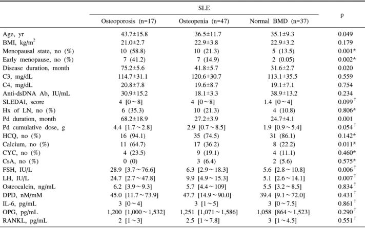
Table 2. Comparison of the clinical and laboratory factors according to the mineral density (T-score) in SLE patients SLE
p Osteoporosis (n=17) Osteopenia (n=47) Normal BMD (n=37)
Age, yr 43.7±15.8 36.5±11.7 35.1±9.3 0.049
BMI, kg/m2 21.0±2.7 22.9±3.8 22.9±3.2 0.179
Menopausal state, no (%) 10 (58.8) 10 (21.3) 5 (13.5) 0.001*
Early menopause, no (%) 7 (41.2) 7 (14.9) 2 (0.05) 0.002*
Disease duration, month 75.2±5.6 41.8±5.7 31.6±2.7 0.020
C3, mg/dL 114.7±31.1 120.6±30.7 113.1±35.5 0.559
C4, mg/dL 20.8±7.8 19.6±8.7 19.1±7.1 0.754
Anti-dsDNA Ab, IU/mL 30.9±15.2 18.1±3.3 38.9±13.2 0.234
SLEDAI, score 4 [0∼8] 4 [0∼8] 1.4 [0∼4] 0.099†
Hx of LN, no (%) 6 (35.3) 10 (21.3) 4 (10.8) 0.806*
Pd duration, month 68.2±18.9 27.2±3.9 24.7±4.1 0.001
Pd cumulative dose, g 4.4 [1.7∼2.8] 2.9 [0.7∼8.5] 1.9 [0.9∼5.4] 0.054†
HCQ, no (%) 16 (94.1) 35 (74.5) 31 (86.1) 0.142*
Calcium, no (%) 11 (64.7) 17 (36.2) 8 (22.2) 0.011*
CYC, no (%) 4 (23.5) 9 (19.1) 4 (11.1) 0.460*
CsA, no (%) 0 (0) 3 (6.4) 2 (5.6) 0.575*
FSH, IU/L 28.9 [3.7∼76.6] 6.3 [2.9∼18.3] 5.6 [2.8∼10.8] 0.006†
LH, IU/L 24.7 [2.7∼47.8] 9.9 [4.9∼15.3] 5.1 [2.6∼14.1] 0.007†
Osteocalcin, ng/mL 6.2 [3.9∼9.3] 5.7 [4.4∼109] 5.5 [3.2∼8.5] 0.834†
DPD, nM/mM 45.0 [11.7∼73.9] 47.7 [14.9∼90.0] 39.4 [9.1∼72.0] 0.431†
IL-6, pg/mL 3 [0∼4] 3 [1∼5] 3 [0∼7.5] 0.861†
OPG, pg/mL 1,200 [1,000∼1,532] 1,251 [1,071∼1,586] 1,058 [864∼1,523] 0.290†
RANKL, pg/mL 2 [1∼3] 2.5 [1∼7.8] 3 [1∼4.5] 0.551†
The data is reported as mean±SD or median [interquartile range: IQR]. BMI: body mass index, SLEDAI: SLE disease activity index, LN: lupus nephritis, HCQ: hydroxychloroquine, CYC: cyclophosphamide, CsA: cyclosporine A, FSH: follicule stimulating hormone, LH:
luteinizing hormone, DPD: deoxypyridinoline, IL-6: interleukin-6, OPG: osteoprotegerin, RANKL: receptor activator NF-kB ligand.
Analysis was done by ANOVA (linearity test), *Pearson Chi-Square, †Kruskal-Wallis test
를 보이는 군 5.6에 비하여 5배 이상 증가되어 있었고, LH 역시 골다공증을 동반한 환자 군에서 24.7로 정상 골밀도 를 보인 환자군 5.1에 비해 4.8배 증가되어 있었다(표 2).
조기폐경이 아닌 환자들을 제외하였을 경우에도 FSH는 골다공증을 보이는 군에서는 20.3로 정상 골밀도를 보이 는 군 11.5에 비하여 1.8배 증가되어 있었고, LH 역시 골다 공증을 동반한 환자 군에서 27.8로 정상 골밀도를 보인 환 자군 10.6에 비해 2.6배 증가되어 있었다. 그러나 골 교체 표지자인 osteocalcin, DPD 농도와, IL-6, OPG, RANKL농도 를 골밀도 검사 시 채취한 환자의 시료(혈청과 소변)에서 측정한 결과 정상 골밀도를 보였던 환자와 골다공증, 골감 소증을 가지고 있는 환자간에 차이를 보이지 않았다.
골밀도 감소와 관련된 독립된 위험인자 분석
다음으로 저자들은 골밀도 검사의 T-점수를 독립변수로 하여 임상적 특징과 검사 결과간의 단변량 분석을 시행하 였다(표 3). 척추 골밀도에서는 체질량계수가 작을 수록, 폐경된 환자일수록, 질병의 이환 기간이 길수록, 당질코르 티코이드 사용기간 및 총 누적 용량이 많을수록 골밀도가
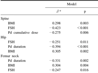
감소하였다. 대퇴골 골밀도에서 시행한 단변량 분석 역시 유사한 결과를 보였으며, 칼슘보충제를 복용하는 경우 척 추 골밀도와 달리 대퇴 골밀도가 증가되는 소견을 보여 칼 슘보충제 사용은 대퇴골 골밀도에 우선적으로 영향을 줌 을 시사하였다. 또한 FSH, LH가 증가할수록 요추 및 대퇴 골 골밀도가 감소하였으나, osteocalcin, DPY, OPG, RANKL, IL-6 등은 골밀도와 뚜렷한 상관관계를 보이지 않았다. 단변량 분석에서 의미 있게 나온 변수들을 중심으 로 다변량 회귀분석을 수행한 결과 척추, 대퇴골, 대퇴경 부 모두에서 FSH농도와 체질량 계수의 감소의 두 인자가 골밀도 감소와 관련된 독립적인 위험인자들이었다. 당질 코르티코이드의 사용과 골밀도 감소와의 관련성에 있어서 는 부위별로 약간의 차이를 보였는데 척추 에서는 누적 사 용량이 대퇴골에서는 총 사용기간이 골밀도감소와 관련된 독립적인 위험인자였다(표 4).
고 찰
본 연구에서는 한국인 루푸스 환자에서 골밀도 감소 정 도와 이에 영향을 미치는 여러 요인들 및 위험인자들을 조
Table 3. Univariate analyses of the clinical, bone turn over markers and hormones in SLE patients according to the bone mineral density
L spine Total Hip Femoral neck
γ* p γ* v γ* p
Age −0.191 0.065 −0.023 0.827 −0.160 0.124
BMI 0.330 0.001 0.393 <0.001 0.277 0.006
Postmenopausal state 0.273 0.007 0.163 0.115 0.267 0.009
Disease duration −0.214 0.038 −0.319 0.002 −0.283 0.006
Hx. of LN −0.133 0.200 −0.063 0.547 −0.057 0.584
SLEDAI −0.197 0.058 −0.114 0.278 −0.086 0.412
Osteocalcin −0.006 0.958 −0.107 0.315 −0.038 0.725
DPD −0.065 0.545 −0.023 0.828 −0.070 0.515
FSH −0.307 0.005 −0.222 0.045 −0.322 0.003
LH −0.242 0.027 −0.212 0.054 −0.297 0.006
IL-6 0.093 0.424 0.033 0.779 0.093 0.423
OPG −0.046 0.696 0.012 0.917 −0.030 0.799
RANKL 0.059 0.612 −0.043 0.710 0.032 0.786
Pd duration −0.225 0.029 −0.280 0.006 −0.264 0.010
Pd cumulative dose −0.236 0.022 −0.213 0.040 −0.191 0.065
CYC 0.175 0.089 0.059 0.567 0.106 0.307
Calcium supplement 0.160 0.121 0.280 0.006 0.268 0.009
BMI: body mass index, LN: lupus nephritis, SLEDAI: SLE disease activity index, DPD: deoxypyridinoline, FSH: follicule stimulating hormone, LH: luteinizing hormone, IL-6: interleukin-6, OPG: osteoprotegerin, RANKL: receptor activator NF-kB ligand, Pd: prednisolone, CYC: cyclophosphamide. *Pearson correlation coefficient
Table 4. Independent association of the spine, femur neck, and hip BMD in SLE patients by linear regression analysis
Model
β* p
Spine
BMI 0.298 0.003
FSH −0.423 <0.001
Pd cumulative dose −0.275 0.006
Hip
FSH −0.251 0.011
Pd duration −0.394 <0.001
BMI 0.305 0.002
Femur neck
Pd duration −0.331 0.002
BMI 0.304 0.004
FSH −0.247 0.016
BMI: body mass index, FSH: follicular stimulating hormone, Pd:
prednisolone. *Untandardized regression coefficient
사해 보고자 하였다. 루푸스 환자들은 대조군에 비하여 골 밀도가 의미 있게 감소되어 있었으며 다양한 위험인자 중 혈청 FSH의 증가, 체질량 계수의 감소, 스테로이드의 사용 기간과 누적용량의 증가가 루푸스 환자에서 골밀도 감소 와 연관된 독립적인 위험인자였다.
폐경기 여성에서는 골소실의 속도가 현저히 증가되어 이 기간 동안 피질골은 10∼15%, 해면골은 25∼30% 소실된 다고 알려져 있다 (30). 가장 주요한 기전 중의 하나가 에
스트로겐의 결핍으로 인한 골재형성의 증가로 생각되나 아직 그 기전은 분명하지는 않다. 최근 연구 결과에 따르 면, 에스트로겐의 효과는 RANKL과 RANK, 그리고 RANKL의 수용성 decoy 수용체인 OPG계를 매개로 하며 에스트로겐 결핍은 전구 조골세포의 OPG를 감소시키고 RANKL 발현이 증가되어 파골세포 발달이 항진되고 골흡 수가 증가하게 된다 (30). 본 연구에서는 직접적으로 혈청 estrogen의 농도를 측정한 것은 아니지만 이를 간접적으로 반영하는 FHS와 LH를 측정하였으며 골밀도가 감소된 루 푸스 환자들에서 골밀도가 정상인 환자들에 비해 혈청 FSH와 LH의 농도가 증가되어 있었다. 이러한 결과는 대상 환자들의 평균 나이가 37살로 비교적 젊은 나이임을 고려 해 볼 때 환자 군에서 비교적 조기에 난소의 기능부전이 수반되었고 이로 인해 골밀도 감소가 초래되었음을 시사 한다.
루푸스 환자에서 조기에 난소부전이 생기는 원인으로는 치료에 사용되는 약제의 영향, 루푸스 자체의 난소침범, 혹은 다른 특발성 원인에 의해 발생한다고 알려져 있다 (31-34). 루푸스 신염의 관해 유도를 위해 흔히 사용되는 cyclophosphamide의 경우 난소 기능부전을 초래한다는 것이 잘 알려져 있다 (31,32). Mok 등에 따르면 cyclophospha- mide의 총 누적용량이 보다 더 중요하며, 치료 시작의 나 이가 많을수록 조기폐경이 초래된다고 보고하였다 (32).
본 연구의 경우 신장염으로 cyclophosphamide를 치료받은
환자 17명이 포함되어 있었으나 cyclophosphamide를 사용 한 경우와 사용하지 않은 경우에 있어 FSH 농도가 차이를 보이지 않았는데 이는 비교적 초기의 신염으로 진단된 환 자들이어서 총 누적 사용용량이 적었고 비교적 젊은 나이 였던 점 때문으로 생각된다. 그 밖에도 황체(corpus luteum) 에 대한 자가항체 존재로 인해 난소기능 부전이 초래된다 는 연구 결과들도 있으나 (33,34), 본 연구에서는 조사되지 않았다. 향후 추가적인 연구가 필요한 부분이라고 하겠다.
루푸스의 치료와 관련하여 골밀도 감소를 일으키는 중요 한 약물로서 당질코르티코이드가 있다. 당질코르티코이드 는 직접적으로 조골세포에 작용하여 골형성을 억제하고 칼슘대사에 영향을 주며 골흡수세포의 활성화를 증가시킴 으로써 골감소를 유발한다 (33). 당질코르티코이드에 의한 골소실은 주로 투여 초기에 현저히 발생하며, glucocorti- coid 1∼5 mg/day의 소량 투여에도 발생하는 것으로 알려 져 있다 (34). 본 연구에서 루푸스의 치료를 위해 사용하였 던 당질코르티코이드 사용과 골밀도 감소가 연관성을 보 였는데 총 누적 용량과 사용기간이 모두 관련이 있었다.
이러한 결과는 루푸스에서 스테로이드는 사용 초기뿐만 아니라 장기적인 사용에 의한 누적 효과 역시 골밀도 감소 에 중요함을 시사한다. 흥미롭게도 해면골의 골밀도를 잘 반영하는 요추는 스테로이드의 총 누적 용량이, 피질골 밀 도를 잘 반영하는 대퇴골의 경우는 총 사용 기간이 골밀도 감소와 연관이 있었는데, 이는 당질코르티코이드 사용할 경우 초기에는 해면골 파괴가 많고 사용 후기로 갈수록 피 질골에서 골밀도 감소가 진행된다는 기존의 발표와 일치 한다 (30,35).
요약하면 루푸스 환자는 건강한 정상인에 비해 골밀도가 감소되어 있는데 이는 조기 난소부전 및 당질코르티코이 드 사용과 같은 질병관련 인자뿐 아니라 저체질량 계수와 같이 질병과 관련되지 않은 인자들과도 관련성을 보이고 있어 루푸스 환자에서 골밀도 감소는 다차원적인 원인에 의한 복합적 과정으로 생각된다. 본 논문의 제한점으로 첫 째, 비교적 환자의 규모가 적었으며, 둘째, FSH와 LH는 생 리주기에 따라 변동을 보이는 호르몬이나 본 연구에서는 생리주기에 따른 채취가 이루어지지는 않았다. 마지막으 로, 단면 연구이기 때문에 기저 원인을 결론 내리는데 한 계가 있다는 점이다. 따라서 향후 보다 많은 환자를 대상 으로 하는 전향적인 연구가 필요하겠다. 또한 루푸스 환자 에서 골다공증에 의한 골절의 위험에 대한 연구가 국내에 서는 아직 미흡한 실정이므로 이에 대한 다기관 연구가 필 요할 것으로 생각된다.
결 론
본 연구를 통하여 루푸스 환자가 건강한 정상인에 비하
여 골밀도가 감소되어 있음을 확인하였고, 혈청 FSH의 증 가, 감소된 체질량 계수와 당질코르티코이드 사용이 이와 관련된 독립적 위험인자이었다. 이는 조절 가능한 요소들 이 골밀도 감소의 주요한 위험인자 임을 시사하는 것으로 치료과정 중 골밀도 감소를 예상하고 관심을 가지고 치료 한다면 상당 부분 예방이 가능할 것으로 생각된다.
참고문헌
1. Urowitz MB, Bookman AA, Koehler BE, Gordon DA, Smythe HA, Ogryzlo MA. The bimodal mortality pattern of systemic lu- pus erythematosus. Am J Med 1976;60:221-5.
2. Ginzler EM, Diamond HS, Weiner M, Schlesinger M, Fries JF, Wasner C, et al. A multicenter study of outcome in systemic lupus erythematosus. I. Entry variables as predictors of prog- nosis. Arthritis Rheum 1982;25:601-11.
3. Ward MM, Pyun E, Studenski S. Mortality risks associated with specific clinical manifestations of systemic lupus erythematosus.
Arch Intern Med 1996;156:1337-44.
4. Ward MM, Pyun E, Studenski S. Causes of death in systemic lupus erythematosus. Long-term followup of an inception cohort.
Arthritis Rheum 1995;38:1492-9.
5. Trager J, Ward MM. Mortality and causes of death in systemic lupus erythematosus. Curr Opin Rheumatol 2001;13:345-51.
6. Gordon C. Long-term complications of systemic lupus erythema- tosus. Rheumatology (Oxford) 2002;41:1095-100.
7. Christian JC, Yu PL, Slemenda CW, Johnston CC Jr. Heritabili- ty of bone mass: a longitudinal study in aging male twins. Am J Hum Genet 1989;44:429-33.
8. Pocock NA, Eisman JA, Hopper JL, Yeates MG, Sambrook PN, Eberl S. Genetic determinants of bone mass in adults. A twin study. J Clin Invest 1987;80:706-10.
9. Johnston CC Jr, Miller JZ, Slemenda CW, Reister TK, Hui S, Christian JC, et al. Calcium supplementation and increases in bone mineral density in children. N Engl J Med 1992;327:82-7.
10. Morrison NA, Qi JC, Tokita A, Kelly PJ, Crofts L, Nguyen TV, et al. Prediction of bone density from vitamin D receptor alleles.
Nature 1994;367:284-7.
11. Dalsky GP. The role of exercise in the prevention of osteo- porosis. Compr Ther 1989;15:30-7.
12. Pacifici R. Estrogen, cytokines, and pathogenesis of postmeno- pausal osteoporosis. J Bone Miner Res 1996;11:1043-51.
13. Anderson GL, Limacher M, Assaf AR, Bassford T, Beresford SA, Black H, et al. Effects of conjugated equine estrogen in postmenopausal women with hysterectomy: the Women's Health Initiative randomized controlled trial. JAMA 2004;291:1701-12.
14. Canalis E. Clinical review 83: Mechanisms of glucocorticoid ac- tion in bone: implications to glucocorticoid-induced osteoporo- sis. J Clin Endocrinol Metab 1996;81:3441-7.
15. Haugeberg G, Ørstavik RE, Kvien TK. Effects of rheumatoid arthritis on bone. Curr Opin Rheumatol 2003;15:469-75.
16. Lacativa PG, Farias ML. Osteoporosis and inflammation. Arq Bras Endocrinol Metabol 2010;54:123-32.
17. Tanaka Y, Watanabe K, Suzuki M, Saito K, Oda S, Suzuki H, et al. Spontaneous production of bone-resorbing lymphokines by
B cells in patients with systemic lupus erythematosus. J Clin Immunol 1989;9:415-20.
18. MacDonald BR, Gowen M. Cytokines and bone. Br J Rheumatol 1992;31:149-55.
19. Report of a WHO Study Group. Assessment of fracture risk and its application to screening for postmenopausal osteoporosis.
World Health Organ Tech Rep Ser 1994;843:1-129.
20. Redlich K, Ziegler S, Kiener HP, Spitzauer S, Stohlawetz P, Bernecker P, et al. Bone mineral density and biochemical param- eters of bone metabolism in female patients with systemic lupus erythematosus. Ann Rheum Dis 2000;59:308-10.
21. García-Carrasco M, Mendoza-Pinto C, Escárcega RO, Jiménez- Hernández M, Etchegaray Morales I, Munguía Realpozo P, et al. Osteoporosis in patients with systemic lupus erythematosus.
Isr Med Assoc J 2009;11:486-91.
22. Kipen Y, Buchbinder R, Forbes A, Strauss B, Littlejohn G, Morand E. Prevalence of reduced bone mineral density in sys- temic lupus erythematosus and the role of steroids. J Rheumatol 1997;24:1922-9.
23. Bhattoa HP, Bettembuk P, Balogh A, Szegedi G, Kiss E. Bone mineral density in women with systemic lupus erythematosus.
Clin Rheumatol 2002;21:135-41.
24. Bhattoa HP, Kiss E, Bettembuk P, Balogh A. Bone mineral den- sity, biochemical markers of bone turnover, and hormonal status in men with systemic lupus erythematosus. Rheumatol Int 2001;21:97-102.
25. Lakshminarayanan S, Walsh S, Mohanraj M, Rothfield N.
Factors associated with low bone mineral density in female pa- tients with systemic lupus erythematosus. J Rheumatol 2001;28:
102-8.
26. Tan EM, Cohen AS, Fries JF, Masi AT, McShane DJ, Rothfield NF, et al. The 1982 revised criteria for the classification of sys- temic lupus erythematosus. Arthritis Rheum 1982;25:1271-7.
27. Park HA, Park JK, Park SA, Lee JS. Age, menopause, and car- diovascular risk factors among korean middle-aged women: the 2005 Korea National Health and Nutrition Examination Survey.
J Womens Health (Larchmt) 2010;19:869-76.
28. Bombardier C, Gladman DD, Urowitz MB, Caron D, Chang CH.
Derivation of the SLEDAI. A disease activity index for lupus patients. The Committee on Prognosis Studies in SLE. Arthritis Rheum 1992;35:630-40.
29. Genant HK, Cooper C, Poor G, Reid I, Ehrlich G, Kanis J, et al. Interim report and recommendations of the World Health Organization Task-Force for Osteoporosis. Osteoporos Int 1999;
10:259-64.
30. Molina B, Lassaletta A, Andion M, Gonzalez-Vicent M, López- Pino MA, et al. A Persistent epidural mass in a child with B-lin- eage ALL. Pediatr Blood Cancer 2010;55:727-9.
31. Warne GL, Fairley KF, Hobbs JB, Martin FI. Cyclophospha- mide-induced ovarian failure. N Engl J Med 1973;289:1159-62.
32. Mok CC, Lau CS, Wong RW. Risk factors for ovarian failure in patients with systemic lupus erythematosus receiving cyclo- phosphamide therapy. Arthritis Rheum 1998;41:831-7.
33. Hofbauer LC, Gori F, Riggs BL, Lacey DL, Dunstan CR, Spelsberg TC, et al. Stimulation of osteoprotegerin ligand and inhibition of osteoprotegerin production by glucocorticoids in human osteoblastic lineage cells: potential paracrine mechanisms of glucocorticoid-induced osteoporosis. Endocrinology 1999;140:
4382-9.
34. Van Staa TP, Leufkens HG, Cooper C. The epidemiology of cor- ticosteroid-induced osteoporosis: a meta-analysis. Osteoporos Int 2002;13:777-87.
35. Dalle Carbonare L, Arlot ME, Chavassieux PM, Roux JP, Portero NR, Meunier PJ. Comparison of trabecular bone micro- architecture and remodeling in glucocorticoid-induced and post- menopausal osteoporosis. J Bone Miner Res 2001;16:97-103.