서론
치조골 결손은 보철 치료 시 심미적인 문제를 야기하고 임플 란트 식립 시에도 골량 부족으로 인한 어려움을 일으킨다. 최근
Correspondence: Dr. Young-Hyuk Kwon
Department of Periodontology, School of Dentistry, Kyung Hee University, 1 Hoeki-dong, Dongdaemoon-gu, Seoul, 130-701, Korea e-mail: [email protected], Tel: 82-2-958-9380, Fax: 82-2-958-9387 접수일: 2008년 2월 20일; 채택일: 2008년 3월 5일
임플란트 치료가 보편화 되고 환자의 심미적인 요구도가 증가 하면서 치조골 결손 부위의 회복이 치료에 중요한 요소가 되었 다. 이에 치조골 재생을 위한 다양한 재료와 술식들이 이용되고 있다. 골형성을 증가시키기 위한 술식들로는 성장인자를 이용
1,2)하거나, 골이식재를 이용한 골유도 및 전도1,3), 인위적인 골 절에 의한 골신장술4,5), 그리고 차폐막을 이용하는 골유도재생
술6,7) 등이 있다. 골재건을 위한 여러 가지 술식 중 골이식술 및
골유도재생술은 예견성이 높고 임상적용이 용이하여 다양한 형
무기인산염이 외방성 수직골 형성에 미치는 영향
이 진
1, 박준봉
1,2, 허 익
1,2, 정종혁
1,2, 권영혁
1,2*
1)1. 경희대학교 치의학전문대학원 치주과학교실 2. 경희대학교 치의학전문대학원 구강생물학연구소
The effect of polyphosphate on exophytic bone formation
Jean Lee
1, Joon-Bong Park
1,2, Yeek Herr
1,2, Jong-Hyuk Chung
1,2, Young-Hyuk Kwon
1,2*
1. Department of Periodontology, School of Dentistry, Kyung Hee University 2. Institute of Oral Biology, School of Dentistry, Kyung Hee University ABSTRACT
Purpose: It has been shown that the inorganic polyphosphate is effective for the regeneration of bones through the preliminary animal test of rabbits. The most effective concentration of the polyphosphate, however, is not known yet. Moreover, the effectiveness of carriers inside human body is not confirmed..
Materials and Methods: In this study, we examined the effect of the concentration of the inorganic polyphosphate on the process of the bone regeneration using the 6 weeks old rabbits with the weight of 2.0 kg in average. We performed the experiment using TR-ePTFE membrane(membrane) filled with collagen immersed in 4%, 8% of inorganic polyphosphate, respectively, following removal of the proper sized cortical bones from the rabbit calvaria.
The experimental results were compared with the one of the following four groups: The negative control group for membrane only, the positive control group for membrane filled with collagen, the first experimental group for membrane filled with collagen immersed in 4% of inorganic polyphosphate, and the second experimental group for membrane filled with collagen immerse in 8%
of inorganic polyphosphate. The fragments of the tissue with membrane obtained from each group of the sacrificed rabbits for 8 or 16 weeks sustained after surgery were then prestained by the Hematoxylin-Eosin stain and coated by resin to form non-decalcified specimens for the histologic examination and analysis. New bone formation was assessed by histomorphometric and statistical analysis.
Results: 1. All groups have shown better bone regeneration at 16weeks than 8weeks. 2. Negative control group has shown more bone regeneration relative to the other groups at 8 and 16 weeks. 3. All experimental groups have shown better bone regeneration relative to positive control group. 4. At 16 weeks, the first experimental group has shown more bone regeneration compared to the second experimental group. Exophytic bone formation is not good at the first and the second experimental groups compared with negative control group. But, the use of 4% inorganic polyphosphate was more effective to bone formation than the use of 8%
inorganic polyphosphate.
Conclusion: With above results, it is suggested the use of inorganic polyphosphate with vehicle under TR-ePTFE membrane.
(J Korean Acad Periodontol 2008;38:59-66)
KEY WORDS: polyphosphate; exophytic bone formation; TR-ePTFE membrane.
태의 골결손부, 치조제증대술 및 임플란트의 식립과 연관된 골 결손부 등에 널리 사용되는 일반적인 치료방법이 되었다.
차폐막 하방에 신생골이 형성될 수 있는 골증강술의 개념 을 Dahlin 등8)이 확립한 이후, Buser 등9)은 차폐막을 적용하 여 골을 형성하는 골유도재생술이라는 용어를 제안하였다. 이 들은 외과적으로 골결손부를 만든 동물 실험을 통해서, 차폐 막의 사용여부에 따라 골결손부의 치유양상이 다른 것을 발견 하였는데 차폐막을 사용하지 않는 골결손부는 상부의 치은 결 합조직에서 유래하는 세포의 증식에 의하여 결합조직으로 채 워졌고 차폐막을 사용한 골결손부는 치은 결합조직세포의 차 단으로 골 조직으로 치유된 결과를 얻을 수 있었다.
골유도재생술에 사용되는 차폐막이 갖추어야 할 조건은 인 체에 무해하고 조직 친화성이 있어야 하며 치은 결합조직의 세포유입을 차단할 수 있는 세포차단성이 있어야 하고 골재생 에 관여하는 세포가 이주, 증식할 수 있는 재생공간을 확보하 고 유지할 수 있는 물리적 성질이 갖추어야 한다11-13). 현재 임 상에서 사용되고 있는 차폐막은 비흡수성과 흡수성으로 분류 되며, 술 후 예견성이 높고 조직유도 능력이 우수한 비흡수성 차폐막이 널리 사용되고 있다. 임상에서 널리 사용되고 있는 비흡수성 차폐막 중 티타늄 강화 차폐막(Titanium re- inforced e-PTFE)은 티타늄 구조로 인해 구부릴 수 있고 조 형이 가능하며 원하는 모양을 유지하는데 유리하다. Jovanovic 등14)은 5마리 성견의 연구에서 티타늄 강화 차폐막을 사용하 여 6개월의 치유기간을 거친 후 4 mm 정도의 골재생을 보고 하였으며 티타늄 강화 차폐막이 부가적인 골이식술 없이도 큰 공간을 잘 유지할 수 있으며 이전의 차폐막과 비교시 더 많은 양의 골을 형성할 수 있다고 하였다. 이처럼 티타늄강화차폐 막은 막과 골결손부 사이의 충분한 공간을 형성하고 보존할 수 있어 결손부의 해부구조가 공간을 형성할 수 없는 상황에 서도 치조제 증강과 재생을 위한 새로운 가능성을 제시한다.
무기인산염(Inorganic polyphosphate)은 인체에 무해한 저 비용의 물질로 골아세포에 고농도로 함유되어 있으며 무기질 침착에 관여하는 것으로 알려져 있다. 또한 여러 세포 실험에서 항생효과 및 골재생에 효과가 있는 것으로 입증되었다25-27). 최근 시험관적 실험을 통하여 무기인산염이 골아세포의 골 형성 지표자로 알려진 오스테오폰틴(osteopontin)과 오스테 오칼신(osteocalcin)의 발현을 증가시켜 골화능력 존재의 사 실이 알려졌다18). 또한 bisphosphonate가 파골기전을 억제하 여 골다공증 치료에 응용하는 단계에 이르렀으며 동일 계열 무기인산염의 임상응용 가능성이 더 높게 되었다. 백묘를 이
용한 예비 동물실험에서 정 등19)은 무기인산염이 골재생 과정 에 미치는 영향을 평가하기 위하여 웅성 백묘 두개관에 일정 크기의 피질골을 제거한 후, 그 상방에 1%, 2%의 무기인산염 에 침적한 탈단백우골을 채운 티타늄 강화 차폐막을 이용하여 골유도재생술을 시행하여 2%의 무기인산염에서 골재생을 확 인하였다. 그러나 아직까지 가장 효과적인 농도를 밝히지 못 하였으며, 어떤 전달체가 가장 효과적이며 체내에서 골재생 능력이 있는지에 대한 연구들이 진행되고 있는 실정이다20-27).
이번 연구는 웅성 백묘 두개관에 피질골을 제거하고 그 상 방에 티타늄 강화 차폐막과 교원질, 그리고 4%, 8% 무기인산 염을 이용하여 골유도재생술을 시행하고 새로 형성된 골을 조 직계측학적으로 분석함으로써 무기인산염이 외방성 수직골 형성에 미치는 효과를 알아보고자 시행하였다.
실험재료 및 방법
1. 실험재료
차폐막으로는 티타늄 강화 차폐막(TR-9W, W.L. Gore &
Associate. Inc, U.S.A)을 사용하였다. 25×20 mm 크기의 티타늄 강화 차폐막을 내면이 가로 8 mm, 세로 5 mm, 높이 4 mm인 직육면체로 만들어 필요한 공간을 확보할 수 있도록 하였다. 차폐막 내부에는 전달체로서 교원질(Colla TapeⓇ, Sulzer Dental Inc. NJ, U.S.A)을 사용하였고, 무기인산염으 로는 Poly P75(Sodium polyphosphate, Sigma, St. Louis, MO, U.S.A)를 사용하였다.
티타늄 강화 차폐막만을 사용한 군을 음성대조군(TG)으로, 교원질과 티타늄 강화 차폐막을 사용한 군을 양성대조군(TG +Col)으로 하고, 4%의 무기인산염에 침적한 교원질과 티타늄 강화 차폐막을 사용한 군을 실험 I 군(TG+Col+P4), 그리고 8% 무기인산염에 침적한 교원질과 티타늄 강화 차폐막을 사 용한 군을 실험 II 군(TG+Col+P8)으로 분류하였다.
2. 실험 동물
생후 6주된 평균 체중 2.0 kg의 웅성 백묘(New Zealand White Rabbit)를 각 군마다 2마리씩 16마리를 사용하였으며, 각 실험동물 당 실험부위는 2부위씩으로 배정하였다. 실험기 간동안 고형사료(축협사료, 축산업협동조합, 한국)를 공급하 였고 분리된 실내 사육실에서 사육하였다.
3. 실험 방법 1) 시술 과정
실험동물에 0.2 ㎖/Kg의 졸레틸(Virbac, France)과 ket- amine을 1:1로 혼합하여 0.5~1 ㎖를 근육 주사하여 전신마취 하고, 수술부위는 리도케인 2%(1:100,000 에피네프린, 광명, 한국)로 국소마취하였다. 두개면 수술부위의 모발을 제거한 후 #15 수술도를 이용하여 시상방향으로 절개, 전층판막을 거 상하였다. 시상 봉합선을 중심으로 좌우측으로 2 mm 떨어진 부위에 차폐막 크기에 맞추어 라운드 카바이드 버(HP long
#6)로 주수하에 5mm×8mm 넓이로 두개관에 약 1~2 mm 깊이로 피질골을 제거하였다. 실험 I, II 군의 경우 미리 직육 면체 모양으로 구부린 2개의 티타늄 강화 차폐막 내부에 4, 8% 농도의 무기인산염에 20분 동안 침적한 교원질 스폰지를 채우고 피질골이 제거된 부위에 차폐막을 위치시키고 4개의 핀으로 고정하였다. 음성대조군의 경우 차폐막만을 핀으로 고 정하였고 양성대조군의 경우 차폐막 내에 교원질 스폰지만을 채워 핀으로 고정하였다. 흡수성 봉합사(4-0 chromic gut, Ailee, Korea)를 이용하여 골막과 표피를 함께 봉합하였다.
2) 술 후 처치
술 후 감염을 방지하기 위하여 수술 당일과 술 후 3일까지 겐타마이신(동화약품, 한국) 0.5 ㎖와 1% 케토프로펜(유니 바 이오테크, 한국) 0.2 ㎖를 근육 주사하였다.
3) 조직준비 및 분석
실험동물들은 술 후 8, 16주에 각 군마다 2마리씩 졸레틸로 근육주사하고 정맥을 통한 이산화탄소로 질식사 시킨 후 두피 를 거상하고 차폐막과 하방 두개골을 포함하는 조직시편을 채 득하였다. 조직학적 관찰을 위해 비탈회골의 표본을 제작하고 자 시편을 중성 포르말린 용액에 1주일 간 고정한 후, 2일 간 70%, 80%, 90%, 95%, 100% 알코올에 단계별로 탈수시켰다.
이후 시편을 technovit 7200VLC 용액(Kultzer & Co.
Germany)에 침적시킨 후 광중합하여 포매하였다. 시편은 EXAKT cutting & grinding system machine(EXAKT Apparaturbau, Germany)을 이용하여 50μm 이하 두께로 절단과 연마를 시행한 뒤 Hematoxylin과 Eosin 중염색을 시 행하여, 조직 슬라이드를 BX-51 Olympus 현미경으로 조직학 적 소견을 관찰하였다. 조직 슬라이드를 DP 71 현미경용 카메 라로 12.5배에서 디지털 사진 촬영 후 Tomoro Scope Eye 프로 그램(Techsan Co. Korea)을 이용하여 조직계측학적 분석을 시행하였다.
4) 조직계측학적 계측 방법
하방의 모식도처럼 신생골의 면적을 계측하였다. 신생골 형성률은 아래의 공식을 적용하여 계산하였다.
New bone formation (%) =
Area of new bone formation (mm
2)
×100
Area of space created by the membrane (mm
2)
Figure 1. A schematic diagram used in histomorphologic analysis.5) 통계학적 분석
실험기간 동안 대조군 및 실험군들의 신생골 형성률에 대 한 기간별 비교는 Mann Whitney U test를 이용하였으며, 실 험 8주, 16주 각 기간에서 각 군들간의 신생골 형성률 비교에 는 Kruskal-Wallis test를 사용하였다. 통계 프로그램은 SPSS ver 12.0을 사용하였다.
실험 성적
1. 조직학적 소견
1) 음성대조군 8주 소견(TG-8w)
핀 고정을 한 차폐막은 형성된 공간 유지가 잘 되었으며 기
존골 표면 위에 외방성으로 신생골이 증식되어 있다(Fig. 2).
2) 음성대조군 16주 소견(TG-16w)
8주에 비하여 기존골 표면으로부터 좀 더 많은 양의 소주골 형성을 관찰할 수 있다(Fig. 3).
3) 양성대조군 8주 소견(TG+Col-8w)
차폐막 하방으로 결합조직이 관찰되며 신생골의 형성은 미 약하였다. 음성대조군에 비해 신생골 형성량은 육안적으로도 적었다(Fig. 4).
4) 양성대조군 16주 소견(TG+Col-16w)
8주째에 비해 신생골 형성이 증가되는 양상을 보이지만,
Figure 2. Trabecular bone formation from the base. H&E Stain, Original magnification x 12.5
Figure 3. More trabecular bone for- mation than that at 8 wks. H&E Stain, Original magnification x 12.5
Figure 4. Slight trabecular bone formation. H&E Stain, Original mag- nification x 12.5
Figure 5. More trabecular bone for- mation than that at 8 wks. H&E Stain, Original magnification x 12.5
Figure 6. Slight trabecular bone formation. H&E Stain, Original mag- nification x 12.5
Figure 7. More trabecular bone for- mation than that at 8 wks. H&E Stain, Original magnification x 12.5
Figure 8. Slight trabecular bone formation. H&E Stain, Original mag- nification x 12.5
Figure 9. More trabecular bone for- mation in height than that at 8 wks.
H&E Stain, Original magnification x 12.5
음성대조군에 비해서 골형성이 적었다(Fig. 5).
5) 실험 I 군 8주 소견(TG+Col+P4-8w)
양성대조군 8주째에 비해 소주골 형성이 미세하게 증가되 는 양상을 보이며, 결합조직은 차폐막 내부의 반 정도만 채워 져 있는 것이 관찰되었다(Fig. 6).
6) 실험 I 군 16주 소견(TG+Col+P4-16w)
실험군 I 8주째에 비해 소주골의 밀도가 뚜렷이 증가하는 것을 볼 수 있었다(Fig. 7).
7) 실험 II 군 8주 소견(TG+Col+P8-8w)
실험군 I과 비교시 신생골의 형성량은 거의 비슷하였고 결 합조직은 차폐막 하방부위를 채우고 있었다(Fig. 8).
8) 실험 II 군 16주 소견(TG+Col+P8-16w)
8주군에 비하여 신생골의 수직적 증식이 증가된 것을 볼 수 있었다(Fig. 9).
2. 조직계측학적 분석
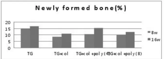
실험 기간에 따른 신생골 형성률을 비교해 보면 음성대조 군 8주에서 15.21±0.11%였고, 음성대조군 16주에서는 16.68
±0.84%로 증가되었다. 양성대조군은 8주와 16주에서 각각 8.37±0.13%, 11.29±0.34%로 음성대조군보다 더 많은 양의 증가를 보였다. 실험 I군은 10.38±0.54%에서 15.34±0.68%
로 가장 증가율이 높았으며, 실험 II군은 9.95±0.89%에서 12.12±1.63%로 증가되었다. Mann Whitney U test를 이용 하여 8주와 16주를 비교 시 모든 군에서 통계학적으로 유의성 있는 증가를 보였다(p<0.05)(Fig. 10). Kruskal-Wallis test
를 이용하여 네 군들을 비교 시 8주와 16주에서 음성대조군, 실험 I군, 실험 II군, 양성대조군 순으로 신생골 형성률이 높 았으며 모두 p<0.05로 통계학적으로 유의성 있는 차이를 보 였다(Fig. 11).
총괄 및 고찰
골유도재생술을 시행한 부위는 웅성 백묘의 두개관을 이용 하였는데 두개관은 형태학적으로나 발생학적으로 막성골이라 는 점에서 안면의 막성골들과 유사성을 가지며 해부학적으로 두개관은 중간에 해면골이 개재되어 있는 두 개의 피질골판으 로 이루어져 있다는 점에서 하악골과 유사하고 생리학적으로 두개관내의 피질골은 퇴축된 하악골과 닮았다고 할 수 있다
33). 이러한 이유로 두개관을 이 실험에서 수술부위로 사용하 였다. 이번 실험에서 1~2 mm 깊이로 피질골을 제거한 것은 골수로부터 유래된 혈액공급 및 골형성인자의 작용을 촉진하 기 위하여34) 시행하였다. 또한 주변이 골로 둘러싸인 와동형 의 골결손부는 주위 골로부터 활발한 골재생이 이루어져 골형 성 촉진 물질의 효과를 구분하기 어렵게 하기 때문에35) 이번 실험에서는 기존 골의 외방으로 골형성을 유도하는 실험모델 을 이용함으로써 다른 골재생에 관여하는 요소들을 배제한 상 태에서 골재생에 미치는 효과를 살펴보려고 하였다.
이번 실험에서는 차폐막을 고정용 핀으로 고정함으로써 차폐막의 동요로 인하여 야기될 수 있는 요인들을 제거하였 다. 차폐막의 고정은 골재생을 유도하는 중요한 요인으로서 차폐막의 미세동요를 방지하기 위하여 고정용 핀을 사용하 여 기저골의 표면을 따라 형성될 수 있는 연조직의 침투를 방지하였다36). 차폐막을 사용하지 않은 골결손부는 상부 치 은결합조직의 빠른 증식 및 유입으로 인하여 골세포의 이동
Figure 10. Mean new bone formation by the period.
*means statistically significant difference between 8 weeks and 16 weeks by Mann Whitney U test(p<0.05).
Figure 11. Mean new bone formation by polyphosphate concentration.
*means statistically significant difference compared to control group by Kruskal-Wallis test(p<0.05).
이 차단됨에 따라서 단순한 결합조직으로 채워지는 반면, 차 폐막을 사용한 골결손부는 상부 치은 결합조직의 유입이 차 단함으로써 하부에서 골조직의 재생을 유도하였다. 아울러 차폐막의 다른 기능은 골결손부 내의 혈병을 보호하고 상부 연조직으로부터의 외력을 차단함으로써 골조직의 재생을 촉 진한다고 하였다37). 전달체로 사용된 교원질 스폰지는 교원 질이 주성분이며 통기성이 좋으며 물질의 90% 이상이 액체 로 채울 수 있는 다공성 구조로 되어있다.
무기인산염(Inorganic polyphosphate, PolyP)은 (NaPO3)n 의 분자식을 갖는 인체에 무해한 물질로서20,21), 수십 또는 수 백 개의 orthophosphate(Pi) residues가 고에너지의 phos- phoanhydride 결합을 형성하고 있는 중합체이다22). 무기인 산염은 미생물 내에서 ATP의 대체물질로서 에너지원으로 작 용하고 인산(Pi)의 저장고 역할을 하며 적은 농도의 무기인산 염이 동물세포에서 관찰되기도 하지만 포유동물에서의 무기 인산염의 기능은 잘 알려져 있지 않다23,24). Fleisch 등25)은 인 (phosphorous)의 합성물질인 bisphosphonate가 골흡수를 막는 강력한 억제물질이라고 보고하였고, Leyhausen 등26)은 많은 양의 무기인산염(Inorganic polyphosphate)이 사람의 하악에서 유래되는 골아 유사세포(human mandibular- derived osteoblast-like cell)에서 관찰되며 골광물화의 조 절자로 작용한다고 하였다. Shiba 등27)은 무기인산염이 사람 의 섬유아세포(human fibroblast cell)의 증식에 관여한다고 하였다. 무기인산염에 대한 연구 중 또한 Kawazoe 등18)은 쥐 를 이용한 세포실험을 통해 무기인산염이 골아세포 분화의 표 지인 osteopontin과 osteocalcin의 발현을 증가시키고 alka- line phosphatase activity도 증가시켰다고 하였으며, 무기 인산염이 polyphosphatase의 활성을 증가시킴으로써 세포의
골화를 촉진시킨다고 보고하였다.
이 등은 토끼의 대퇴골에 교원질과 탈염골분(demineralized human bone powder)을 매개체로 2% 무기인산염의 효과에 관한 실험을 통해, 무기인산염이 매개체의 골전도능을 향상시 키거나 손상된 골조직의 골유도능을 증가시킴으로써 골재생 을 촉진한다고 보고하였다.
이번 실험에서는 무기인산염의 농도와 치유기간을 달리하 여 그 양상과 골재생 효과를 알아보고자 하였다. 각 군 모두 8주보다 16주에서 더 많은 신생골 형성을 보임으로써 치유기 간이 긴 쪽에서 신생골 형성률이 좋았다. 양성 대조군과 실험 군 I, II를 비교 시 두 실험군 모두에서 골형성 양이 양성대조 군보다 좋았고 이는 무기인산염이 골재생에 효과가 있음을 보 여주었다. 하지만 예상과는 달리 8주와 16주 모두에서 음성 대조군이 다른 군들에 비해 더 많은 신생골 형성을 보였다. 기 존의 여러 연구에서 다양한 전달체를 이용한 무기인산염의 골 재생 효과가 보고되었으나 본 실험에서 실험군보다 음성 대조 군의 신생골 형성률이 오히려 높게 나온 것은 전달체의 문제 가 아니었나 추측한다. 이에 무기인산염의 골재생 효과를 높 이기 위해서는 전달체의 종류와 형태가 무엇보다 중요한 것 같다.
실험 I군과 II군을 비교하였을 때 4%의 무기인산염을 적용 한 실험 I군이 8%를 적용한 실험 II군보다 골 형성면에서 효 과적이었다. 본 실험의 결과에서 무기인산염의 농도와 골재생 효과는 서로 비례하지 않았다. 이번 실험을 통해 적절한 무기 인산염의 농도가 오히려 고농도의 무기인산염보다 골재생에 효과적이었음을 알 수 있었다. 향후 전달체를 달리하여 골재 생에 보다 효과적인 최적의 무기인산염 농도를 찾는 연구가 계속되어야 하겠다.
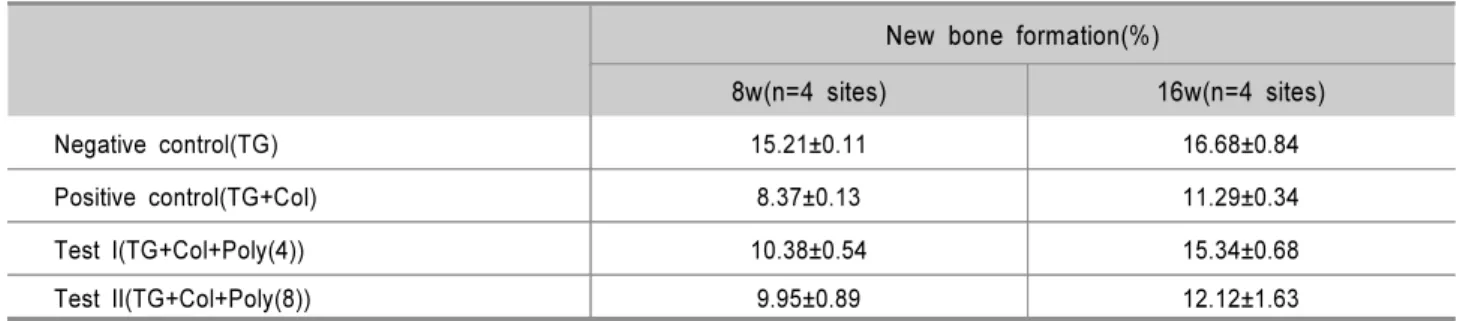
Table 1. New Bone Formation(%)
New bone formation(%)
8w(n=4 sites) 16w(n=4 sites)
Negative control(TG) 15.21±0.11 16.68±0.84
Positive control(TG+Col) 8.37±0.13 11.29±0.34
Test I(TG+Col+Poly(4)) 10.38±0.54 15.34±0.68
Test II(TG+Col+Poly(8)) 9.95±0.89 12.12±1.63
* Statistically significant difference between 8 weeks and 16 weeks (p<0.05).
참고문헌
1. Reddi AH, Weintroub S, Muthukumaran N. Biologic prin- ciples of bone induction. Orthop Clin North Am 1987;
18:207-212.
2. Urist MR. Bone formation by autoinduction. Science 1965;
150:893-899.
3. Burchardt H. The biology of bone graft repair. Clin Orthop Rel Res 1983;174:28-42.
4. Ilizarov GA. The tension-stress effect on the genesis and growth of tissues: Part I. The influence of stability of fix- ation and soft tissue preservation. Clin Orthop 1989;238:
249-281.
5. Ilizarov GA. The tension-stress effect on the genesis and growth of tissues: Part II. The influence of rate and fre- quency of distraction. Clin Orthop 1989;239:263-285.
6. Kostopoulos L, Karring T. Augmentation of the rat man- dible using guided tissue regeneration. Clin Oral Impl Res 1994;5:75-82.
7. Dahlin C, Linde A, Gottlow J, Nyman S. Healing of bone defects by guided tissue regeneration. Plastic Reconst Sur 1988;81:672-677.
8. Dahlin C, Sennerby L, Leckholm U, Lindhe A, Nyman S.
Generation of new bone around titanium implants using a membrane technique: An experimental study in rabbits. Int J Oral Maxillofac Implants 1989;4:19-25.
9. Buser D, Dura K, Belser UC, Hirt HP, Berthold H.
Localized ridge augmentaion using guided bone re- generation: Surgical procedure in the maxilla. Int J Periodont Rest Dent 1993;13:29-45
10. Dahlin C, Sennerby L, Leckholm U, Lindhe A, Nyman S.
Generation of new bone around titanium implants using a membrane technique: An experimental study in rabbits. Int J Oral Maxillofac Implants 1989;4:19-25.
11. Siebert J, Nyman S. Localized ridge augmentation in dogs:
A pilot study in using membranes and hydroxyapatite. J Periodontol 1990;61:157-165.
12. Becker W, Becker BE, Handlesman M, Celletti R, Ochsenbein C, Hardwick R, Langer B. Bone formation at dehisced dental implant sites treated with implant augmen- tation material: A pilot study in dogs. Int J Periodont Rest Dent 1990;10:93-101.
13. Werrer K, Gotfredsen K, Hjrting-Hansen E, Karring T.
Guided tissue regeneration of dental implants placed into extraction sockets: An experimental study in monkeys. Clin Oral Impl Res 1991;2:166-171.
14. Jovanovic SA, Schenk RK, Orsini M, Kenney Eb.
Supracrestal bone formation around dental implants: An ex- perimental dog study. Int J Oral Maxillofac implants 1995;10:23-31.
15. Becker W, Dahlin C, Becker BE. The use of e-PTFE bar- rier membranes for bone promotion around titanium im- plants placed into extraction socket: A prospective multi- center study. Int J Oral Maxillofac Implants 1994;9:31-40.
16. Niederman R, Savitt ED, Heeley JD, Duckworth JE.
Regeneration of furca bone using Gore-Tex periodontal material. Int J Periodont Res Dent 1989;9:468-480.
17. Pontoriero R, Lindhe J, Nyman S, Karring T, Rosenberg E, Sanavi F. Guided tissue regeneration in the treatment of furcation defects in mandibular molars. A clinical study of degree III involvements. J Clin Periodontol 1989;16:
170-174.
18. Kawazoe Y, Shiba T, Nakamura R, Mizuno A, Tsutsumi L, Uematsu T, Yamaoka M, Shindoh M, Kohgo T. Induction of calcification in MC3T3-E1 cells by inorganic poly- phosphate. J Dent Res 2004;83:613-618.
19. Chung JH, Kwon YH, Park JB, Herr Y. Effect of inorganic polyphosphate on guided bone regeneration. J Korean Acad Periodontol 2005;35:491-510.
20. The United States Department of Agriculture. Meat and poultry products: phosphates and sodium hydroxide. Fed Regist 1982;47:10779.
21. Lanigan RS. Final report on the safety assessment of so- dium metaphosphate, sodium trimetaphosphate, and sodium hexametaphosphate. Int J Toxicol 2001;20(Suppl.3):75-89.
22. Kornberg A. Inorganic polyphosphate: toward making a for- gotten polymer unforgettable. J Bacteriol 1995;177:491-496.
23. Kumble KD, Kornberg A. Inorganic polyphosphate in mam- malian cells and tissues. J Biol Chem 1995;270:5818-5822.
24. Gabel NW, Thomas V. Evidence for the occurrence and distribution of inorganic polyphosphate in vertebrae tissues.
J Neurochem 1971;18:1229-1242.
25. Fleisch H. Mechanisms of action of the bisphosphonates.
Medicina(B Aires) 1997;57(Suppl.):65-75.
26. Leyhausen G, Lorenz B, Zhu H,Geurtsen W, Bohnensack R, Müller WE, Schröder HC. Inorganic polyphosphate in human osteoblast-like cells. J Bone Miner Res 1998;13:
803-812.
27. Shiba T, Nishimura D, Kawazoe Y, Onodera Y, Tsutsumi K, Nakamura R, Ohshiro M. Modulation of mitogenic activ- ity of fibroblast growth factors by inorganic polyphosphate.
J Bio Chem 2003;278:26788-26792.
28. Jovanovic SA, Spiekermann H, Richter EJ. Bone re- generation around titanium implants in dehiscenced defect sites: A clinical study. Int J Oral Maxillofac Implants 1992;
7:233-245.
29. Becker W, Becker B, Handelsman M, Ochsenbein C, Albrektsson T. Guided tissue regeneration for implant placed into extraction sockets: A study in dogs. J Periodontol 1991;62:703-709.
30. Brägger U, Hammerle CHF, Lang NP. Immediate trans- mucosal implants using the principle of guided tissue re- generation II: A cross-sectional study comparing the clin- ical outcome 1 year after immediate to standard implant placement. Clin Oral Impl Res 1997;7:268-276.
31. Buser D, Brägger U, Land NP, Nyman S. Regeneration and enlargement of jaw bone using guided tissue regeneration. Clin Oral Impl Res 1990;1:22-32.
32. Schenk RK, Hardwick WR, Dahlin C. Healing pattern of bone regeneration in membrane-protected defects. A histo- logic study in the canine mandible. Int J Oral Maxillofac Implants 1994;9:13-29.
33. Lee KH, Kwon YH, Park JB, Herr Y. Effect of pore num- ber of titanium mesh on bone formation in the procedure
of GBR. J Korean Acad Periodontol 2004;34:411-424.
34. Majzoub Z, Berengo M, Giardino R, Cordioli G. Role of intramarrow penetration in osseous repair: A pilot study in the rabbit calvaria. J Periodontol 1999;70:1501-1510.
35. Lundgren D, Lundgren AK, Sennerby L, Nyman S.
Augmentation of intramembranous bone beyond the skeletal envelope using an occlusive titanium barrier. Clin Oral Impl Res 1995;6:67-72.
36. Simon M, Trisi P, Piattellli A. Vertical ridge augmentation using a membrane technique associated with osseointe- grated implants. Int J Periodont Rest Dent 1994;14:496- 511.
37. Jovanovic SA, Schenk RK, Orsini M, Kenney Eb.
Supracrestal bone formation around dental implants : an experimental dog study. Int J Oral Maxillofac Implants 1995;10:23-31.
38. Jovanovic SA, Nevins M. Bone formation utilizing tita- nium-reinforced barrier membranes. Int J Periodont Rest Dent 1995;15:57-69.
39. Tinti C, Parma-Benfenati S, Pollizzi G. Vertical ridge aug- mentation: what is the limit? Int J Periodont Rest Dent 1996;16:220-229.
40. Tinti C, Vincenzi GP. Expanded polytetrafluoroethylene ti- tanium -reinforced membrane for regeneration of mucogin- gival recession defects: A 12-cases report. J Periodontol 1994;65:1088-1094.