Clinical Significance of Serum Autoantibodies in Idiopathic Interstitial Pneumonia
Although autoantibodies are routinely screened in patients with idiopathic interstitial pneumonia, there are no reliable data on their clinical usefulness. The aim of this study was to investigate the prognostic value of autoantibodies for predicting the development of new connective tissue disease in these patients and also mortality. We conducted retrospective analysis of the baseline, and follow-up data for 688 patients with idiopathic interstitial pneumonia (526 with idiopathic pulmonary fibrosis, 85 with nonspecific interstitial pneumonia, and 77 with cryptogenic organizing pneumonia) at one single tertiary referral center. The median follow-up period was 33.6 months. Antinuclear antibody was positive in 34.5% of all subjects, rheumatoid factor in 13.2%, and other specific autoantibodies were positive between 0.7%-6.8% of the cases. No significant difference in patient survival was found between the autoantibody-positive and -negative groups. However, the presence of autoantibodies, especially antinuclear antibody with a titer higher than 1:320, was a significant predictor for the future development of new connective tissue diseases (relative risk, 6.4), although the incidence was low (3.8% of all subjects during follow-up). In conclusion, autoantibodies are significant predictors for new connective tissue disease development, although they have no prognostic value.
Key Words: Idiopathic Interstitial Pneumonias; Idiopathic Pulmonary Fibrosis; Nonspecific Interstitial Pneumonia; Cryptogenic Organizing Pneumonia; Antinuclear Antibody;
Rheumatoid Factor; Autoantibody; Connective Tissue Disease; Incidence; Predictive Factor Bo Hyoung Kang,1,* Jin Kyeong Park,1,*
Jae Hyung Roh,1 Jin Woo Song,1 Chang Keun Lee,2 Miyoung Kim,3 Se Jin Jang,4 Thomas V Colby,5 and Dong Soon Kim1
Departments of 1Pulmonary and Critical Care Medicine, 2Rheumatology, 3Radiology, and
4Pathology, University of Ulsan, College of Medicine, Asan Medical Center, Seoul, Korea; 5Department of Laboratory Medicine and Pathology, Mayo Clinic, Scottsdale, AZ, USA
*Bo Hyoung Kang and Jin Kyeong Park contributed equally to this work.
Received: 21 June 2012 Accepted: 28 February 2013 Address for Correspondence:
Dong Soon Kim, MD
Department of Pulmonary and Critical Care Medicine, Asan Medical Center, University of Ulsan College of Medicine, 88 Olympic-ro 43-gil, Songpa-gu, Seoul 138-736, Korea Tel: +82.2-3010-3132, Fax: +82.2-3010-6968 E-mail: [email protected]
http://dx.doi.org/10.3346/jkms.2013.28.5.731 • J Korean Med Sci 2013; 28: 731-737 Respiratory Diseases
INTRODUCTION
The diagnosis of idiopathic interstitial pneumonia (IIP) requires exclusion of other known causes of interstitial pneumonia, in- cluding drugs or other environmental exposures and connec- tive tissue disease (CTD) (1). All types of IIP, except respiratory bronchiolitis associated with interstitial lung disease (RB-ILD), can occur in CTDs and the prognosis of CTD-related interstitial pneumonia (CTD-IP) is reported to be better than that of IIP (2). Therefore, most clinicians routinely do serologic testing for CTD such as antinuclear antibody (ANA) and rheumatoid fac- tor (RF), in addition to detailed history taking and physical ex- amination at the time of diagnosis (3). Recently, American Tho- racic Society (ATS)/European Respiratory Society (ERS)/Japa- nese Respiratory Society (JRS)/Latin American Thoracic Society (LATS) guidelines for idiopathic pulmonary fibrosis (IPF) have recommended the testing of autoantibodies as an initial diag- nostic procedure. However, there are no reliable data on the role of screening autoantibodies in patients with suspected IIP.
Furthermore, some patients with CTD-IP may present as IIP
without any clinical signs of CTD at the time of initial diagnosis and the manifestations of CTD will be apparent during a later follow-up (4). However there are no data on the incidence of new CTD arising in IIP cases, except in the case of nonspecific interstitial pneumonia (NSIP), and it is not yet clear whether the presence of autoantibodies anticipates the evolution of overt CTD or predicts future prognosis.
The aim of this study was to investigate whether the presence of autoantibodies has: 1) any prognostic value for mortality;
and 2) predictive value for the development of overt CTD in pa- tients with IIP.
MATERIALS AND METHODS Subjects
The present retrospective study included 688 patients (526 with IPF, 85 with NSIP, and 77 with cryptogenic organizing pneumo- nia [COP]) diagnosed from January 1995 to December 2009 at Asan Medical Center, Seoul, Korea according to the ATS/ESR classification (1). Usual interstitial pneumonia (UIP) patterns
were confirmed by surgical lung biopsy (294 patients, 55.9%) and/or high-resolution computed tomography (HRCT). Both NSIP and COP were diagnosed by surgical lung biopsy. Patients with a history of drug toxicity, or exposure to environmental agents known to cause interstitial lung disease, or overt CTDs were excluded. Diagnosis of rheumatoid arthritis (RA) (5), and systemic lupus erythematosus (SLE) (6) were based on the ACR criteria. Dermatomyositis (DM) and polymyositis (PM) were diagnosed according to the Bohan-Peter criteria (7). The LeRoy and Medsger criteria, the American-European criteria and the Alarcon-Segovia and Cardiel criteria were used for the diagno- sis of systemic sclerosis, Sjogren’s syndrome, and mixed CTD respectively (8-10). Undifferentiated connective tissue disease (UCTD) was diagnosed, if the patients had suggestive symp- toms or signs with positive autoantibody result but did not ful- fill the diagnostic criteria for a specific rheumatic disease (11).
Anti-neutrophil cytoplasmic antibody (ANCA)-associated vas- culitides was diagnosed based on the ACR classification and the Chapel Hill consensus (12-14). Although this was a retro- spective study, a thorough systematic history (see Table E1, on- line supplemental data), physical examination, and serological testing for CTD were performed at the time of initial diagnosis in all patients suspected of CTD and also intermittently during follow-up.
Methods
The biopsy slides were reviewed independently by at least two pathologists who were blind to the clinical findings. The HRCT images were reviewed by radiologists also in a blind manner.
All diagnoses were made using a multidisciplinary approach that included experienced clinicians, radiologists, and patholo- gists. The majority of the patients with IIP in our current study cohort have been analyzed in our previous studies (2, 15-17).
All of the current data were obtained from medical records, and the survival status was obtained from hospital medical records, the records of National Health Insurance of Korea, and/or through telephone interviews. Most of the clinical parameters were ob- tained within one month of surgical lung biopsy or HRCT.
Antinuclear antibody
ANA was tested in the serum using a commercially available pre-standardized kit (ANA/HEp-2 Test System; Zeus Scientific, Inc., Raritan, NJ, USA). If the serum tested positive in the initial 1:40 dilution, it was serially titrated to 1:1,280. Autoantibodies against extractable nuclear antigens (ENAs) were tested using an ENA Combi ELISA kit (BL Diagnostika, Mainz, Germany). A signal-to-cut-off ratio greater than 1.0 was considered positive.
Rheumatoid factor
RF was measured using a commercially available kit (RapiTex RF; Dade Behring Inc., Deerfield, IL, USA) that uses slide latex
agglutination for qualitative measurements. A positive aggluti- nation reaction indicated the presence of at least 20 IU/mL in the serum.
Anti-citrullinated protein and anti-neutrophil cytoplasmic antibody
Anti-citrullinated protein (anti-CCP) was measured using a commercially available kit (EliA CCP; Phadia inc., Uppsala, Sweden) that uses an enzyme immunoassay. A positive reac- tion was indicated by at least 10 U/mL CCP in serum. ANCA was measured using a commercially available kit (EliA Well;
Phadia inc., Uppsala, Sweden). A positive reaction for myelo- peroxidase (MPO)-ANCA was considered to be at least 3.5 IU/
mL, whereas a minimum reading of 2.0 IU/mL was required for proteinase-3 (PR3)-ANCA positivity.
Statistical analysis
All values are described as the mean ± standard deviation. A chi-square test or Fisher’s exact test was used for categorical data, and an unpaired Student’s t-test or Mann-Whitney test was used for continuous data. P values less than 0.05 were con- sidered statistically significant (two-tailed). Statistical analyses were done using SPSS version 18.0 (SPSS, Chicago, IL, USA).
Ethics statement
This study was approved by the institutional review board of Asan Medical Center (2009-0283). Since this was a retrospec- tive observational study, and the serologic tests were done as diagnostic procedures, the need to obtain written consent of the individual patients was waived.
RESULTS
Frequency of autoantibodies detected in patients with IIP The mean age was 61 yr and 68.0% were male (Table 1). The me- dian follow-up period was 33.6 months (IQR, 16.3-62.1 months).
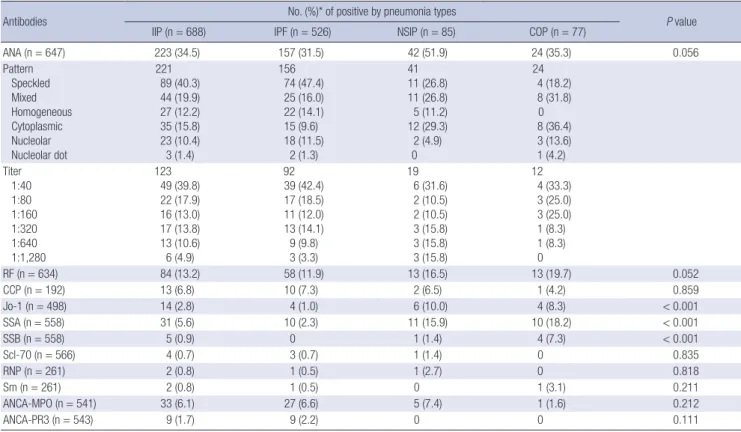
ANA and RF were evaluated in more than 90% of the subject patients and most of the specific antibodies were also tested in the majority of the patients, with exception of anti-CCP anti- body which was measured in just 192 subjects (27.9%). Approx- imately one-third of the patients (223, 34.5%) were positive for ANA and 13.2% had positive RF results. However, the preva- lence of most of the specific autoantibodies was low (between 0.7% and 6.8%). ANA positivity was more frequent in the NSIP group compared with the other groups (Table 2).
In patients with IPF, a speckled pattern was the most com- mon. The ANA titer was available in 547 patients, including ANA-negative (< 1:40) patients. The majority of the patients had a low ANA titer (less than 1:80), and only 30% had a titer higher than 1:320 (Table 2).
Table 1. Baseline clinical and demographic features of all patients
Baseline features Findings
Total number 688
Age (yr) 61.3 ± 9.4
Males 468 (68.0%)
Never smoking 264 (38.4%)
Pulmonary function FVC (% predicted) FEV1 (% predicted) TLC (% predicted) DLco (% predicted)
72.3 ± 19.5 83.8 ± 20.2 75.9 ± 17.1 63.6 ± 20.8 6MWT
Distance, meters
Lowest SpO2 (%) 452.7 ± 110
89.2 ± 7.1 BAL
Macrophage (%) Lymphocyte (%) Neutrophil (%)
65.5 ± 21.6 17.3 ± 14.9 11.2 ± 15.9 IIP type
IPF NSIP COP
526 (76.5%) 85 (12.4%) 77 (11.2%)
FVC, forced vital capacity; FEV1, forced expiratory volume in one second; TLC, total lung capacity; DLco, diffusing capacity of the lungs for carbon monoxide; 6MWT, 6- minute walk test; SpO2, oxygen saturation; BAL, bronchoalveolar lavage; IIP, idiopathic interstitial pneumonia; IPF, idiopathic pulmonary fibrosis; NSIP, nonspecific interstitial pneumonia; COP, cryptogenic organizing pneumonia.
Table 2. Frequency of autoantibodies detected in patients with IIP
Antibodies No. (%)* of positive by pneumonia types
P value
IIP (n = 688) IPF (n = 526) NSIP (n = 85) COP (n = 77)
ANA (n = 647) 223 (34.5) 157 (31.5) 42 (51.9) 24 (35.3) 0.056
Pattern Speckled Mixed Homogeneous Cytoplasmic Nucleolar Nucleolar dot
221 89 (40.3) 44 (19.9) 27 (12.2) 35 (15.8) 23 (10.4) 3 (1.4)
156 74 (47.4) 25 (16.0) 22 (14.1) 15 (9.6) 18 (11.5)
2 (1.3)
41 11 (26.8) 11 (26.8) 5 (11.2) 12 (29.3) 2 (4.9) 0
24 4 (18.2) 8 (31.8) 0
8 (36.4) 3 (13.6) 1 (4.2) Titer
1:40 1:80 1:160 1:320 1:640 1:1,280
123 49 (39.8) 22 (17.9) 16 (13.0) 17 (13.8) 13 (10.6) 6 (4.9)
92 39 (42.4) 17 (18.5) 11 (12.0) 13 (14.1) 9 (9.8) 3 (3.3)
19 6 (31.6) 2 (10.5) 2 (10.5) 3 (15.8) 3 (15.8) 3 (15.8)
12 4 (33.3) 3 (25.0) 3 (25.0) 1 (8.3) 1 (8.3) 0
RF (n = 634) 84 (13.2) 58 (11.9) 13 (16.5) 13 (19.7) 0.052
CCP (n = 192) 13 (6.8) 10 (7.3) 2 (6.5) 1 (4.2) 0.859
Jo-1 (n = 498) 14 (2.8) 4 (1.0) 6 (10.0) 4 (8.3) < 0.001
SSA (n = 558) 31 (5.6) 10 (2.3) 11 (15.9) 10 (18.2) < 0.001
SSB (n = 558) 5 (0.9) 0 1 (1.4) 4 (7.3) < 0.001
Scl-70 (n = 566) 4 (0.7) 3 (0.7) 1 (1.4) 0 0.835
RNP (n = 261) 2 (0.8) 1 (0.5) 1 (2.7) 0 0.818
Sm (n = 261) 2 (0.8) 1 (0.5) 0 1 (3.1) 0.211
ANCA-MPO (n = 541) 33 (6.1) 27 (6.6) 5 (7.4) 1 (1.6) 0.212
ANCA-PR3 (n = 543) 9 (1.7) 9 (2.2) 0 0 0.111
*Data are presented as number (% of examined patients). IIP, idiopathic interstitial pneumonia; IPF, idiopathic pulmonary fibrosis; NSIP, nonspecific interstitial pneumonia; COP, cryptogenic organizing pneumonia; ANA, antinuclear antibody; RF, rheumatoid factor; CCP, citrullinated protein; Jo-1, anti-Jo1 antibody; SSA, anti-SSA antibody (anti-Ro anti- body); SSB, anti-SSB antibody (anti-La antibody); Scl 70, anti-topoisomerase antibody; RNP, anti-ribonucleoprotein antibody; Sm, anti-Smith antibody; ANCA, anti-neutrophil cy- toplasmic antibody; MPO, myeloperoxidase; PR3, proteinase-3.
Comparisons of the clinical features of IIP patients according to the presence of autoantibodies
Among the patients who were positive for ANA, females and never smokers were predominant (Table E2, online supplemen- tal data). Patients with positive ANA titers had a lower lung func- tion and a tendency towards a higher lymphocyte percentage in bronchoalveolar lavage (BAL) fluid than ANA-negative cases.
There were no significant differences between the RF (+) and RF (-) groups other than a higher percentage of neutrophils in the BAL fluid of RF (+) patients.
Because the prognostic value of autoantibodies is more im- portant in IPF than in any other types of IIP, we only analyzed and compared the outcome for IPF. The median survival out- come was not significantly different between the ANA-positive and ANA-negative groups (40.6 vs 46.2 months) (Table E2, on- line supplemental data). The one- and three-year survival rates for ANA-positive patients (83.9% and 67.0%, respectively) were also not found to be significantly different from those of ANA- negative patients (1-yr, 85.4%; 3-yr, 65.2%; P = 0.155). The result was the same when only the patients with higher titers of ANA were categorized as the positive group. Similarly, in all patients including those with NSIP and COP, no significant difference in survival was found between the ANA-positive and –negative patients (data not shown).
Table 3. Development of CTD during follow-up
Diseases CTD (%) ANA
P value RF
P value
(+) (-) (+) (-)
IIP (n = 688) 26 (3.8) 19 (8.5) 7 (1.7) < 0.001 10 (11.9) 16 (2.9) 0.001
IPF (n = 526) 13 (2.5) 10 (6.4) 3 (0.9) 0.001 3 (5.2) 10 (2.3) 0.192
NSIP (n = 85) 8 (9.4) 5 (11.9) 3 (7.7) 0.715 4 (30.8) 4 (6.0) 0.021
COP (n = 77) 5 (6.5) 4 (16.7) 1 (2.3) 0.049 3 (23.1) 2 (3.8) 0.048
ANA, antinuclear antibody; RF, rheumatoid factor; IIP, idiopathic interstitial pneumonia; IPF, idiopathic pulmonary fibrosis; NSIP, nonspecific interstitial pneumonia; COP, crypto- genic organizing pneumonia.
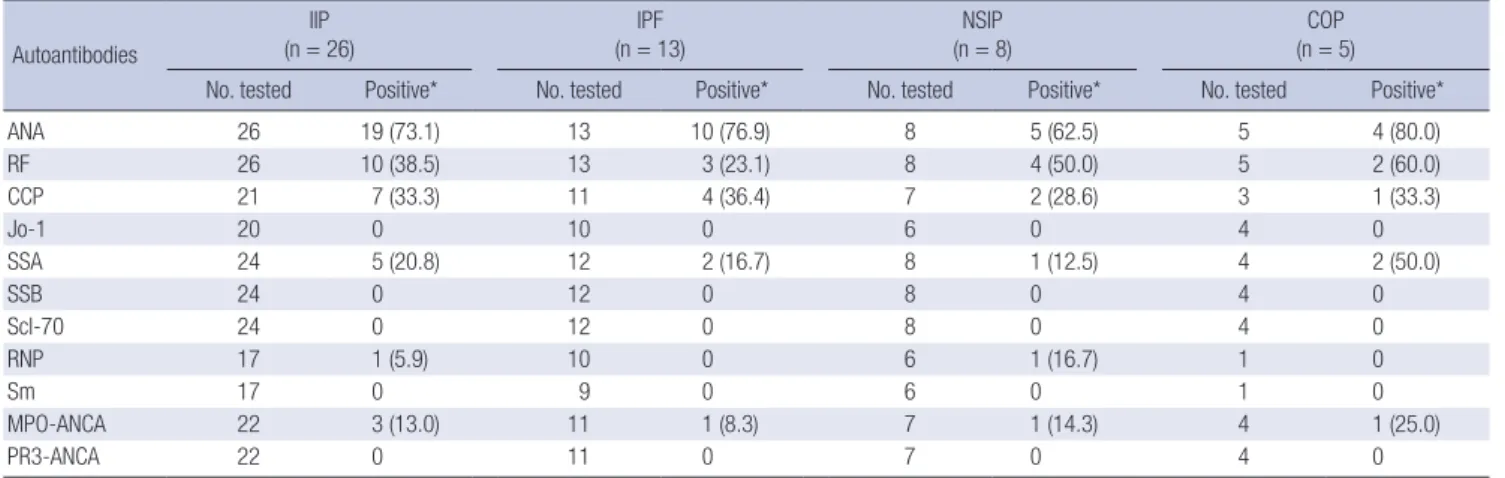
Table 4. Initial prevalence of autoantibodies in patients who developed CTD
Autoantibodies
IIP (n = 26)
IPF (n = 13)
NSIP (n = 8)
COP (n = 5)
No. tested Positive* No. tested Positive* No. tested Positive* No. tested Positive*
ANA 26 19 (73.1) 13 10 (76.9) 8 5 (62.5) 5 4 (80.0)
RF 26 10 (38.5) 13 3 (23.1) 8 4 (50.0) 5 2 (60.0)
CCP 21 7 (33.3) 11 4 (36.4) 7 2 (28.6) 3 1 (33.3)
Jo-1 20 0 10 0 6 0 4 0
SSA 24 5 (20.8) 12 2 (16.7) 8 1 (12.5) 4 2 (50.0)
SSB 24 0 12 0 8 0 4 0
Scl-70 24 0 12 0 8 0 4 0
RNP 17 1 (5.9) 10 0 6 1 (16.7) 1 0
Sm 17 0 9 0 6 0 1 0
MPO-ANCA 22 3 (13.0) 11 1 (8.3) 7 1 (14.3) 4 1 (25.0)
PR3-ANCA 22 0 11 0 7 0 4 0
*Data are presented as number (% of examined patients). IIP, idiopathic interstitial pneumonia; IPF, idiopathic pulmonary fibrosis; NSIP, nonspecific interstitial pneumonia; COP, cryptogenic organizing pneumonia; ANA, antinuclear antibody; RF, rheumatoid factor; CCP, citrullinated protein; Jo-1, anti-Jo1 antibody; SSA, anti-SSA antibody (anti-Ro anti- body); SSB, anti-SSB antibody (anti-La antibody); Scl 70, anti-topoisomerase antibody; RNP, anti-ribonucleoprotein antibody; Sm, anti-Smith antibody; ANCA, anti-neutrophil cy- toplasmic antibody; MPO, myeloperoxidase; PR3, proteinase-3.
Table 5. Relative risk of an ANA titer higher than 1:320 for the development of CTD
RR 95% CI P value
IIP 6.413 2.340-17.569 0.002
IPF 6.318 1.597-24.994 0.024
NSIP 4.625 1.094-15.246 0.067
IIP, idiopathic interstitial pneumonia; IPF, idiopathic pulmonary fibrosis; NSIP, nonspe- cific interstitial pneumonia; COP, cryptogenic organizing pneumonia; RR, relative risk;
95% CI, 95% confidential interval.
Development of overt CTD during follow-up
Of the 688 patients in our current study cohort with IIP, 26 cases (3.8%) developed overt CTD: 2.5% in IPF, 6.5% in COP, and 9.4%
in NSIP (Table 3). Rheumatologic consultation was done for all patients at the time of CTD diagnosis but not initially, because they did not have any symptoms suggestive of CTDs. RA was the most common CTD (all in the IPF group), followed by Sjo- gren’s syndrome and PM/DM (Table E3, online supplemental data). Two patients who were positive MPO-ANCA (one with IPF and one with COP) developed vasculitis (microscopic poly- angiitis). CTD development was higher in the ANA-positive group (8.5% in IIP, 6.4% in IPF) than the ANA-negative group (1.7% of IIP, 0.9% of IPF; P = 0.001). Most of the patients (73.1%) who developed CTD had positive ANA titers at the time of the initial diagnosis of IIP (Table 4), which was significantly higher than the prevalence in all subjects (34.5%)(Table 2). Moreover, the frequency of CTD development correlated with an increas- ing ANA titer (data not shown): the relative risk for a titer higher than 1:320 was 6.431 (95% CI, 2.340-17.569; P = 0.002) (Table 5).
Regarding specific autoantibodies, the frequency of positivity of anti-SSA (20.8%), anti-CCP (33.3%), and MPO-ANCA (13.0%) was significantly higher in the patients who developed CTD. In addition, the frequency of CTD development was higher in the
patients who were positive for specific autoantibodies (16.3 vs 2.1%; P < 0.001), especially in cases positive for anti-CCP and anti-SSA (anti-CCP, 53.8%; anti-SSA, 16.1%), when compared with the no CTD group (7.8%, and 3.6%, respectively; Table E4, online supplemental data).
DISCUSSION
The results of our current study show that in IPF, the presence of autoantibodies has no significant predictive value for survival.
However, autoantibodies were found to be predictive for the fu- ture development of CTD, although the incidence of newly de- veloped CTD was low in our patient series. The majority of our patients who developed CTD had a positive ANA titer, and the frequency of new CTD was significantly higher among patients
who were positive for ANA (> 1:320), anti-CCP, and anti-SSA.
Although it is well known that CTD-IP has a better prognosis than IPF and that some patients with IIP develop CTD during follow-up, the true relationship between autoantibodies and an IIP prognosis has not been clear to date. This relationship is more critical in patients with a UIP pattern because of the poor prognosis associated with IPF and the much better prognosis for cases of CTD-related UIP. Recently, we reported that the prognosis was similar in IPF patients with and without autoan- tibodies, although the pathological features of patients with IPF and positive autoantibodies were closer to those of CTD-UIP cases than to IPF cases without autoantibodies (17). Because of the small number of subjects in that study (n = 100), we recom- mended further investigation with a larger cohort. In our pres- ent study, which analyzes a much larger number of patients, no relationship between survival and autoantibodies could be con- firmed. Although the result was negative, this is the first reliable data obtained from large number of the patients with a relative- ly long-term follow-up period.
Although conducted before the ATS/ERS consensus classifi- cation was developed, many previous studies have reported a high prevalence of ANA and RF positivity (6, 18-22), and which was also recently confirmed by Fischer et al. (23) for surgical lung biopsy proven IPF. The prevalence was higher in idiopath- ic NSIP (24) and similar results were obtained in our present study. ANA is also present in healthy individuals (25-27); 1:40 in 25%-30%, 1:80 in 10%-15%, and 1:160 or greater in 5% (27). In our current study, the prevalence of a low titer of ANA was simi- lar but the prevalence of a high titer of ANA seemed to be high- er than previously reported value (Table 2). Moreover, whereas Fischer et al. (23) have reported that a nucleolar pattern is pre- dominant (26%) in IIP with a frequent development of sclero- derma. Mitoo et al. (24) reported that a speckled pattern was predominant in these cases without any development of sclero- derma, similar to the findings in our present study.
The incidence of CTD in our patients series was found to be low, with only 2.5% in IPF patients (3.8% in IIP), compared with the 19.1% reported by Homma et al. (4) and the 17.5% reported by Mittoo et al. (24). However, the subject numbers in those two studies were relatively small (n = 13) (4), or (n = 97) (24) and they included other types of interstitial lung disesase with sig- nificant referral bias by study design. Despite the low incidence of CTD, our study results show that CTD-development is close- ly associated with ANA; not only was there a higher incidence of CTD in the ANA-positive group than in the ANA-negative group (8.5% vs 1.7%, respectively; P = 0.001), but there was also a higher initial positivity for ANA in the CTD-development group compared with the non-CTD group. Our results also provide supporting evidence for accepting an ANA titer > 1:320 as a provisional criterion for lung dominant CTD, as proposed pre- viously by Fischer et al. (28).
Mittoo et al. (24) have reported that inflammatory myositis is the most common new CTD. In our present study however, this condition developed in only three patients; one with NSIP and two with COP, and no patients with in IPF. Because of the un- availability of anti-synthetase antibody test for our present anal- yses, we cannot exclude the possibility of misdiagnosis. How- ever, we paid particular attention to the clinical features of that disease in concert with our rheumatology colleagues and con- sidering rapid (less than one year) development of inflamma- tory myositis reported by Mittoo et al. (24) the chance of misdi- agnosis is likely to be low. In our present study, the most com- mon type of subsequent CTD was found to be RA in IPF, con- sistent with the observation that the UIP pattern is the most fre- quent pathological pattern in RA-IP patients. The anti-CCP an- tibody test was introduced late into our study and was therefore performed in only one-third of the patients. However, about half of the patients who developed RA were positive for the anti- CCP antibody at the initial evaluation without showing any clin- ical features of RA. Hence, our results also indicate that anti- CCP positivity is a predictor for RA (29-31).
Classically, specific autoantibodies are considered to be high- ly specific for the diagnosis of certain rheumatologic diseases (32, 33). A few of our patients were positive for specific autoan- tibodies but without any evidence of CTD or vasculitis (Table 4).
Four of these cases developed CTD that was specific for the cor- responding autoantibody, such as anti-RNP for MCTD or anti- SSA for Sjogren’s syndrome, and two patients with positive MPO- ANCA developed vasculitis (microscopic polyangiitis). In con- trast to ANA (24-26), only a few previous studies have reported the prevalence of specific autoantibodies in non-CTD individu- als; anti-Ro, 2.7%-10.2% (33, 34), anti-La, 0.9% (34), anti-Scl70, 0%-3% (35, 36), anti-Sm, 0%-0.5% (34, 35), which are similar to our results.
Another value of autoantibody testing is in providing clue to a possible unrecognized CTD. Our study was not designed to evaluate this possibility. However, Mittoo et al. (24) have report- ed that 71% of the patients they analyzed with a newly diagnosed CTD had a positive ANA titer, in contrast to a 45% positivity lev- el in non-CTD patients. Related to this, Castelino et al. (37) have reported that among 15 patients referred to as IPF, seven were diagnosed as CTD and all were ANA-positive. Despite being limited by the small numbers of patients examined, these earli- er reports showed that a significant number of patients with IIP may have an unrecognized CTD and that a positive ANA result may have utility as a warning signal for CTD.
A positive RF result has been previously reported in 4% of young healthy individuals (38) and in 3%-25% of elderly sub- jects without rheumatologic diseases (39, 40). Considering the mean age of our patients, the prevalence of RF positivity in our patients is similar to that of elderly people. Although we observ- ed that CTD development was higher in patients who were pos-
itive RF, most of the patients were in the NSIP group.
Our present study has several limitations. Because this is a retrospective study, and despite of all the efforts to exclude CTD at the initial diagnosis, there is still a possibility that CTD was missed in some cases, especially inflammatory myositis and UCTD. Although we used a checking assessment protocol for CTD, there remains the possibility that minor symptoms and/
or signs had been missed by either the patients themselves or their physicians. In addition, we could not check the presence of myositis associated specific antibodies. However, a thorough systematic review of patient symptoms, physical examination, and serological testing for CTD with frequent rheumatologic consultation were performed in the majority of the patients at the time of initial diagnosis and again during follow-up. There- fore the possibility that we failed to exclude CTD patients is less likely. Another possibility is that the development of CTD was masked by immunosuppressive therapies administered after the initial diagnosis. However, most of the patients with IPF in our present study were not treated, and if treated, the duration was only briefly. Therefore this possibility that CTD develop- ment was masked by immunosuppressive therapies is again less likely. Another noteworthy possible limitation is the rela- tively short follow-up duration (median, 33.6 months). Howev- er, considering the short survival period associated with IPF and the high proportion of IPF case in our cohort, our follow- up period may be reasonable for the purpose of these analyses.
Nonetheless, it may be too short for the development of CTD, as it usually takes more than 10 yr in patients with autoantibod- ies to develop overt CTD. This may be one possible explanation for the low incidence of CTD development in patients with au- toantibodies in our study. Despite these limitations, our study provides a robust analyseis of the incidence of newly arising CTD in patients with IIP, particularly IPF, and also the clinical significance of autoantibodies.
In conclusion, our current findings shows that the presence of autoantibodies, especially ANA with a titer > 1:320, and anti- CCP and anti-Ro antibody positivity is clinically useful for pre- dicting the future development of CTD in patients with IIP, al- though theses factors have no significant predictive value for mortality.
ACKNOWLEDGMENTS
The authors thank Min Ju Kim for assistance with the statistical analysis.
DISCLOSURE
The authors have no conflicts of interest to disclose.
REFERENCES
1. American Thoracic Society; European Respiratory Society. American Thoracic Society/European Respiratory Society International Multidisci- plinary Consensus Classification of the Idiopathic Interstitial Pneumo- nias: this joint statement of the American Thoracic Society (ATS), and the European Respiratory Society (ERS) was adopted by the ATS board of directors, June 2001 and by the ERS Executive Committee, June 2001.
Am J Respir Crit Care Med 2002; 165: 277-304.
2. Park JH, Kim DS, Park IN, Jang SJ, Kitaichi M, Nicholson AG, Colby TV.
Prognosis of fibrotic interstitial pneumonia: idiopathic versus collagen vascular disease-related subtypes. Am J Respir Crit Care Med 2007; 175:
705-11.
3. Raghu G, Collard HR, Egan JJ, Martinez FJ, Behr J, Brown KK, Colby TV, Cordier JF, Flaherty KR, Lasky JA, et al. An official ATS/ERS/JRS/ALAT statement: idiopathic pulmonary fibrosis: evidence-based guidelines for diagnosis and management. Am J Respir Crit Care Med 2011; 183: 788- 824.
4. Homma Y, Ohtsuka Y, Tanimura K, Kusaka H, Munakata M, Kawakami Y, Ogasawara H. Can interstitial pneumonia as the sole presentation of collagen vascular diseases be differentiated from idiopathic interstitial pneumonia? Respiration 1995; 62: 248-51.
5. Arnett FC, Edworthy SM, Bloch DA, McShane DJ, Fries JF, Cooper NS, Healey LA, Kaplan SR, Liang MH, Luthra HS, et al. The American Rheu- matism Association 1987 revised criteria for the classification of rheu- matoid arthritis. Arthritis Rheum 1988; 31: 315-24.
6. Hochberg MC. Updating the American College of Rheumatology revised criteria for the classification of systemic lupus erythematosus. Arthritis Rheum 1997; 40: 1725.
7. Bohan A, Peter JB. Polymyositis and dermatomyositis (first of two parts).
N Engl J Med 1975; 292: 344-7.
8. LeRoy EC, Medsger TA Jr. Criteria for the classification of early systemic sclerosis. J Rheumatol 2001; 28: 1573-6.
9. Vitali C, Bombardieri S, Jonsson R, Moutsopoulos HM, Alexander EL, Carsons SE, Daniels TE, Fox PC, Fox RI, Kassan SS, et al. Classification criteria for Sjögren’s syndrome: a revised version of the European criteria proposed by the American-European Consensus Group. Ann Rheum Dis 2002; 61: 554-8.
10. Alarcón-Segovia D, Cardiel MH. Comparison between 3 diagnostic cri- teria for mixed connective tissue disease: study of 593 patients. J Rheu- matol 1989; 16: 328-34.
11. Mosca M, Tani C, Bombardieri S. Undifferentiated connective tissue dis- eases (UCTD): a new frontier for rheumatology. Best Pract Res Clin Rheu- matol 2007; 21: 1011-23.
12. Jennette JC, Falk RJ, Andrassy K, Bacon PA, Churg J, Gross WL, Hagen EC, Hoffman GS, Hunder GG, Kallenberg CG, et al. Nomenclature of systemic vasculitides: proposal of an international consensus conference.
Arthritis Rheum 1994; 37: 187-92.
13. Lightfoot RW Jr, Michel BA, Bloch DA, Hunder GG, Zvaifler NJ, McShane DJ, Arend WP, Calabrese LH, Leavitt RY, Lie JT, et al. The American Col- lege of Rheumatology 1990 criteria for the classification of polyarteritis nodosa. Arthritis Rheum 1990; 33: 1088-93.
14. Masi AT, Hunder GG, Lie JT, Michel BA, Bloch DA, Arend WP, Calabrese LH, Edworthy SM, Fauci AS, Leavitt RY, et al. The American College of
Rheumatology 1990 criteria for the classification of Churg-Strauss syn- drome (allergic granulomatosis and angiitis). Arthritis Rheum 1990; 33:
1094-100.
15. Kim DS, Yoo B, Lee JS, Kim EK, Lim CM, Lee SD, Koh Y, Kim WS, Kim WD, Colby TV, et al. The major histopathologic pattern of pulmonary fi- brosis in scleroderma is nonspecific interstitial pneumonia. Sarcoidosis Vasc Diffuse Lung Dis 2002; 19: 121-7.
16. Jegal Y, Kim DS, Shim TS, Lim CM, Do Lee S, Koh Y, Kim WS, Kim WD, Lee JS, Travis WD, et al. Physiology is a stronger predictor of survival than pathology in fibrotic interstitial pneumonia. Am J Respir Crit Care Med 2005; 171: 639-44.
17. Song JW, Do KH, Kim MY, Jang SJ, Colby TV, Kim DS. Pathologic and radiologic differences between idiopathic and collagen vascular disease- related usual interstitial pneumonia. Chest 2009; 136: 23-30.
18. Scadding JG, Hinson KF. Diffuse fibrosing alveolitis (diffuse interstitial fibrosis of the lungs): correlation of histology at biopsy with prognosis.
Thorax 1967; 22: 291-304.
19. Turner-Warwick M, Haslam P, Weeks J. Antibodies in some chronic fi- brosing lung diseases: II. immunofluorescent studies. Clin Allergy 1971;
1: 209-19.
20. Nagaya H, Sieker HO. Pathogenetic mechanisms of interstitial pulmo- nary fibrosis in patients with serum antinuclear factor: a histologic and clinical correlation. Am J Med 1972; 52: 51-62.
21. Chapman JR, Charles PJ, Venables PJ, Thompson PJ, Haslam PL, Maini RN, Turner Warwick ME. Definition and clinical relevance of antibodies to nuclear ribonucleoprotein and other nuclear antigens in patients with cryptogenic fibrosing alveolitis. Am Rev Respir Dis 1984; 130: 439-43.
22. Shmerling RH, Delbanco TL. The rheumatoid factor: an analysis of clin- ical utility. Am J Med 1991; 91: 528-34.
23. Fischer A, Pfalzgraf FJ, Feghali-Bostwick CA, Wright TM, Curran-Ever- ett D, West SG, Brown KK. Anti-th/to-positivity in a cohort of patients with idiopathic pulmonary fibrosis. J Rheumatol 2006; 33: 1600-5.
24. Mittoo S, Gelber AC, Christopher-Stine L, Horton MR, Lechtzin N, Da- noff SK. Ascertainment of collagen vascular disease in patients present- ing with interstitial lung disease. Respir Med 2009; 103: 1152-8.
25. Tan EM, Feltkamp TE, Smolen JS, Butcher B, Dawkins R, Fritzler MJ, Gordon T, Hardin JA, Kalden JR, Lahita RG, et al. Range of antinuclear antibodies in “healthy” individuals. Arthritis Rheum 1997; 40: 1601-11.
26. Kavanaugh A, Tomar R, Reveille J, Solomon DH, Homburger HA. Guide- lines for clinical use of the antinuclear antibody test and tests for specific autoantibodies to nuclear antigens: American College of Pathologists:
American College of Pathologists. Arch Pathol Lab Med 2000; 124: 71-81.
27. Solomon DH, Kavanaugh AJ, Schur PH; American College of Rheuma-
tology Ad Hoc Committee on Immunologic Testing Guidelines. Evi- dence-based guidelines for the use of immunologic tests: antinuclear an- tibody testing. Arthritis Rheum 2002; 47: 434-44.
28. Fischer A, West SG, Swigris JJ, Brown KK, du Bois RM. Connective tissue disease-associated interstitial lung disease: a call for clarification. Chest 2010; 138: 251-6.
29. Rantapää-Dahlqvist S, de Jong BA, Berglin E, Hallmans G, Wadell G, Stenlund H, Sundin U, van Venrooij WJ. Antibodies against cyclic citrul- linated peptide and IgA rheumatoid factor predict the development of rheumatoid arthritis. Arthritis Rheum 2003; 48: 2741-9.
30. Nielen MM, van Schaardenburg D, Reesink HW, van de Stadt RJ, van der Horst-Bruinsma IE, de Koning MH, Habibuw MR, Vandenbroucke JP, Dijkmans BA. Specific autoantibodies precede the symptoms of rheu- matoid arthritis: a study of serial measurements in blood donors. Arthri- tis Rheum 2004; 50: 380-6.
31. Van Venrooij WJ, van Beers JJ, Pruijn GJ. Anti-CCP Antibody, a Marker for the Early Detection of Rheumatoid Arthritis. Ann N Y Acad Sci 2008;
1143: 268-85.
32. Von Mühlen CA, Tan EM. Autoantibodies in the diagnosis of systemic rheumatic diseases. Semin Arthritis Rheum 1995; 24: 323-58.
33. Reveille JD, Solomon DH; American College of Rheumatology Ad Hoc Committee of Immunologic Testing Guidelines. Evidence-based guide- lines for the use of immunologic tests: anticentromere, Scl-70, and nucle- olar antibodies. Arthritis Rheum 2003; 49: 399-412.
34. Conrad K, Mehlhorn J. Diagnostic and prognostic relevance of autoanti- bodies in uranium miners. Int Arch Allergy Immunol 2000; 123: 77-91.
35. Hayashi N, Koshiba M, Nishimura K, Sugiyama D, Nakamura T, Mori- nobu S, Kawano S, Kumagai S. Prevalence of disease-specific antinuclear antibodies in general population: estimates from annual physical ex- aminations of residents of a small town over a 5-year period. Mod Rheu- matol 2008; 18: 153-60.
36. Parker JC, Bunn CC. Sensitivity of the Phadia EliA connective tissue dis- ease screen for less common disease-specific autoantibodies. J Clin Pathol 2011; 64: 631-3.
37. Castelino FV, Goldberg H, Dellaripa PF. The impact of rheumatological evaluation in the management of patients with interstitial lung disease.
Rheumatology (Oxford) 2011; 50: 489-93.
38. Newkirk MM. Rheumatoid factors: what do they tell us? J Rheumatol 2002; 29: 2034-40.
39. Litwin SD, Singer JM. Studies of the incidence and significance of anti- gamma globulin factors in the aging. Arthritis Rheum 1965; 8: 538-50.
40. Cammarata RJ, Rodnan GP, Fennell RH. Serum anti-gamma-globulin and antinuclear factors in the aged. JAMA 1967; 199: 455-8.