Omental Infarction Associated with Rib Cage Anomaly
in Achondroplasia: Report of a Rare Case
늑골 기형이 동반된 연골무형성증 환자에게서 발생한 대망 경색: 드문 증례 보고
Tae Hyung Kim, MD , You Sung Kim, MD*
Department of Radiology, Ilsan Paik Hospital, Inje University College of Medicine, Goyang, Korea
Omental infarction, a rare cause of acute abdominal pain, is usually difficult to diagnose before surgery. Several studies have shown that CT is useful in diagnosing an omental infarction. We report the first case of an omental infarction secondary to rib cage anomaly associated with achondroplasia. Preoperative CT revealed a fatty mass in the anterior perihepatic space and anterior flaring of the ribs. The patient, a 41-year-old man, was diagnosed with omental infarc- tion in the anterior perihepatic space and treated with intravenous painkillers. After discharge, because of recurrent abdominal pain, he was readmitted and successfully underwent laparo- scopic partial omentectomy. The mass was confirmed to be an infarcted omentum with fat ne- crosis and hemorrhage. Thus, omental infarction should be considered as a differential diagno- sis for acute abdominal pain, especially in patients with achondroplasia. Contrast-enhanced abdominal CT can help in correctly diagnosing a suspected omental infarction.
Index terms Omentum; Infarction; Acute Abdomen; Computed Tomography, X-Ray;
Achondroplasia
INTRODUCTION
Omental infarction rarely causes acute abdominal pain. On contrast-enhanced CT of the abdomen, omental infarction presents as a well-defined solitary ovoid or triangular fatty mass adjacent to the large bowel loops, with whorled patterns of the heteroge- neous or linear fat strands, and fat infiltration can be seen around the infarction (1, 2).
A whorled vascular pedicle may also be identified. Even though omental infarction is seldom diagnosed preoperatively, awareness of this condition is important to clinicians and radiologists because it can easily mimic the common causes of acute abdominal
Received November 13, 2018 Revised January 9, 2019 Accepted January 23, 2019
*Corresponding author You Sung Kim, MD Department of Radiology, Ilsan Paik Hospital, Inje University College of Medicine,
170 Juhwa-ro, Ilsanseo-gu, Goyang 10380, Korea.
Tel 82-31-910-7628 Fax 82-31-910-7369 E-mail youskim@gmail.com This is an Open Access article distributed under the terms of the Creative Commons Attribu- tion Non-Commercial License (https://creativecommons.org/
licenses/by-nc/4.0) which permits unrestricted non-commercial use, distribution, and reproduc- tion in any medium, provided the original work is properly cited.
ORCID iDs You Sung Kim https://
orcid.org/0000-0003-4388-2659 Tae Hyung Kim
https://
orcid.org/0000-0001-5426-4739
pain that need surgical treatment.
We report the first case of a surgically and histopathologically proven omental infarction associated with rib cage anomaly in an achondroplasia patient, which was diagnosed by pre- operative CT.
CASE REPORT
A 41-year-old man visited our emergency department with acute abdominal pain which had started 5 days ago in the right upper quadrant. He had been diagnosed with achondro- plasia and had no other associated symptoms. His vital signs were immediately assessed and were in the normal range. Physical examination showed tenderness at the right upper quad- rant without definite Murphy’s sign. Clinical laboratory examination showed increased erythrocyte sedimentation rate (28 mm/hr) and C-reactive protein (1.5 mg/dL).
Abdominal radiography showed a nonspecific distribution of bowel gas. Abdominal CT was performed immediately. The CT examination revealed a 4.2 × 1.4 × 4.4 cm fatty mass with central mixed soft-tissue attenuation in the anterior aspect of the liver, IV and VIII seg- ments (Fig. 1A, B). Both ribs of the patient showed anterior flaring, which formed a space in the anterior perihepatic space for omentum to be caught in. The CT also showed an ill-de- fined arterial enhancing lesion and indentation at the adjacent liver parenchyma. Based on these findings, he was diagnosed with omental infarction in the anterior perihepatic space.
After applying fluid therapy with an intravenous painkiller, the pain in his right upper quad- rant pain improved. After discharge from our hospital, he complained of recurrent abdomi- nal pain and decided to undergo surgical resection. Two weeks later, he underwent surgery for resection of the infarcted omentum.
During surgery, the greater omentum was found between the anterior abdominal wall and the liver surface. The trapped segment was grossly necrotic and had adhered to the abdomi- nal wall and the adjacent liver surface (Fig. 1C, D). Therefore, laparoscopic partial omentecto- my was performed and the pathological results showed necrosis and hemorrhage in the re- sected omentum. There was no specific event after surgery, and he was discharged from the hospital 2 days postoperatively.
DISCUSSION
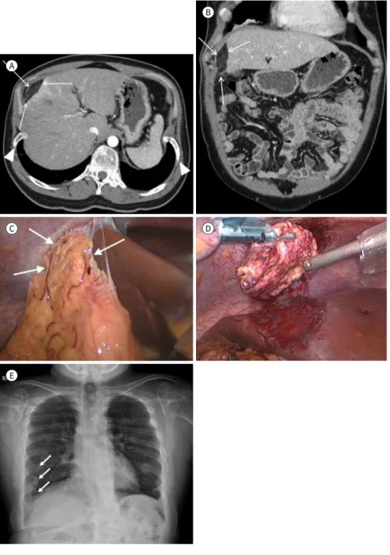
Fig. 1. Omental infarction in a 41-year-old achondroplasia patient, complaining with acute abdominal pain.
A-E. Axial (A) and coronal (B) CT scans show a fatty mass (arrows) with central mixed soft-tissue attenuation in the anterior aspect of the liver (segments IV and VIII). CT scans also show an ill-defined arterial-enhancing le- sion and indentation at the adjacent liver parenchyma. The axial (A) CT scan also shows anterior flaring of both ribs (arrowheads). The intraoperative image (C) shows the greater omentum found between the anterior ab- dominal wall and liver surface; the trapped segment is grossly necrotic and is adhered to the abdominal wall and adjacent liver surface (arrows). The surgeon successfully resected the trapped and infarcted omentum and controlled liver surface bleeding with argon plasma coagulation (D). The posteroanterior chest radiograph (E) shows shortening of anterior ribs and anteriorly flared ribs (arrows). A horizontal arrangement of rib is also seen, probably due to kyphoscoliosis, which is accompanied by achondroplasia.
A
C
E
D B
venous stasis and thrombosis, progresses to hemorrhagic necrosis, which leads to extravasa- tion of serosanguineous peritoneal fluid (7, 8).
In our case, the patient was diagnosed with achondroplasia, and he showed anterior flar- ing of both ribs on chest radiograph and CT. We assume that the anterior flaring of the ribs made space between the anterior abdominal wall and the anterior surface of the liver, and interposition of the omentum occurred due to this free space. This looked similar to the pathophysiology of Chilaiditi sign, which is characterized by colonic interposition between the liver and the diaphragm. Hepatic, intestinal, and/or diaphragmatic etiologies contribute to the pathogenesis of Chilaiditi sign. Variations in normal anatomy or an existing anatomi- cal anomaly can lead to the pathological interposition of the colon (9). We also think that this trapped omentum underwent a similar pathology of omental infarction which mentioned above. Considering the pathophysiology of omental infarction, we expected that the omen- tum would easily get caught in the free space if the entrance was narrow.
To our knowledge, our report is the first case of an omental infarction associated with rib cage anomaly in an achondroplasia patient, which correlates with both radiological and his- topathological findings. The management of omental torsion is debatable. Some authors support conservative management, whereas others advocate that surgical treatment is ad- vantageous, especially considering the advanced laparoscopic technologies (10). Our patient’
s symptoms improved immediately with painkillers via intravenous fluid therapy, so he wanted to observe the progress. Finally, he decided to have surgical resection due to recurrent severe abdominal pain despite the use of oral analgesic.
In conclusion, although omental infarction is a rare cause of acute abdominal pain, it should be considered as one of the differential diagnoses for acute abdomen, especially in pa- tients with achondroplasia. In some cases of suspected omental infarction, contrast-enhanced abdominal CT can assist in the diagnosis.
Conflicts of Interest
The authors have no potential conflicts of interest to disclose.
REFERENCES
1. Grattan-Smith JD, Blews DE, Brand T. Omental infarction in pediatric patients: sonographic and CT find- ings. AJR Am J Roentgenol 2002;178:1537-1539
2. Singh AK, Gervais DA, Hahn PF, Sagar P, Mueller PR, Novelline RA. Acute epiploic appendagitis and its mim- ics. Radiographics 2005;25:1521-1534
ol Hepatol (N Y) 2012;8:276-278
10. Nubi A, McBride W, Stringel G. Primary omental infarct: conservative vs operative management in the era of ultrasound, computerized tomography, and laparoscopy. J Pediatr Surg 2009;44:953-956
늑골 기형이 동반된 연골무형성증 환자에게서 발생한 대망 경색: 드문 증례 보고
김태형 · 김유성*
대망 경색은 급성 복통의 드문 원인 중 하나로, 수술 전 진단이 어렵다. 이전의 여러 연구를 통해 컴퓨터단층촬영(CT)이 대망 경색을 진단하는 데 유용하다는 것이 알려져 있다. 저자들 은 늑골 기형이 동반된 연골무형성증을 진단받은 41세 남자 환자에게서 발생한 속발성 대망 경색을 보고하고자 한다. 환자는 급성 복통을 주소로 응급실 방문 후 시행한 조영증강 CT에 서 그물막 경색을 진단받았다. 환자는 통증 조절 후 귀가하였으나, 통증이 지속되어 재내원 하여 수술적 제거를 시행하였고 최종 병리검사에서 확진되었다. 드문 원인이기는 하지만 급 성 복통을 호소하는 환자, 특히 연골무형성증이 있는 환자에게서 대망 경색을 감별진단 중 하나로 고려해야 한다는 점과, 조영증강 CT가 대망 경색을 정확히 진단하는 데 도움이 될 수 있다는 점을 함께 보고하고자 한다.
인제대학교 의과대학 일산백병원 영상의학과