677
The Usefulness of P504S/34βE12 Immunostaining for the Detection of Prostate Cancer
Jong-hyun Yun, Hyun-deuk Cho1, Doo-sang Kim, Chang-ho Lee From the Departments of Urology and 1Pathology, Soonchunhyang University Che- onan Hospital, Soonchunhyang University College of Medicine, Cheonan, Korea Purpose: We evaluated the significance of the P504S expression in prostate cancer and also the usefulness of the P504S/34βE12 combined immuno- staining method for diagnosing prostate cancer, and we did this by per- forming histological analysis of needle biopsy specimens.
Materials and Methods: Prostate tissue specimens were obtained from 83 patients with clinically suspected prostate cancer. A total of 54 prostate needle biopsy specimens were immunostained with an enzyme commonly overexpressed in prostate cancer (P504S) and also with an antibody against a basal cell marker (34βE12). A total of 83 cases were immunostained with 34βE12, including 29 cases that were stained with only with HMW- CK (34βE12).
Results: P504S immunostaining was positive in 96.3% (26 of 27 cases) of the prostate cancer specimens. 34βE12 immunostaining was positive in 97.2% (35 of 36 cases) of the benign prostate tissues. Of the 30 P504S posi- tive immunostaining cases, 26 cases were prostate cancers, 3 cases were ASAP and 1 case was ASAP+PIN. Of the 36 34βE12 positive immuno- stained cases, 35 cases were benign and 1 case was ASAP. In the P504S (+)/34βE12 () cases, there are no benign prostate lesions. There are no benign prostate lesions in the P504S ()/34βE12 (+) cases, and all the cases were benign. There were no statistical correlations between the grade of P504S staining and the clinical parameters such as serum PSA, the clinical stage and the Gleason scores.
Conclusions: Combining P504S as a positive marker for prostate cancer with 34βE12 as a negative marker might improve the diagnostic perfor- mance. (Korean J Urol 2007;48:677-683)
Key Words: Prostate cancer, Needle biopsy, P504S protein
대한비뇨기과학회지 제 48 권 제 7 호 2007
순천향대학교 의과대학 순천향대학교
천안병원 비뇨기과학교실, 1병리과
윤종현ㆍ조현득1ㆍ김두상ㆍ이창호
접수일자:2007년 4월 18일 채택일자:2007년 5월 16일
교신저자: 이창호
순천향대학교 천안병원 비뇨기과
충남 천안시 봉명동 23-20
330-721
TEL: 041-570-2275 FAX: 041-574-6248 E-mail: [email protected]
서 론
침생검으로 얻은 전립선조직으로 전립선암 진단을 할 경 우 검체의 양이 적어 검체의 국소부위에서만 비정상적인 선조직이 관찰될 때는 병리진단이 어려운 경우가 있다. 이 때 보조적인 방법으로 면역염색을 시행한다.
전립선암은 기저세포층이 소실되는 반면 양성 전립선질 환은 기저세포층이 유지된다. 따라서 기저세포를 특이 염 색하면 전립선암과 양성 전립선 질환을 구분할 수 있다. 기 저세포를 염색하는 항체 및 이를 이용한 면역염색의 유용
성은 여러 문헌에 보고되어 있으며,1 여러 기저세포 표지자 중 34βE12는 가장 먼저 개발된 기저세포의 표지자이다.
최근 전립선암의 진단에 유용한 표지자로 α-methyl- acyl-Co A racemase (AMACR)가 있다. AMACR은 전립선암 조직에서 발현이 증가하는 반면에 양성전립선조직에서는 거의 발현되지 않는다. 세포 내에서 AMACR의 발현을 확 인하는 monoclonal 항체로는 P504S가 있다.2,3
본 연구에서는 임상적으로 전립선이 의심되는 환자의 침 생검 조직에서 P504S의 발현양상과 진단의 보조도구로서 의 유용성을 알아보고자 하였다. 또한 P504S 면역염색을 전 립선암 진단의 양성표지자로, 34βE12 면역염색을 전립선
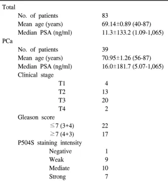
Table 1. Patients and their parameter characteristics Total
No. of patients 83
Mean age (years) 69.14±0.89 (40-87) Median PSA (ng/ml) 11.3±133.2 (1.09-1,065) PCa
No. of patients 39
Mean age (years) 70.95±1.26 (56-87) Median PSA (ng/ml) 16.0±181.7 (5.07-1,065) Clinical stage
T1 4 T2 13 T3 20 T4 2 Gleason score
≤7 (3+4) 22 ≥7 (4+3) 17 P504S staining intensity
Negative 1 Weak 9 Mediate 10 Strong 7
PSA: prostate-specific antigen, PCa: prostate cancer 대상 및 방법
1. 전립선 조직 생검
2003년 1월부터 2006년 3월까지 본원 비뇨기과에서 전립 선 침생검을 시행한 83명의 전립선 생검 조직을 대상으로 하였다. 전립선 침생검은 초음파 유도하 여섯 부위 또는 8, 10, 12 부위 생검을 시행하였다. 환자는 40-87세 (평균 69.14±
0.89세)였고 전립선특이항원 (prostate-specific antigen; PSA) 은 1.09-1,065ng/dl (중앙값 11.3±133.2ng/ml)이었다 (Table 1).
83례 침생검조직의 hematoxylin-eosin 염색에 기초한 병리 진단은 39례가 전립선암, 7례가 비전형적 작은꽈리증식증 (atypical small acinar proliferation; ASAP), 1례가 ASAP+전립 선상피내종양 (prostatic intraepithelial neoplasia; PIN), 그리고 36례가 양성전립선질환이었다.
2. 면역염색
1) P504S 면역염색: 파라핀 포매 조직은 xylene으로 탈파 라핀 처리하였고 고농도 알콜에서 저농도 알콜로 함수과정 을 거쳤다. P504S (α -methylacyl-Co A racemase: clone: 13H4 1,2,3 monoclonal rabbit anti-human P504S; code M3616, DakoCytomation)의 항원부활을 위해 극초단파 (microwave)
munostain (Bond-X)으로 염색하였다. 증류수로 수세 후 저 농도 알콜에서 고농도 알콜로 탈수과정을 거치고 봉입한 후 검경하였다.
P504S에서 양성 면역 염색의 기준은 저배율로 보았을 때 내강을 가지는 환상 선구조로 세포 내 세포질에 전반적으 로 염색이 되었을 때를 양성으로 판정하였고, 염색의 강도 를 전체 슬라이드의 가장 진한 염색이 전체 조직 슬라이드 의 30% 이하 세포 내 세포질에 염색이 되었을 때를 약한 염색, 전체 슬라이드의 가장 진한 염색이 전체 조직 슬라이 드의 30%에서 60% 이하의 세포 내 세포질에 염색이 되었 을 때를 중등도 염색, 전체 슬라이드의 가장 진한 염색이 전체 조직 슬라이드의 60% 이상 세포 내 세포질에 염색이 되었을 때를 강한 염색으로 나누었다 (Fig. 1). 83례의 침생 검 조직 중 54례에서 P504S 면역염색을 시행하였다.
2) 34βE12 면역염색: 파라핀 포매 조직을 4-μm 두께로 잘라 절편을 얻은 후, 조직 슬라이드를 펩신처리하고 0.1%
proteinase에서 30분 동안 37oC에서 전처치를 했다. Keratin 903 1:100 희석 용액에 3시간 처리 후 cytokine용액 처리하 였다. 수세와 버퍼처리 후 hematoxylin으로 대조 염색하였 다 (Fig. 2). 양성 면역 염색의 기준은 선의 변연부를 따라서 잘 염색이 된 경우로 하였다.
83례의 침생검 조직 전 예에서 34βE12 면역염색을 시행 하였고, 이중 54례는 P504S 면역염색을 함께 시행하였다.
3) 통계분석: 통계학적 검정은 면역염색의 강도와 임상지 표와의 연관성을 파악하기 위해 Fisher's exact test를 이용하 였고 p값이 0.05 미만인 경우 의의가 있는 것으로 판정하였 다.
결 과
1. 전립선 침생검조직에서 P504S, 34βE12 면역염색
총 83례의 침생검조직은 전립선암 39례, ASAP 7례, ASAP+PIN 1례 그리고 양성전립선질환 36례였다. 이 중 P504S 면역염색을 시행한 54례는 전립선암 27례, ASAP 4 례, ASAP+PIN 1례 그리고 양성전립선질환 22례였다.
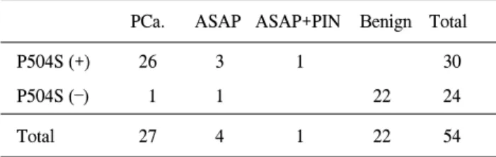
전립선암으로 진단되고 P504S 면역염색을 시행한 27례 중 P504S 면역염색에서 양성인 경우는 26례로 전립선암 진 단에 대한 P504S 면역염색의 민감도는 96.3%, 특이도는 85.2%였다. P504S 면역염색을 시행한 54례 중 P504S 양성 인 경우는 30례로 이 중 26례는 전립선암, 3례는 ASAP, 1례 는 ASAP+PIN으로 양성전립선질환인 경우는 없었다. P504S 면역염색의 양성예측률은 86.7%, 음성예측률은 95.8%였다
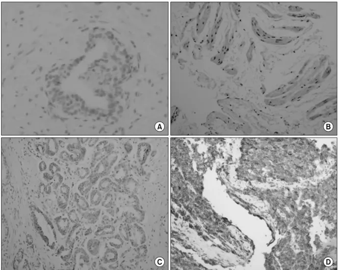
Fig. 1. P504S staining of the human prostate (A) P504S negative staining (x400) specimens at the intracytoplasm, (B) P504S weak staining (x200) of the intracytoplasm, (C) P504S moderate staining (x200) of the intracytoplasm and (D) P504S strong staining (x100) of the intra- cytoplasm.
Fig. 2. 34βE12 staining of the human prostate (A) 34βE12 negative staining (x100) specimens at the basement membrane and (B) 34βE12 positive staining (x200) of the basement mambrane.
Table 3. Correlation between the grade of P504S staining and the clinical parameters A: PSA level and P504S staining intensity (p=0.386)
PSA level
n=27 Total
0-4 4-10 10-20 20-
Staining intensity Negative 0 0 1 0 1
Weak 0 1 2 6 9
Moderate 0 2 5 3 10
Strong 0 3 3 1 7
Total 0 6 11 10 27
B: Primary Gleason score and P504S staining intensity (p=0.175)
Gleason score
n=27 Total
4 5 6 7 8 9
Staining intensity Negative 1 0 0 0 0 0 1
Weak 0 1 0 1 6 1 9
Moderate 0 0 5 5 0 0 10
Strong 1 0 3 3 0 0 7
Total 2 1 8 9 6 1 27
C: Clinical stage and P504S staining intensity (p=0.088)
Clinical stage
n=27 Total
T1c T2a T2b T3a T3b T4a
Staining intensity Negatve 0 0 0 0 1 0 1
Weak 0 0 0 5 3 1 9
Moderate 0 4 2 0 4 0 10
Strong 2 1 1 1 1 1 7
Total 2 5 3 6 9 2 27
PSA: prostate-specific antigen
P504S (+) 26 3 1 30
P504S () 1 1 22 24
Total 27 4 1 22 54
PCa.: prostate cancer, ASAP: atypical small acinar proliferation, PIN: prostate intraepithelial neoplasia
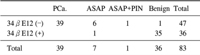
양성전립선질환으로 진단된 36례 중 34βE12 면역염색에 서 양성인 경우는 35례로 양성전립선질환 진단에 대한 34 βE12 면역염색의 민감도는 97.3%, 특이도는 97.9%였다.
34βE12 면역염색을 시행한 83례 중 34βE12 양성인 경우 는 36례로 이 중 35례는 양성전립선질환, 1례는 ASAP였다.
34βE12 면역염색의 양성예측률은 97.2%, 음성예측률은 97.9%였다 (Table 4).
Table 5. Combined analysis of the P504 and 34βE12 immuno- staining results
PCa. ASAP ASAP+PIN Benign Total P504S (+)
26 2 1 29
/34βE12 () P504S ()
22 22
/34βE12 (+)
Harmonious immunostaining results 51 cases P504S (+)
1 1
/34βE12 () P504S ()
1 1 2
/34βE12 (+)
Conflicting immunostaining results 3 cases
Total 27 4 1 22 54
PCa.: prostate cancer, ASAP: atypical small acinar proliferation, PIN: prostate intraepithelial neoplasia
Table 4. Results of 34βE12 immunostaining
PCa. ASAP ASAP+PIN Benign Total
34βE12 () 39 6 1 1 47
34βE12 (+) 1 35 36
Total 39 7 1 36 83
PCa.: prostate cancer, ASAP: atypical small acinar proliferation, PIN: prostate intraepithelial neoplasia
2. P504S 면역염색과 34βE12 면역염색의 결과의 조합
P504S 면역염색 양성, 34βE12 면역염색 음성 [P504S (+)/
34βE12 ()]인 경우는 모두 29례였는데 전립선암 26례, ASAP 2례, ASAP+PIN 1례로 양성전립선질환인 경우는 없 었다. P504S 면역염색 음성, 34βE12 면역염색 양성[P504S ()/34βE12 (+)]인 경우는 모두 22례였는데 모두 양성전립 선질환이었다 (Table 5).
P504S (+)/34βE12 () 또는 P504S ()/34βE12 (+)로 전립 선암에 대한 양성표지염색인 P504S와 음성표지염색인 34 βE12이 조화로운 결과를 보이는 경우에 민감도와 특이도 는 각각 100%와 88%였고, 양성예측률과 음성예측률은 각 각 89.7%와 100%였다.
P504S (+)/34βE12 (+) 또는 P504S ()/34βE12 ()로 전립 선암에 대한 양성표지염색인 P504S와 음성표지염색인 34 βE12이 대립하는 결과를 보인 경우는 전체 54례 중 3례로 5.6%였다. P504S (+)/34βE12 (+)인 경우는 1례였는데 ASAP 였고, P504S ()/34βE12 ()인 경우는 2례였는데, 전립선암 1례, ASAP 1례였다. 모두 양성 또는 모두 음성으로 대립하
는 결과를 보이는 경우 양성전립선질환인 경우는 없었다 (Table 5).
고 찰
면역염색은 검체의 제한된 부위에서 의심스러운 소견이 있을 때 유용한 진단 보조 도구이다. 양성 선조직은 기저세 포층이 보존되어 있으나, 악성 선조직은 기저세포층이 소 실되어 있어 기저세포층에 염색을 시도하는 방법을 사용해 왔다.4 이 중 본 연구에서 사용한 34βE12는 가장 먼저 개발 된 기저세포 특이 항체로 단일복제 항케라틴 항체 (mono- clonal anti-keratin antibody)이다.
기저세포를 염색하는 방법은 관찰하는 조직이 암이 아닌 것을 확인하는 일종의 음성 표지자로 암조직을 확인하는 양성 표지자인 AMACR에 대한 항체인 P504S를 사용할 수 있다.
AMACR은 전립선암 조직에 대한 유전자 검색을 통해 확 인되었는데, 양성 전립선조직에 비해 전립선암 조직에서 발현이 현저히 증가한다.5 AMACR 유전자는 염색체 5번에 위치하며 이의 인체에서의 역할, 전립선암에서의 역할에 대해서는 명확히 알려진 바는 없으나, 포화지방산 (branched chain fatty acid)과 지방산 유도체 (fatty acid derivative)의 산 화 (oxidation)에 관여한다.3
전립선암 조직에서 AMACR 단백 발현 민감도는 97%, 특 이도는 100%이다.5 따라서 전립선암의 진단에 민감하고 특 이적인 검사이나 PIN, ASAP 등 전립선암 유사조직에서도 염색이 되고 일부 양성 전립선조직에서도 발현이 된다. 또 한 전립선암이어도 AMACR의 염색이 되지 않는 경우도 있 다. 본 논문의 경우, 27례의 전립선암 환자 중 1례의 전립선 암 환자는 P504S 염색에 음성이었고 (3.7%, 1/27), 30명의 P504S 양성 환자 중 26명의 전립선암이었으나, 3명의 ASAP 환자와 1명의 ASAP+PIN 환자 (13.3%, 4/30)도 P504S 양성 이었다.
그러므로, P504S 면역염색은 단독으로 진단을 내리기보 다는 H&E 염색을 통해 세포 형태, 그리고 의심스러운 경우 에는 기저세포 면역염색 결과와 연관지어서 진단에 참고 하는 것이 중요하다. AMACR의 염색결과는 주의깊게 판독 해야 한다. AMACR, HMWCK, p63, H&E 염색에 의해 전립 선암으로 진단된 215례의 조직 중 18%는 AMACR 음성이 었다. 본 연구에서는 P504S 양성이면서 양성전립선질환인 경우는 없었다. PIN은 전립선암의 전구병변으로 생각되고, ASAP는 병리조직의 특성상 추적검사와 재생검이 요구된 다. PIN 또는 ASAP는 재생검 시 대략 40%의 환자에서 전립 선암이 발견된다.6 본 연구의 P504S 면역염색에서 양성인
또는 추적 및 재생검이 요구되는 전립선질환으로 추정 진 단할 수 있다.
P504S 면역염색을 통해 확인한 AMACR의 발현 정도와 임상에서 사용하는 전립선암의 일반적인 지표인 PSA, Glea- son score 그리고 임상병기와의 관련성을 알아보았다. 이는 전립선암에서 AMACR의 역할을 추정하는 한편, 이의 임상 적 유용성을 알 수 있는 방도가 될 수 있기 때문이다. 기존 의 보고에서는 Gleason's score, pathologic stage, pre-operative PSA와 P504S와의 중요한 연관성을 얻지 못하고 있다.5,7 연 구결과, P504S 면역염색의 강도는 PSA 치, Gleason score, 임상병기와 연관성이 없었다. 이러한 결과는 AMACR이 전 립선암의 초기 단계에서부터 발현이 증가하는 경향이 있으 며, 이는 예후 인자로서의 의미가 없으나 조기 진단에 유용 하다.5,7
전립선암은 기저 세포층이 없어서 기저 세포의 존재는 전립선암 진단을 제외하는 데 도움을 준다. P504S가 개발되 고 사용되기 전까지 전립선암 진단을 보조하기 위한 면역 염색 표지자들은 본 연구에서의 34βE12를 포함하여 모두 양성전립선 질환을 확인하기 위한 일종의 음성표지자였다.
Gown과 Vogel8이 기저 세포층 표지자인 고분자량 CK903 (34βE12)으로 첫 결과를 보고한 이후 많은 연구가 있었다.
34βE12는 단일복제 항케라틴 항체 (monoclonal anti-keratin antibody)인 고분자량 싸이토케라틴 (high molecular weight- cytokeratin; HMW-CK)이며 악성 질환에 대해 음성 염색 표 지자 (negative staining marker)이다.9
HMW-CK [keratin-903 anti-CYTOKERATIN antibody, clone 34βE12–.Cat. No. C34903 (concentrated antibody), Enzo Life Science]은 특징적으로 “complex” epithelium에서 발견되고 전립선의 양성 small-acinar lesion과 반응하여 전립선암과 구별하는 데 유용하다.8,10-14 Hedrick과 Epstein14은 전립선암 환자 66례 중 34βE12 양성일 경우 전립선암 환자가 한 예 도 없어서 전립선암을 진단하는 데 있어 유용하다고 보고 했다. 그러나 기저세포층 염색은 양성전립선 조직에서도 자주 탈락하거나 소실되는 경우가 발생하고,15 민감도와 특 이도가 낮은 편이다.16
전립선암 진단의 양성 표지자 P504S와 음성 표지자 34β E12 이 두 가지 면역조직화학 염색을 같이 사용하는 것은 전립선암을 분석하는 데 민감도를 높이는 것이다.
본 연구에서 두 면역염색의 결과가 조화로운 경우, 즉 P504S가 양성이면서 34βE12가 음성으로, P504S가 음성이 면서 34βE12가 양성으로 조화로운 결과를 보이는 경우 민 감도와 특이도는 100%와 88%였다.
않은 조직의 진단에 도움이 될 것이라 기대한다.
결 론
P504S 면역염색은 전립선 침생검 조직에서 전립선암의 진단에 민감하고 특이적이며, 면역 염색 강도와 전립선암 의 임상지표들 사이에 연관성은 없었다. 전립선 침생검 조 직의 검사 시에 전립선암의 양성 표지로 P504S 면역염색, 음성 표지로 34βE12 면역염색을 조합하면 전립선암의 진 단에 큰 도움을 받을 수 있다. 두 면역염색이 조화로운 결과 를 보이지 않는 경우에는 재생검이 요구된다.
REFERENCES
1. Green R, Epstein JI. Use of intervening unstained slides for immunohistochemical stains for high molecular weight cyto- keratin on prostate needle biopsies. Am J Surg Pathol 1999;
23:567-70
2. Zhou M, Jiang Z, Epstein JI. Expression and diagnostic utility of alpha-methylacyl- CoA-racemase (P504S) in foamy gland and pseudohyperplastic prostate cancer. Am J Surg Pathol 2003;27:772-8
3. Jiang Z, Woda BA, Rock KL, Xu Y, Savas L, Khan A, et al. P504S: a new molecular marker for the detection of pro- state carcinoma. Am J Surg Pathol 2001;25:1397-404 4. Park WH, Lee SL, Gong GY, Ahn HJ. Role of basal cell and
secretory cell in benign prostate hyperplasia and prostatic cancer. Korean J Urol 1997;38:386-92
5. Rubin M, Zhou M, Dhanasekaran SM, Varambally S, Barrette TR, Sanda MG, et al. Alpha-methylacyl coenzyme A racemase as a tissue biomarker for prostate cancer. JAMA 2002;
287:1662-70
6. Leite KR, Mitteldorf CA, Camara-Lopes LH. Repeat prostate biopsies following diagnoses of prostate intraepithelial neo- plasia and atypical small gland proliferation. Int Braz J Urol 2005;31:131-6
7. Luo J, Zha S, Gage WR, Dunn TA, Hicks JL, Bennett CJ, et al. Alpha-methylacyl-CoA racemase: a new molecular marker for prostate cancer. Cancer Res 2002;62:2220-6 8. Gown AM, Vogel AM. Monoclonal antibodies to human
intermediate filament proteins. II. Distribution of filament proteins in normal human tissues. Am J Pathol 1984;114:
309-21
9. McCulloch DR, Opeskin K, Thompson EW, Williams ED.
BM18: a novel androgen-dependent human prostate cancer xenograft model derived from a bone metastasis. Prostate 2005;65:35-43
10. Battifora H, Kopinski M. The influence of protease digestion and duration of fixation on the immunostaining of keratins. A comparison of formalin and ethanol fixation. J Histochem Cytochem 1986;34:1095-100
11. Gown AM, Vogel AM. Monoclonal antibodies to intermediate filament proteins of human cells: unique and cross-reacting antibodies. J Cell Biol 1982;95:414-24
12. Gown AM, Vogel AM. Monoclonal antibodies to human intermediate filament proteins. III. Analysis of tumors. Am J Clin Pathol 1985;84:413-24
13. Grignon DJ, Ro JY, Ordonez NG, Ayala AG, Cleary KR.
Basal cell hyperplasia, adenoid basal cell tumor, and adenoid
cystic carcinoma of the prostate gland: an immunohisto- chemical study. Hum Pathol 1985;19:1425-33
14. Hedrick L, Epstein JI. Use of keratin 903 as an adjunct in the diagnosis of prostate carcinoma. Am J Surg Pathol 1989;
13:389-96
15. Fleshman RL, MacLennan GT. Immunohistochemical markers in the diagnosis of prostate cancer. J Urol 2005;173:1759 16. Molinie V, Fromont G, Sibony M, Vieillefond A, Vassiliu V,
Cochand-Priollet B, et al. Diagnostic utility of a p63/alpha- methyl-CoA-racemase (P504S) cocktail in atypical foci in the prostate. Mod Pathol 2004;17:1180-90