Image Findings of Sarcomatous Intrahepatic Cholangiocarcinoma Focused on Gd-EOB-DTPA Enhanced MRI: A Case Report
INTRODUCTION
Sarcomatoid changes of epithelial neoplasms are rare but found occasionally in renal cell carcinoma, squamous cell carcinoma (involving the lungs, esophagus, and skin), and adenocarcinoma of the lung (1). In the liver, sarcomatous transformation is very rare and has been mainly reported in hepatocellular carcinoma (about 3.9 to 9.4% in autopsy) and rarely in cholangiocarcinoma (about 4.5% in autopsy) (2, 3).
Sarcomatous changes of cholangiocarcinoma are defined as "sarcomatous intrahepatic cholangiocarcinoma (ICC)" in the World Health Organization (WHO) classification of tumor. Sarcomatous ICC is a extremely rare disease and only a few cases are reported in the English literature (3). It has been reported that prognosis for sarcomatous ICC is worse than that for an ordinary ICC (4). So it is important for the radiologist to recognize and differentiate sarcomatous change from the ordinary ICC. In the literature, sarcomatous ICC have been reported as hypointensity on T1 weighted image (WI) and hyperintensity relative to liver parenchyma on T2WI. On dynamic contrast-enhanced magnetic resonance (MR) imaging, peripheral contrast enhancement is frequently seen in the arterial phase and progressive concentric filling pattern is seen in the late venous phase (5). But these findings are similar to ordinary ICC and characteristic radiologic differences are not identified yet (5). We report a case of sarcomatous ICC in a 79-year- old male and describe the findings of serial computed tomography (CT), EOB-DTPA
This is an Open Access article distributed under the terms of the Creative Commons Attribution Non-Commercial License (http://creativecommons.org/licenses/
by-nc/3.0/) which permits unrestricted non-commercial use, distribution, and reproduction in any medium, provided the original work is properly cited.
Received: January 8, 2015 Revised: March 9, 2015 Accepted: March 10, 2015 Correspondence to:
Seong Hoon Kim, M.D.
Department of Radiology, Daegu Fatima Hospital, 99 Ayang-ro, Dong-gu, Daegu 701-724, Korea.
Tel. +82-53-940-7165 Fax. +82-53-954-7417 Email: nosmokeman@naver.com
Copyright © 2015 Korean Society of Magnetic Resonance in Medicine (KSMRM)
Case Report
Sarcomatous Intrahepatic cholangiocarcinoma is a rare but an aggressive variant of cholangiocarcinoma with a very poor prognosis. A 79-year-old man was admitted to our hospital because of incidentally found liver mass. Magnetic resonance imaging (MRI) revealed well-defined hypointense mass on T1WI and heterogeneous hyperintense mass on T2WI. Gd-EOB-DTPA enhanced study shows peripheral rim- like enhancement in arterial phase and progressive concentric filling of contrast in delayed phase. And mass shows significant enhancement in hepatobiliary phase. The pathologic diagnosis was intrahepatic cholangiocarcinoma with sarcomatous change.
Keywords: Sarcomatous intrahepatic cholangiocarcinoma; Sarcomatous trans- formation; Cholangiocarcinoma; Computed tomography; Magnetic resonance imaging;
Hepatobiliary phase
Ki Beom Kim, Seong Hoon Kim
Department of Radiology, Daegu Fatima Hospital, Daegu, Korea
enhanced MRI and positron emission tomography (PET)-CT.
CASE REPORT
A 79-year-old man was incidentally found to have a liver tumor. Biochemical indices of liver function showed mild elevation of bilirubin, with a total bilirubin level of 1.5 mg/dL (< 1.2 mg/dL) and direct bilirubin level of 0.5 mg/
dL (< 0.4 mg/dL). Tumor marker such as carcinoembryonic antigen (CEA), carbohydrate antigen (CA) 125, and alpha- fetoprotein (AFP) were within normal range, except elevated CA19-9 level, 413.1 U/mL (< 39 U/mL). Serum markers for hepatitis B were negative.
CT examination was performed and precontrast scan
showed 6.0 × 4.2 cm sized low attenuated mass lesion in right posterior segment of the liver (Fig. 1a). On contrast- enhanced CT, the mass had thin peripheral enhancement in arterial phase (Fig. 1b) and subtle concentric filling of contrast in portal phase (Fig. 1c).
On MR imaging, an axial T1-weighted image (T1WI) showed a well-defined hypointense mass (Fig. 2a) and T2WI showed heterogeneous hyperintense mass in right posterior segment of the liver (Fig. 2b). Gd-EOB-DTPA enhanced study obtained during arterial phase revealed peripheral rim- like enhancement of the mass (Fig. 2c). And progressive concentric filling of contrast is observed in delayed phase (Fig. 2d). The mass showed significant enhancement in hepatobiliary phase (20 min after injection) (Fig. 2e) and increased signal intensity (SI) on high-b-value diffusion-
c
a b
Fig. 1. A hypoattenuating mass in right posterior segment of the liver is shown on abdominal dynamic computed tomography pre (a), arterial (b) and portal phase (c) images.
The mass has thin peripheral enhancement in arterial phase and concentric filling of contrast media in portal phase.
a b
c d
Fig. 2. Magnetic resonance (MR) imaging. An axial T1-weighted image (WI) shows a well-defined hypointense mass (a) and T2WI shows heterogeneous hyperintense mass (b) in right posterior segment of the liver. Gd-EOB-DTPA enhanced MRI obtained during the arterial phase reveals peripheral rim-like enhancement (c). The mass demonstrates progressive concentric filling of contrast in delayed phase (d) and significant enhancement in hepatobiliary phase (20 min after injection) (e). The mass shows increased SI on high-b-value diffusion-weighted image (f).
e f
weighted image (Fig. 2f).
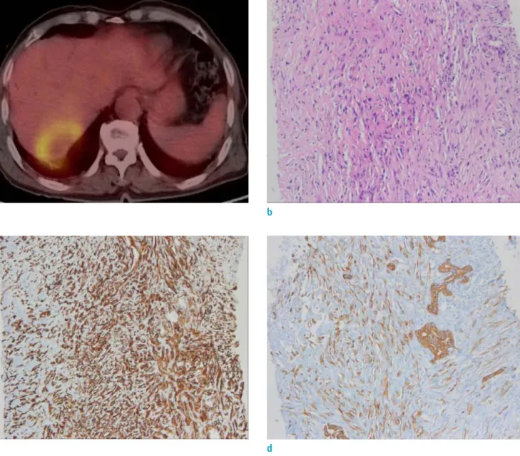
Positron emission tomography (PET)-CT scan showed intense fluorodeoxyglucose (FDG) uptake in the mass, indicating possibility of malignant lesion (Fig. 3a).
The patient underwent an ultrasonography-guided liver biopsy. A histological examination showed some parts of the tumor appeared sarcomatous and were composed of pleomorphic spindle cells with numerous mitoses, whereas other parts were composed of adenocarcinoma cells (Fig.
3b). An immunochemical examination of the sarcomatous
components showed positive staining for vimentin (Fig.
3c) and low molecular cytokeratin (CK7) (Fig. 3d). Based on these immunohistochemical findings, the tumor was diagnosed as a sarcomatous ICC. The patient was inoperable state with poor liver function and concurrent chemoradiation therapy (CCRT) was planned. But after 3 months, the patient was expired with massive pleural effusion and multiple lung metastasis.
a b
Fig. 3. Positron emission tomography (PET)-CT scan shows intense fluorodeoxyglucose (FDG) uptake in the mass, indicating possibility of malignant lesion (a). Photomicrograph (original magnification, × 100; H&E stain) shows some parts of the tumor appeared sarcomatous and were composed of pleomorphic spindle cells with numerous mitoses, whereas other parts were composed of adenocarcinoma cells (b). An immunochemical examination of the sarcomatous components showed positive staining for vimentin (c) and low molecular cytokeratin (CK7) (d).
c d
DISCUSSION
Sarcomatous change of the epithelial neoplasm is a rare condition with a poor clinical course (1). Especially, sarcomatous change of the liver is very rare and mostly reported in hepatocellular carcinoma. Cholangiocarcinoma with sarcomatoid transformation is a extremely rare disease and have been reported in only 4.5% in autopsy (2, 3).
The typical MR signal characteristics of cholangio- carcinoma are hypointense on T1WI, and hyperintense on T2WI relative to liver parenchyma (6). In the enhancement study with hepatocyte-specific contrast, strong arterial peripheral rim-like enhancement is often observed due to neoangiogenesis in the periphery of the tumor (7).
Approximately 10 minutes after contrast injection, the desmoplastic fibrous components of the tumor demonstrate progressive and concentric filling of contrast (8). And in hepatobiliary phase, the tumor shows low signal intensity due to strong enhancement of the surrounding liver parenchyma (6, 9). In the literature, radiologic findings of sarcomatous ICC are similar to ordinary ICC and characteristic radiologic differences are not identified yet (5).
Interestingly, our case of sarcomatous ICC showed significant enhancement on 20 min high blood pressure (HBP) and this is not common finding in ordinary ICC.
As the signal intensity of ICC displayed by MRI depends on the tissue components (10), significant enhancement on HBP means abundant fibrotic tissue components in sarcomatous transformation. Unfortunately, ordinary ICC with extensive fibrosis can reveal a certain enhancement on HBP, too. So this finding cannot be specific for sarcomatous transformation of ICC. And weather this finding would be useful in differentiating sarcomatous ICC from ordinary ICC is questionable. To determine the specific imaging findings of ICC with sarcomatoid transformation, further evaluation with a large number of cases will be warranted.
In the literature, radiologic findings of sarcomatous ICC are similar to ordinary ICC and characteristic radiologic differences are not identified yet (5). Since there is no characteristic clinico-radiological findings yet, the diagnosis of sarcomatous ICC is based on histopathologic and immunohistochemical examinations. The sarcomatous components show immune reaction with vimentin and cytokeratin and our case was based on the same method.
This feature is an indicator of sarcomatous transformation in a neoplasm of epithelial origin (1, 3, 4).
It has been reported that prognosis for sarcomatous ICC is worse than that for a ordinary ICC. The factors that account
for the poor prognosis may be ascribed to the remarkable intrahepatic development, especially the high potential of the sarcomatous component to metastasize. Vascular invasion has been shown to be more frequently present if there is a sarcomatous component (4).
In conclusion, we report a case of sarcomatous ICC that is very rare but aggressive malignancy. Early rim- like enhancement and progressive concentric contrast filling pattern are similar to ordinary ICC. The mass shows significant enhancement in HBP. Further reports are needed to determine the specific imaging findings of sarcomatous ICC in various cases including findings in hepatobiliary phase of enhanced MRI.
REFERENCES
1. Haratake J, Horie A. An immunohistochemical study of sarcomatoid liver carcinomas. Cancer 1991;68:93-97 2. Kakizoe S, Kojiro M, Nakashima T. Hepatocellular
carcinoma with sarcomatous change. Clinicopathologic and immunohistochemical studies of 14 autopsy cases.
Cancer 1987;59:310-316
3. Nakajima T, Tajima Y, Sugano I, Nagao K, Kondo Y, Wada K. Intrahepatic cholangiocarcinoma with sarcomatous change. Clinicopathologic and immunohistochemical evaluation of seven cases. Cancer 1993;72:1872-1877 4. Tsou YK, Wu RC, Hung CF, Lee CS. Intrahepatic sarcomatoid
cholangiocarcinoma: clinical analysis of seven cases during a 15-year period. Chang Gung Med J 2008;31:599-605 5. Bilgin M, Toprak H, Bilgin SS, Kondakci M, Balci C. CT and
MRI findings of sarcomatoid cholangiocarcinoma. Cancer Imaging 2012;12:447-451
6. Jeong WK, Kim YK, Song KD, Choi D, Lim HK. The MR imaging diagnosis of liver diseases using gadoxetic acid: emphasis on hepatobiliary phase. Clin Mol Hepatol 2013;19:360-366
7. Frydrychowicz A, Lubner MG, Brown JJ, et al. Hepatobiliary MR imaging with gadolinium-based contrast agents. J Magn Reson Imaging 2012;35:492-511
8. Vanderveen KA, Hussain HK. Magnetic Resonance Imaging of cholangiocarcinoma. Cancer Imaging 2004;4:104-115 9. Campos JT, Sirlin CB, Choi JY. Focal hepatic lesions in Gd-
EOB-DTPA enhanced MRI: the atlas. Insights Imaging 2012;3:451-474
10. Chong YS, Kim YK, Lee MW, et al. Differentiating mass- forming intrahepatic cholangiocarcinoma from atypical hepatocellular carcinoma using gadoxetic acid-enhanced MRI. Clin Radiol 2012;67:766-773