Received on March 7, 2015. Revised on May 12, 2015. Accepted on May 20, 2015.
CC This is an open access article distributed under the terms of the Creative Commons Attribution Non-Commercial License (http://creativecommons.org/licenses/by-nc/4.0) which permits unrestricted non-commercial use, distribution, and reproduction in any me- dium, provided the original work is properly cited.
*Corresponding Author. Jae Seung Kang, Department of Anatomy, Seoul National University College of Medicine, 28 Yongon-dong, Chongno-gu, Seoul, Korea. Tel: 82-2-740-8132; Fax: 82-2-741-8202; E-mail: genius29@snu.ac.kr
#The authors are equally contributed to this work.
Abbreviations: CD, Crohn's disease; DAI, disease activity index; DSS, dextran sulfate sodium; HPV, human papillomavirus; IBD, inflammatory bowel disease; KSHV, Kaposi’s sarcoma-associated herpesvirus; UC, ulcerative colitis
Alloferon Alleviates Dextran Sulfate Sodium-induced Colitis
Hyemin Kim1,2#, Jong Pil Im3#, Joo Sung Kim3, Jae Seung Kang1,2* and Wang Jae Lee1
1Department of Anatomy, Seoul National University College of Medicine, 2Institute of Allergy and Clinical Immunology, Seoul National University Medical Research Center, 3Department of Internal Medicine and Liver Research Institute, Seoul National University College of Medicine, Seoul 110-799, Korea
Dysfunction of gut immune regulation is involved in mu- cosal damage in inflammatory bowel disease (IBD). How- ever, there is still no efficacious immune-regulator for the treatment of IBD. Alloferon is a novel immune-modu- latory peptide that was originally isolated from infected insects. It shows anti-inflammatory effects by the regu- lation of cytokine production by immune cells and their activities. Therefore, we investigated the effect of alloferon in a mouse model of colitis using dextran sulfate sodium (DSS). Colitis was induced by administration of DSS in drinking water for 7 consecutive days. It was confirmed by the presence of weight loss, diarrhea, hematochezia, and colon contraction. Alloferon was injected 4 days after DSS administration. We found that alloferon improved the pathogenesis of IBD based on the reduced disease activity in- dex (DAI) and colon contraction. Edema, epithelial ero- sion, and immune cell infiltration were found in mice ad- ministered DSS, but the phenomena were reduced follow- ing alloferon treatment. The plasma level of IL-6, a classi- cal pro-inflammatory cytokine in colitis, was also de- creased by alloferon. Moreover, alloferon inhibited the TNF-α-induced degradation and phosphorylation of IκB in Colo205 colon cancer cells. Taken together, these results show that alloferon has anti-inflammatory effects and at- tenuates DSS-induced colitis.
[Immune Network 2015;15(3):135-141]
Keywords: Alloferon, Inflammatory bowel disease (IBD), Anti-inflammation, DSS-induced colitis
INTRODUCTION
Inflammatory bowel disease (IBD) is a term that describes a group of inflammatory conditions of the gastrointestinal tract. This chronic disease is characterized by abdominal pain, vomiting, diarrhea, rectal bleeding, and weight loss.
The major forms of idiopathic IBD are Crohn's disease (CD) and ulcerative colitis (UC). UC is characterized by diffused mucosal inflammation with extensive superficial mucosal ulceration. Histopathological features include a significant number of neutrophils in the lamina propria and the crypts as well as the depletion of goblet cells. CD is characterized by the aggregation of macrophages that fre- quently form non-caseating granulomas. Unlike UC, CD may be patchy, segmental, and typically transmural (1,2).
The onset of IBD typically occurs in the second and third decades of life and the majority of affected individual progress to relapses and a chronic condition. A rising trend has been reported in the incidence and prevalence of IBD in Asia and it is postulated that this increase may be re- lated to the westernized lifestyle including changes in diet and environment, such as improved sanitation and in- dustrialization (3). During the past two decades in Korea,
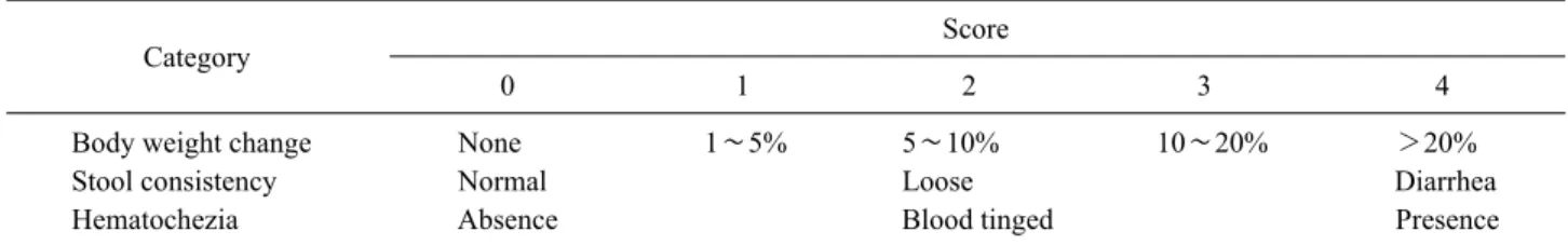
Table I. Disease activity index (DAI)
Category Score
0 1 2 3 4
Body weight change None 1∼5% 5∼10% 10∼20% >20%
Stool consistency Normal Loose Diarrhea
Hematochezia Absence Blood tinged Presence
Stool consistency, gross bleeding (hematochezia), and weight loss (body weight change) were scored after DSS treatment. A specialist scored each group in the three categories shown in the table and then divided thesum of each score by three
the mean annual incidence rates of CD and UC increased significantly from 0.05 and 0.34 per 100,000 inhabitants, respectively, in 1986∼1990 to 1.34 and 3.08 per 100,000 inhabitants, respectively, in 2001∼2005. In addition, the adjusted prevalence rates of CD and UC per 100,000 in- habitants were 11.24 and 30.87, respectively in 2005.
Although the incidence and prevalence of CD and UC in Korea are still lower than those in Western countries, they are rapidly increasing (4). The cause of IBD is known to be an inadequate and exaggerated immune response against commensal microbiota, especially in genetically suscep- tible individuals (1,5). There is no well-established therapy for IBD and, therefore, the quality of life and expectancy are compromised in patients with IBD.
Alloferon is a bioactive, slightly cationic peptide origi- nally isolated from an experimentally infected blowfly, Calliphora vicina. It consists of 13 amino acids with the following sequence: HGVSGHGQHGVHG (6). Alloferon has stimulatory effects on natural killer (NK) cells and in- duces in vivo interferon (IFN) production in mice, which indicates the anti-viral and anti-tumor capabilities of allo- feron as an immunomodulatory peptide (6). The tumori- static and tumoricidal activities of alloferon were also re- ported in DBA/2 mice grafted with syngeneic P388 murine leukemia cells (7). We recently reported that alloferon has dual functions; one involves direct inhibition of the repli- cation of the Kaposi's sarcoma-associated herpesvirus (KSHV) and the other is the effective eradication of vi- rus-infected cells by the activation of NK cells. Alloferon regulated the KSHV life cycle by the down-regulation of activator protein (AP)-1 activity and enhanced anti-viral immunity by up-regulation of NK cell cytotoxicity (8). It was also found that alloferon has anti-tumor effects medi- ated by the up-regulation of the NK-activating receptor 2B4 and enhancement of granular exocytosis from NK
cells (9). In addition, the anti-inflammatory effects of allo- feron on UVB-induced inflammation was observed in the human keratinocyte HaCaT cell line and hairless mouse skin (10).
In the present study, we examined the anti-inflammatory effect of alloferon and its possible therapeutic efficacy in a murine colitis model induced with DSS.
MATERIALS AND METHODS Animals and induction of colitis
C57BL/6 mice were maintained under specific pathogen- free conditions at the animal facility of the Seoul National University College of Medicine. Eight- to ten-week-old male mice were used in the experiments. All animal ex- periments were reviewed and approved by the Institutional Animal Care and Use Committee of the Seoul National University. Mice were administered 3% DSS (MP Bio- medicals Irvine, CA, USA) dissolved in drinking water for 7 days to induce colitis. Alloferon was kindly provided by EntoPharm (Seoul, Korea). Alloferon (50 μg) in normal saline was intraperitoneally (i.p.) injected to mice. On day 4 after DSS administration, alloferon was injected for an- other 4 consecutive days.
Evaluation of colitis
Disease activity index (DAI): The mice were monitored daily for behavior, water and food consumption, body weight, stool consistency, and the presence of gross blood in the stool or anus, which were presented as the disease activity index (DAI). The DAI was calculated as follows, (stool consistency+gross bleeding+weight loss)/3, based on Table I.
Measurement of colon length: After the mice had been eu- thanized, the colon was excised, and the length was meas- ured from the anus to the ileocecal valve.
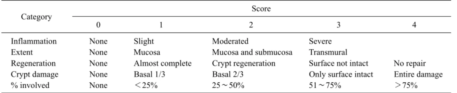
Table II. Histological grade
Category Score
0 1 2 3 4
Inflammation None Slight Moderated Severe
Extent None Mucosa Mucosa and submucosa Transmural
Regeneration None Almost complete Crypt regeneration Surface not intact No repair
Crypt damage None Basal 1/3 Basal 2/3 Only surface intact Entire damage
% involved None <25% 25∼50% 51∼75% >75%
After DSS and alloferon treatment, colons were collected, fixed, and stained with H&E. A specialist scored each group in the five categories shown in the table and then the divided the sum of each score by five. DSS, dextran sulfate sodium
Figure 1. Experimental design of dextran sulfate sodium (DSS) and alloferon treatment. Mice were treated with 3% DSS admin- istered in drinking water for 7 days. Alloferon (50 μg/mouse, i.p.) was daily injected to mice four times from day 4 of DSS treatment.
Histological evaluation: The colon was washed in PBS, fixed in 4% paraformaldehyde (PFA), paraffin-embedded, sectioned, and then stained with H&E. The histological as- sessment was performed by a trained pathologist who was blinded to the treatment. All histological quantification was performed in a blinded fashion using a scoring system.
The histological grade was calculated based on Table II.
The sum of each score in the five denoted categories was divided by five.
Measurement of cytokines
The blood was collected from the orbital plexus using hep- arinized capillary tubes under anesthesia with zoletil and xylazine (25 and 10 mg/kg, respectively). After centri- fugation at 14,000 rpm for 30 min at 4oC, the plasma ob- tained was stored at −70oC until used. The plasma IL-6 concentration measured using an ELISA kit according to the manufacturer’s instruction (R&D Systems, Minneap- olis, MN, USA).
Cell culture
The Colo205 (human colon cancer cell line) was main- tained in Roswell Park Memorial Institute (RPMI)) 1640 medium (Welgene, Seoul, Korea) supplemented with 10%
heat-inactivated FBS (HyClone, Logan, UT, USA), 100 U/ml penicillin, and 100 μg/ml streptomycin (Welgene, Seoul, Korea) at 37oC in an atmosphere of 5% CO2. Immunoblotting
The Colo205 cells (1×10⁶) were seeded on a 6-well plate and treated with alloferon (1 μg) for 24 h. Then, TNF-α (10 ng/ml) was added for 10 min. After washing the cells with PBS, they were homogenized with lysis buffer and
total proteins were quantified using the bicinchoninic acid (BCA) assay. Equal amounts of protein were resolved us- ing a 10% SDS-PAGE and then transferred to a nitro- cellulose membrane. After blocking with 5% nonfat milk, the membranes were incubated overnight with antibodies for anti-phospho (p)-Iκ B, anti-Iκ B (Cell Signaling, Danvers, MA, USA), or β-actin (Sigma, St. Louis, MO, USA) at 4oC. After incubating with HRP-conjugated anti-rabbit or anti-mouse IgG (Cell Signaling, Danvers, MA, USA), the immunoreactive proteins were visualized with an ECL de- tection system (Amersham Biosciences Corp., Piscataway, NJ, USA).
Statistics
The data were expressed as mean±SD for each group of independent experiments. For the comparison of three or more groups, the data were analyzed using the Student’s t-test or one-way ANOVA followed by the Newman-Keuls multiple comparison test. A p-value<0.05 was considered
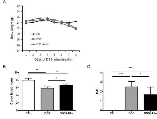
Figure 2. Alloferon decreased the severity of dextran sulfate sodium (DSS)-induced colitis. (A) Body weight was monitored for 7 days following 3% DSS and allo- feron (50 μg/mouse) treatment.
(B) After DSS and alloferon treatment for 7 days, colon length of each experimental group was measured (n=three, *p<0.05,
**p<0.01). (C) Body weight loss, stool consistency, and bloody stool were presented as DAI according to the criteria described in the Materials and Methods. DAI calculated as, (stool consistency+
gross bleeding+weight loss)/3,
*p<0.05, ***p<0.001. DAI, disease activity index; ctl, control;
Allo, alloferon.
statistically significant. The statistical analyses were car- ried out using the GraphPad InStat (GraphPad Software, San Diego, CA, USA).
RESULTS
Alloferon decreased the severity of DSS-induced colitis
To induce colitis, mice were treated with 3% DSS in their drinking water for 7 days. To evaluate the effect of allofer- on, it was injected into mice once a day for 4 days (Fig. 1).
The body weight was decreased (Fig. 2A) and the length of the colon from the anus to the ileocecal valve contracted in all mice with DSS treatment (Fig. 2B). To evaluate the severity of the colitis, the stool consistency, gross bleed- ing, and weight loss were scored and represented the re- sults were expressed as the DAI. DSS administration sig- nificantly increased the disease activity (Fig. 2C).
Alloferon treatment did not reduce the weight loss caused by DSS treatment (Fig. 2A). However, alloferon effec- tively decreased the colon contraction, and thereby in- creased the colon length (Fig. 2B). In addition, the bloody stool and diarrhea were reduced, and these effects collec- tively decreased the DAI (Fig. 2C). Therefore, it appears that alloferon had a mitigating effect on the DSS-induced
colitis.
Alloferon decreased inflammation in DSS-induced colitis
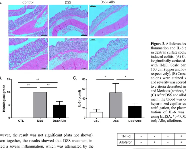
The colon of the DSS-treated mice showed more severe destruction of the epithelial architecture with a loss of crypts and epithelial integrity, submucosal edema, and in- tense inflammatory cellular infiltration in all layers than that of the controls did (Fig. 3A). The histological grade indicated the presence of severe inflammation in the DSS- treated mice (Fig. 3B). The alloferon- and DSS-treated mice also showed severe inflammation, which included edema and epithelial erosion. However, the epithelial integrity was less disrupted, and the number of infiltrated cells was reduced more following the alloferon injection than it was without treatment (Fig. 3A). Interleukin (IL-6) is a pro-in- flammatory cytokine that is increased by DSS treatment (11-13). To evaluate the inflammation, the level of IL-6 was measured in the plasma of mice after DSS or alloferon treatment. The result showed that DSS administration con- siderably increased the production of IL-6, and this effect was significantly decreased by alloferon (Fig. 3C). In addi- tion, one of the immunoregulatory cytokines, IL-10 (14,15) was measured, and the level was increased following DSS treatment and this effects was decreased by alloferon.
Figure 3. Alloferon decreased in- flammation and IL-6 production in dextran sulfate sodium (DSS)- induced colitis. (A) Colons were longitudinally sectioned and stained with H&E. Scale bar, 200 and 100 μm (upper and lower panels, respectively). (B) Cross-sectioned colons were stained with H&E and severity was scored according to criteria described in Materials and Methods (n=three, **p<0.01.
(C) After DSS and alloferon treat- ment, the blood was collected in heparinized capillaries. After ce- ntrifugation, the plasma concen- tration of IL-6 was measured using ELISA, *p<0.05; ctl, con- trol; Allo, alloferon.
p-Iκ B Iκ B
β-actin
Figure 4. Alloferon inhibited Iκ B degradation. Colo205 cells (1×10⁶) were treated with TNF-α (10 ng/ml) for 10 min after alloferon pre-treatment (1 μg) for 24 h. The expression of Iκ B and phospho-(p)-Iκ B was determined using immunoblotting.
β-actin was used as a loading control.
However, the result was not significant (data not shown).
Taken together, the results showed that DSS treatment in- duced a severe inflammation, which was attenuated by the injection of alloferon.
Alloferon inhibited Iκ B degradation by TNF-α in colon cancer cells
To determine the possible mechanism underlying the allo- feron-induced decrease of the inflammation in colitis, we examined the degradation and phosphorylation of IκB us- ing immunoblotting. It has been reported that DSS-induced murine colitis activates Nuclear factor-kappaB (NF-kB) signaling, which is critical in the pathophysiology of IBD (16-18). The key step in NF-κB activation is the release of the NF-κB dimers from their inhibitory proteins, which is mediated via proteolysis of Iκ B (19). Tumor nerosis fac- tor-alpha (TNF-α) is known to initiate signal transduction via NF-κB (17,20). TNF-α increases the phosphorylation of Iκ B and activates NF-κB to transcribe numerous in- flammatory genes. In Fig. 4, TNF-α treatment was shown to increase the expression of p-Iκ B and decrease that of Iκ B in the Colo205 colon cancer cell line. The increased
degradation of IκB was remarkably reduced following al- loferon treatment.
DISCUSSION
Although the exact pathogenesis of IBD remains unclear, recent evidence suggests that IBD results from an in-
appropriate inflammatory response to intestinal microbes in genetically susceptible individuals (1,21). The incidence and prevalence of IBD are increasing in different regions of the world, indicating its emergence as a global disease (22). However, there is no ideal therapeutic agent and re- search focused on developing a more in-depth and in- tegrated understanding of the mechanisms mediating in- testinal immune homeostasis and therapy are needed. In the present study, the immunomodulatory peptide alloferon was shown to decreased the DSS-induced inflammation, infiltration of immune cells, and IL-6 production, which eventually reduced the severity of colitis. This effect was evidenced by a decrease in the DAI level and histological grade.
Alloferon was first isolated from the bacterial- chal- lenged larvae of the blowfly C. vicina based on the fact that insects can rapidly eliminate microbes or fungi by pro- ducing a variety of anti-bacterial and anti-fungal sub- stances (6). The pharmaceutical value of the peptide has been mainly related to its ability to not only stimulate NK cytotoxicity and IFN production in animal and human mod- els, but to also enhance anti-viral and anti-tumor activities in mice (7-9). The antiviral and immunomodulatory effects of alloferon have also been clinically proved in patients infected with herpes simplex virus (HSV) and the human papillomavirus (HPV) (23,24). We recently reported that alloferon decreased the production of UVB-induced pro-in- flammatory cytokines such as IL-1α , IL-1β , IL-6, and IL-18 by the inhibition of p38 mitogen-activated protein kinase (MAPK) inactivation (10). The present study also verified the anti-inflammatory effect of alloferon in a murine colitis model using DSS (Fig. 2 and 3).
In IBD, the levels of various pro-inflammatory cyto- kines such as IL-1, IL-6, TNF-α, and IFN-γ are known to be increased. NF-κB activation stimulates the ex- pression of IL-1, IL-6, IL-8, and TNF-α (25). Among these cytokines, the level of IL-6 was investigated follow- ing DSS and alloferon treatment (Fig. 3C). IL-6 plays an important role in enhancing T-cell survival and apoptosis resistance in the lamina propria at the site of inflammation (26). In addition, it promotes the survival of intestinal epi- thelial cells (27,28). The concentration of IL-6 was in- creased by DSS treatment, and this effect was decreased following alloferon treatment (Fig. 3C). This suggests that alloferon efficaciously decreased the inflammation and se- verity of colitis. Because IL-6 activates Signal transducer
and activator of transcription 3 (STAT3) for epithelial sur- vival, the changes in STAT3 signaling and IL-6 production in the inflamed colonic tissue need to be further studied.
Patients with chronic UC and CD have an increased risk of developing colorectal cancer (29). Therefore, we also examined the effect of alloferon on colon cancer cells.
Alloferon is known to activate NF-κB signaling by down- regulating antioxidant proteins and Iκ Bα (30). However, alloferon treatment decreased the degradation and phos- phorylation of Iκ B in colon cancer cells (Fig. 4), indicating the inactivation of NF-κB signaling. NF-κB activation by alloferon was reported to stimulate the synthesis of IFN, thereby inducing anti-viral function in human Burkitt’s lymphoma cells containing HPV (30). There has been no report determining the signaling mechanism induced by al- loferon in any tumor cells. Therefore, the effect of allofer- on including its dephosphorylation of Iκ B in tumor cells should be further investigated. Furthermore, the immunor- egulatory effects of alloferon should be investigated in- tensely in a cancer model as a research subject because alloferon enhances NK cell activity, which play a critical role in host immunity against cancer (31).
In summary, alloferon, a peptide consisting of 13 amino acids showed anti-inflammatory effects in an inflamed colon. Further studies are warranted to elucidate the under- lying mechanisms and to intensively evaluate its ther- apeutic potential in colitis and colon cancer treatment.
ACKNOWLEDGEMENTS
This work is supported by the grants (04-2011-1030) from Seoul National University Hospital to Jong Pil Im.
CONFLICTS OF INTEREST
The authors have no financial conflict of interest.
REFERENCES
1. Xavier, R. J., and D. K. Podolsky. 2007. Unravelling the patho- genesis of inflammatory bowel disease. Nature 448: 427-434.
2. Baumgart, D. C., and W. J. Sandborn. 2007. Inflammatory bowel disease: clinical aspects and established and evolving therapies.
Lancet 369: 1641-1657.
3. Thia, K. T., E. V. Loftus, Jr., W. J. Sandborn, and S. K. Yang.
2008. An update on the epidemiology of inflammatory bowel dis- ease in Asia. Am. J .Gastroenterol. 103: 3167-3182.
4. Yang, S. K., S. Yun, J. H. Kim, J. Y. Park, H. Y. Kim, Y. H.
Kim, D. K. Chang, J. S. Kim, I. S. Song, J. B. Park, E. R. Park, K. J. Kim, G. Moon, and S. H. Yang. 2008. Epidemiology of in- flammatory bowel disease in the Songpa-Kangdong district, Seoul, Korea, 1986-2005: a KASID study. Inflamm. Bowel. Dis. 14:
542-549.
5. Kraus, T. A., L. Toy, L. Chan, J. Childs, A. Cheifetz, and L.
Mayer. 2004. Failure to induce oral tolerance in Crohn's and ulcer- ative colitis patients: possible genetic risk. Ann. N. Y. Acad. Sci.
1029: 225-238.
6. Chernysh, S., S. I. Kim, G. Bekker, V. A. Pleskach, N. A.
Filatova, V. B. Anikin, V. G. Platonov, and P. Bulet. 2002.
Antiviral and antitumor peptides from insects. Proc. Natl. Acad.
Sci. U. S. A. 99: 12628-12632.
7. Chernysh, S., K. Irina, and A. Irina. 2012. Anti-tumor activity of immunomodulatory peptide alloferon-1 in mouse tumor trans- plantation model. Int. Immunopharmacol. 12: 312-314.
8. Lee, N., S. Bae, H. Kim, J. M. Kong, H. R. Kim, B. J. Cho, S.
J. Kim, S. H. Seok, Y. I. Hwang, S. Kim, J. S. Kang, and W.
J. Lee. 2011. Inhibition of lytic reactivation of Kaposi's sarco- ma-associated herpesvirus by alloferon. Antivir. Ther. 16: 17-26.
9. Bae, S., K. Oh, H. Kim, Y. Kim, H. R. Kim, Y. I. Hwang, D.
S. Lee, J. S. Kang, and W. J. Lee. 2013. The effect of alloferon on the enhancement of NK cell cytotoxicity against cancer via the up-regulation of perforin/granzyme B secretion. Immunobiology 218: 1026-1033.
10. Kim, Y., S. K. Lee, S. Bae, H. Kim, Y. Park, N. K. Chu, S. G.
Kim, H. R. Kim, Y. I. Hwang, J. S. Kang, and W. J. Lee. 2013.
The anti-inflammatory effect of alloferon on UVB-induced skin inflammation through the down-regulation of pro-inflammatory cytokines. Immunol. Lett. 149: 110-118.
11. Lee, M. J., J. K. Lee, J. W. Choi, C. S. Lee, J. H. Sim, C. H.
Cho, K. H. Lee, I. H. Cho, M. H. Chung, H. R. Kim, and S. K.
Ye. 2012. Interleukin-6 induces S100A9 expression in colonic epi- thelial cells through STAT3 activation in experimental ulcerative colitis. PLoS One 7: e38801.
12. Sander, L. E., F. Obermeier, U. Dierssen, D. C. Kroy, A. K. Singh, U. Seidler, K. L. Streetz, H. H. Lutz, W. Muller, F. Tacke, and C. Trautwein. 2008. Gp130 signaling promotes development of acute experimental colitis by facilitating early neutrophil/macro- phage recruitment and activation. J. Immunol. 181: 3586-3594.
13. Lin, Y., X. Yang, W. Yue, X. Xu, B. Li, L. Zou, and R. He.
2014. Chemerin aggravates DSS-induced colitis by suppressing M2 macrophage polarization. Cell. Mol. Immunol. 11: 355-366.
14. Li, B., R. Alli, P. Vogel, and T. L. Geiger. 2014. IL-10 modulates DSS-induced colitis through a macrophage-ROS-NO axis. Mucosal Immunol. 7: 869-878.
15. Jarry, A., C. Bossard, C. Bou-Hanna, D. Masson, E. Espaze, M.
G. Denis, and C. L. Laboisse. 2008. Mucosal IL-10 and TGF-beta play crucial roles in preventing LPS-driven, IFN-gamma-mediated epithelial damage in human colon explants. J. Clin. Invest. 118:
1132-1142.
16. Marrero, J. A., K. A. Matkowskyj, K. Yung, G. Hecht, and R.
V. Benya. 2000. Dextran sulfate sodium-induced murine colitis ac- tivates NF-kappaB and increases galanin-1 receptor expression.
Am. J. Physiol. Gastrointest. Liver Physiol. 278: G797-G804.
17. Andresen, L., V. L. Jorgensen, A. Perner, A. Hansen, J. Eugen-
Olsen, and J. Rask-Madsen. 2005. Activation of nuclear factor kappaB in colonic mucosa from patients with collagenous and ul- cerative colitis. Gut 54: 503-509.
18. Rogler, G., K. Brand, D. Vogl, S. Page, R. Hofmeister, T. Andus, R. Knuechel, P. A. Baeuerle, J. Scholmerich, and V. Gross. 1998.
Nuclear factor kappaB is activated in macrophages and epithelial cells of inflamed intestinal mucosa. Gastroenterology 115: 357-369.
19. Kanarek, N., N. London, O. Schueler-Furman, and Y. Ben-Neriah.
2010. Ubiquitination and degradation of the inhibitors of NF- kappaB. Cold Spring Harb. Perspect. Biol. 2: a000166.
20. Chandel, N. S., W. C. Trzyna, D. S. McClintock, and P. T.
Schumacker. 2000. Role of oxidants in NF-kappa B activation and TNF-alpha gene transcription induced by hypoxia and endotoxin.
J. Immunol. 165: 1013-1021.
21. Hanauer, S. B. 2006. Inflammatory bowel disease: epidemiology, pathogenesis, and therapeutic opportunities. Inflamm. Bowel. Dis.
12 Suppl 1: S3-S9.
22. Molodecky, N. A., I. S. Soon, D. M. Rabi, W. A. Ghali, M. Ferris, G. Chernoff, E. I. Benchimol, R. Panaccione, S. Ghosh, H. W.
Barkema, and G. G. Kaplan. 2012. Increasing incidence and preva- lence of the inflammatory bowel diseases with time, based on sys- tematic review. Gastroenterology 142: 46-54.
23. Ershov, F., A. Kubanova, B. Pinegin, A. Shulzhenko, G. Bekker, and S. Chernysh. 2003. Allokine-α effect on the course of chronic genital herpes relapses. Materia Medica 40: 103-111.
24. Chernysh, S., and N. Gordja. 2011. The immune system of mag- gots of the blow fly (Calliphora vicina) as a source of medicinal drugs. J. Evol. Biochem. Physiol. 47: 524-533.
25. Taylor, B. S., M. E. de Vera, R. W. Ganster, Q. Wang, R. A.
Shapiro, S. M. Morris, Jr., T. R. Billiar, and D. A. Geller. 1998.
Multiple NF-kappaB enhancer elements regulate cytokine in- duction of the human inducible nitric oxide synthase gene. J. Biol.
Chem. 273: 15148-15156.
26. Neurath, M. F., S. Finotto, I. Fuss, M. Boirivant, P. R. Galle, and W. Strober. 2001. Regulation of T-cell apoptosis in inflammatory bowel disease: to die or not to die, that is the mucosal question.
Trends Immunol. 22: 21-26.
27. Grivennikov, S., E. Karin, J. Terzic, D. Mucida, G. Y. Yu, S.
Vallabhapurapu, J. Scheller, S. Rose-John, H. Cheroutre, L.
Eckmann, and M. Karin. 2009. IL-6 and Stat3 are required for survival of intestinal epithelial cells and development of col- itis-associated cancer. Cancer Cell 15: 103-113.
28. Jin, X., T. A. Zimmers, Z. Zhang, R. H. Pierce, and L. G.
Koniaris. 2010. Interleukin-6 is an important in vivo inhibitor of intestinal epithelial cell death in mice. Gut 59: 186-196.
29. Triantafillidis, J. K., G. Nasioulas, and P. A. Kosmidis. 2009.
Colorectal cancer and inflammatory bowel disease: epidemiology, risk factors, mechanisms of carcinogenesis and prevention strategies.
Anticancer Res. 29: 2727-2737.
30. Ryu, M. J., V. Anikin, S. H. Hong, H. Jeon, Y. G. Yu, M. H.
Yu, S. Chernysh, and C. Lee. 2008. Activation of NF-kappaB by alloferon through down-regulation of antioxidant proteins and IkappaBalpha. Mol. Cell. Biochem. 313: 91-102.
31. Cheng, M., Y. Chen, W. Xiao, R. Sun, and Z. Tian. 2013. NK cell-based immunotherapy for malignant diseases. Cell. Mol.Immunol.
10: 230-252.