Occlusal Analysis of the Patients with Temporomandibular Disorders Using T-Scan II System
Dong-Hyo Yang, D.D.S., Won-Seop Lee, D.D.S.,M.S.D., Mee-Eun Kim, D.D.S.,M.S.D.,Ph.D,
Department of Oral Medicine, Dankook University School of DentistryCorrelation between occlusal contact pattern and TMD have been hypothesized and partially investigated but results are controversial and not conclusive. The purposes of this study were to compare right-to-left difference of occlusal contact pattern, through contact points, contact force and occlusal balance, in the patients with unilateral TMD and also to evaluate its change related with TMD treatment.
36 patients with unilateral TMD from Department of Oral Medicine in Dankook University Dental Hospital were selected in this study (M:F=7:29, mean age of 29.2±14.8 years). A computerized T-Scan II system (Tekscan, INC., USA) was employed for occlusal analysis and the simultaneity and occlusal balance through the number of tooth contact and magnitude of contact force were determined before and after TMD treatment.
The number of contact points and contact force was more on the unaffected side than the affected side before treatment (p=0.056 and p=0.060, respectively) while significant difference between both sides was not found after treatment. The number of contact points and contact force on the affected sides significantly increased after treatment (p=0.038 and p=0.052), but the unaffected sides exhibited no significant difference between before and after treatment.
In addition, sides difference in relative contact force decreased from about 27% to about 12% after TMD treatment (p=0.001).
According to the results of this study, it is likely that unilateral TMD impairs right-to-left occlusal balance and that conservative TMD treatment alleviates the imbalance, subsequently leading to more symmetrical occlusal condition with increased contact points and force.
Key words : T-Scan II, TMD, Contact point, Contact force, Occlusal balance.
1)
I. INTRODUCTION
Temporomandibular disorders (TMD) cover a
Corresponding author: Prof. Mee-Eun Kim, Department of Oral Medicine,
Dankook University, School of Dentisty Sinbu-dong San 7-1 Cheonan 330-716 Tel. 041-550-1915
Fax. 041-556-9665
E-mail. [email protected] Received : 2007-01-04 Accepted : 2007-03-11
*The present research was conducted by the research fund of Dankook University in 2005.
wide range of abnormal and pathologic conditions
caused by physical strain and primary disease of
the muscles of mastication and temporomandibular
joints, which are accompanied by headache and
orofacial pain, and impaired mandibular function
1-3).
It is critical, for management of TMD, to
appreciate the major causes that may be associated
with the condition and a review of scientific
literature reveals five major factors associated with
TMD including trauma, emotional stress, deep pain
input and parafunctional activities and occlusal
condition
4). Occlusal condition is one contributing
factor to TMD that has been strongly debated for
many years. In recent years, the acceptance of
theories about the multifactorial etiology of TMD has resulted in less emphasis being placed on occlusion as a TMD-related factor
7)although the presence of some occlusal abnormalities has been considered a major factor in the etiology
5-6). Aside from occlusal condition as one contributing factor to TMD, change in occlusal contact pattern, which is either temporary or permanent, is one of common complaints from the patients with TMD in clinical setting. Correlation between occlusal contacts and signs and symptoms of TMD have been hypothesized and partially investigated but results are controversial and not conclusive
7-12). Naeije et al
13)indicated that asymmetry of bilateral distribution of occlusal contact in TMD patients whereas Visser et al
14)showed no significant differences in the electromyographic asymmetries of masticatory muscles. Therefore, assessment of relationships between TMD and occlusal contact patterns could be still of importance.
The purposes of this study were to compare right-to-left difference of occlusal contact pattern, through contact points, contact force and occlusal balance, in the patients with unilateral TMD and also to evaluate effect of TMD treatment. A computerized T-Scan II system was employed for occlusal analysis.
Ⅱ. MATERIALS AND METHODS
A computerized occlusal analysis system used for this study was T-ScanTMII (Tekscan, INC., USA).
T-Scan II system consists of a piezoelectric foil sensor, sensor support, scanning handle, parallel hardware, and software for recording, analyzing, and viewing the data. The sensor is 60 ㎛-thick and made of a polyester film. The film is covered by a silver thread grid, the intersecting points of which are bathed by conductive ink. When the patient closes firmly on the sensor, the resultant reduction in electric resistance is translated into an image on the screen
15).
A 0.01 second real-time occlusal contact recording and 0.01 second incremental playback of
the tooth contact timing data can illustrate the order of tooth contacts, as well as their force content. The combination of contact order, contact duration that precedes the next occlusal contact, contact location within the arch, and magnitude of contact force determine the degree of contact simultaneity and the occlusal balance that is present or absent in a particular occlusal scheme
16). 36 subjects in this study were selected from patients with unilateral TMD examined and diagnosed in the department of Oral Medicine, Dankook University Dental Hospital. Ratio of male to female was 7:29 and their mean age was 28.9±14.7 years. None of them had any serious illness and excluded were those who had extensive prosthesis of more than 3-unit bridgework, missing more than single tooth except third molar, or significant dental and periodontal lesions. All of them gave informed consent prior to the examination.
Occlusal analysis using the T-Scan II system was carried out at the first visit prior to treatment and was repeated after complete or substantial relief of their signs and symptoms. Therapeutic modalities provided to them employed variable conservative treatment including behavioral modification, pharmacological therapy, physical therapy and exercise, and occlusal splint therapy.
None of them were involved with any irreversible therapy such as prosthetic, orthodontic and surgical treatment.
During the T-Scan II recordings, the subjects were seated in a dental chair in upright position.
Once the foil sensor was correctly inserted between
upper and lower arches in the subject’s mouth, he
or she was asked to close the mouth and occlude
the sensor in the habitual intercuspal position with
normal pressure. After several practices were made
until a repeatable pattern of tooth contacts was
produced on the computer monitor to verify the
reliability of the data, a representative force movie
was recorded and printed. The simultaneity and
occlusal balance through the number of tooth
contact and magnitude of contact force were
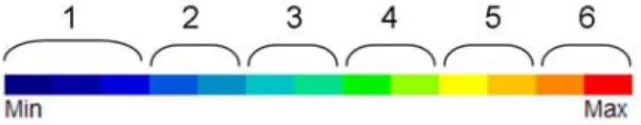
Fig. 1. The legend (color scale) of contact force.
determined. With this system, each test sheet was divided into right and left sides by the midline of the dental arch. The balance of contact force was expressed as the percentage of total contact force on the side, either left or right, which was defined as ‘relative contact force (%)’ here.
Magnitude of contact force was relatively displayed by color scale on the screen (that is, blue is the lowest and red the highest), so that contact force in this study was obtained by multiplying the number of contact points by intensity of each point.
The force intensity was determined from 1 (the lowest) to 6 (the highest) according to the legend (Fig. 1).
Number of Contact points
Pre-Tx Post-Tx Paired t-tests
Affected side 51.94±36.24 65.32±42.12 p=0.038
Unaffected side 59.74±36.40 71.55±46.10 p=0.136
Paired t-tests p=0.056 p=0.153
Contact force (=number of contact point x intensity of each point)
Pre-Tx Post-Tx Paired t-tests
Affected side 95.07±87.13 133.26±111.35 p=0.052
Unaffected side 112.52±80.93 142.51±121.24 p=0.172
Paired t-tests p=0.060 p=0.295
Relative contact force (%)
Pre-Tx Post-Tx Paired t-tests
Affected side(A) 50.06 ± 18.08 49.17 ± 9.57
Unaffected side(B) 49.94 ± 18.08 50.83 ± 9.57
Absolute sides difference (¦A-B¦) 27.38 ± 23.15 12.62 ± 14.32 p=0.001
(N=36) Table 1. Number of occlusal contact points, contact force and relative contact force (mean±SD) between
before and after treatment.
Collected data was processed using SPSS Window program ver 12.0. Paired t-test was used to compare any difference of occlusal contact pattern between the affected and unaffected sides of the TMD patients, as well as between before and after treatment.
Ⅲ. RESULTS
Table 1 represents the number of contact points,
contact force and relative contact force in the
patients with unilateral TMD. The number of
contact points and contact force was more on the
unaffected side than the affected side before
treatment (p=0.056 and p=0.060, respectively) while
significant difference between both sides was not
found after treatment.(Fig. 2 & 3). The number of
contact points and contact force on the affected
sides significantly increased after treatment
(p=0.038 and p=0.052), but the unaffected sides
exhibited no significant difference between before
Fig. 2. Number of contact points for the unilateral TMD patients before and after treatment.
Fig. 3. Contact force for the unilateral TMD patients before and after treatment.
and after treatment.
The mean relative contact force (%) of total contact force before treatment was 50.06±18.08% on the affected sides and 49.94% on the unaffected, symptom-free sides and, after TMD treatment, they were changed into 49.17±9.57% and 50.83±9.57%, respectively. It is unlikely that their values showed noticeable difference before and after treatment. However, when comparing absolute sides difference of the relative contact force ( relative force of the affected side - relative force of the unaffected side ), the sides difference significantly decreased from 27.38% before treatment to 12.67% after treatment.(p=0.001, Fig. 4)
Fig. 4. Absolute sides difference of the relative contact force for the unilateral TMD patients before and after treatment.
Ⅳ. DISCUSSION
Diagnosis of occlusion for TMD patients is important and variable methods have been used for examination of occlusal contact. T-Scan II system was used for the patients with unilateral TMD in this study. Conventional registration materials such as inked marking strips
17), waxes
18), ribbons and silicone
19)and other impression materials such as plaster have been used for occlusal analysis.
Although these materials are preferred primarily because of their low cost and easy application, they are affected by the presence of saliva
20)and show low reproducibility and high variability
21). T-Scan system registers and depicts a measure of the dynamics of occlusion
22,23). The sensitivity of this system is not changed whether the saliva is present or not
23). In a few study about comparing the T-Scan system with another registration material in vivo and in vitro, this system showed high accuracy and validity
24,25). Moreover, T-Scan can be used to objectively and quantitatively analyze the balance of occlusal contact force expressed by percentage value although complicated calculations by hand were required to evaluate number and force of occlusal contact.
The results from this study showed that the
number of contact points and contact force was
more on the unaffected side than the affected side
before treatment (p=0.056 and p=0.060, respectively) and that the difference decreased after treatment (Fig. 2 & 3). These findings are closely associated with significant decrease in the sides difference of relative contact force between before and after treatment (Fig. 4), which likely means alleviation of deteriorated right-to-left occlusal balance with TMD treatment.
Ciancaglini et al
26)studied on occlusal contacts of 25 TMD patients and 25 healthy controls in the intercuspal position through wax registration and showed no difference between the two groups for the overall number and distribution of contacts or for any side and intensity of contact. However, considering intra-subject occlusal contact distribution, they confirmed significant positive association of TMD with bilateral asymmetry of the total number of contact points and of the number of posterior contacts. Mizui et al
27)also demonstrated a marked difference in occlusal balance between TMD patients and the normal controls by mean of T-Scan system. While the controls had bilateral balance and an anteroposterior center of force in the first molar region, TMD patients showed asymmetrical time moment and occlusal force parameters. In addition, it was observed that the center of effort anteroposteriorly was not always located in the 1st molar region.
Kurita et al
28)evaluated the effect of splint on occlusal force in patients with masticatory muscle disorders using the computerized system with Dental Prescale
Ⓡand presented that there was no significant changes in the number of occlusal points, mean occlusal pressure, and asymmetry in occlusal balance between before and after the use of the splint. However, the study showed that the integrated occlual load converged to the normal value with the use of the splint, exhibiting normalizing effect of splint on the occlusal force.
The occlusal loads in the higher level decreased and, in contrast, those in the lower level increased with the use of the splint. They suggested that the use of splint has the effect of reducing the hyperactivity and the asymmetry in the activity of
the jaw elevator muscles, and consequently brings a stable and physiologically optimal occlusal force from the muscles. Shoji
29)evaluated treatment efficacy in the TMD patients through occlusal force with Dental Prescale
Ⓡand exhibited that occlusal force was significantly increased after treatment.
These findings in part agree with ours exhibiting manifest difference between the affected and unaffected sides before TMD treatment and decrease of the difference after TMD treatment, subsequently leading to more symmetrical occlusal condition with increased contact points and force.
However, there still exist several studies indicating approximately the same number of contact points on the right and left sides and bilateral almost balanced occlusion
30-32).
The study by Ciancaglini et al
26)also indicated significant concordance (88.9%) between the side of disorder and the side of higher number of contacts in 9 patients with unilateral TMD. This result is in contrast with ours demonstrating the less contact points and force on the affected side compared to the unaffected side. This discrepancy may be in part explained by selection of TMD patients and methodology used for occlusal analysis. As TMD embraces variable conditions related with masticatory muscles and TMJ, it is therefore needed to evaluate occlusal contact pattern in large population with TMD divided more specific subgroups.
Conclusively, based on our results, it is likely that unilateral TMD impairs right-to-left balance of contact force and conservative TMD treatment without any irreversible occlusal therapy alleviates the imbalance, leading to more symmetrical occlusal condition with increased contact points and force.
REFERENCES
1. Griffiths TH. American Dental Association: report of the president s conference on the examination, diagnosis and management of temporomandibular disorders. J Am Dent Assoc 1983;66:75-77.
2. McNeill C, ed. Craniomandibular disorders. Guidelines for evaluation, diagnosis, and management. American
Academy of Craniomandibular Disorders. Chicago, Qunitessence 1990, pp21-24.
3. Bakke M, Möller E. Craniomandibular disorders and masticatory muscle function. Scand J Dent Res 1992;100:32-38.
4. Okeson JP. Management of Temporomandibular Disorders and Occlusion. 5th ed. St. Louis, Mosby 2003.
5. Thilander B, Rubio G, Pena L, de Mayorga C. Pre- valence of temporomandibular dysfunction and its association with malocclusion in children and adolescents: an epidemiologic study related to specified stages of dental development. Angle Orthod 2002;72:146-154.
6. Kirveskari P. The role of occlusal adjustment in the management of temporomandibular disorders. Oral Surg Oral Med Oral Pathol Oral Radio Endod.
1997;83:87-90.
7. Droukas B, Lindee C, Carlsson GE. Occlusion and mandibular dysfunction: a clinical study of patients referred for functional disturbances of the masticatory system. J Prosthet Dent 1985;54:402-406 8. Ingervall B, Hahner R, Kessi S: Pattern of tooth contacts in eccentric mandibular positions in young adults. J Prosthet Dent 1991;66:169-176
9. Takenoshita Y, Kebe T, Yamamoto M, Oka M.
Occlusal contact area and temporomandibular joint symptoms. Oral Surg Oral Med Oral Pathol. 1991;
72:388-394
10. Wanman A, Agerberg G. Etiology of cranioman- dibular disorders: evaluation of some occlusal and psychosocial factors in 19-year-olds. J Craniomandib Prac 1991;5:35-44
11. Watanabe EK, Yatani H, Kuboki T et al. The relationship between signs and symptoms of temporomandibular disorders and bilateral occlusal contact patterns during lateral excursions. J Oral Rehabil 1998;25:409-415
12. Yamashita S, Ai M, MIzutani H. Tooth contacts patterns in patients with temporomandibular dysfunction. J Oral Rehabil 1991;18:431-437.
13. Naeije M, McCarroll RS, Weijs WA. Electromyo- graphic activity of the human masticatory muscles during submaximal clenching in the inter-cuspal position. J Oral Rehabil 1989;16:63-70
14. Visser A, McCarroll RS, Oosting J, Naeije M.
Masticatory electromyographic activity in healthy young adults and myogenous craniomandibular disorder patients. J Oral Rehabil 1994;21:67-76.
15. Dawson PE. Evaluation, diagnosis and treatment of occlusal problems. 1st ed., St. Louis, 1974, The CV Mosby Company, pp. 99.
16. Kerstein RB. Combining technologies: A computerized occlusal analysis system synchronized with a computerized electromyography system. Cranio 2004;22:96-109.
17. Schelb E, Kaiser D, Brukl C. Thickness and marking characteristics of occlusal registration strips. J Prosthet Dent 1982;48:575-578.
18. Dawson PE. Evaluation, diagnosis and treatment of occlusal problems. 1st ed., St. Louis, 1974, The CV Mosby Company, pp. 99.
19. Millstein P. An evaluation of occlusal contact marking indicators: a descriptive, qualitative method.
Quintessence Int 1983; 14:813-836.
20. Saraçoğlu A, Őzpinar B. In vivo and in vitro evaluation of occlusal indicator sensitivity. J Prosthet Dent 2002; 88:522-526.
21. Muller J, Gotz G, Horz W, Kraft E. An experimental study on the influence of the derived casts on the accuracy of different recording materials. Part II:
Polyether, acrylic resin and corrected wax wafer. J Prosthet Dent 1990;63:389-395.
22. Ahlgren J. Pattern of chewing and malocclusion of teeth. A clinical study. Acta odontol Scand 1967;25:3-13.
23. Wilding RJ, Lewin A. A model for optimum functional human jaw movements based on values associated with preferred chewing patterns. Arch Oral Biol 1991:36;519-523.
24. Reza Moini M, Neff PA. Reproducibility of occlusal contacts utilizing a computerized instrument.
Quintessence Int 1991;22:357-360.
25. Maness WL. Laboratory comparison of three occlusal registration methods for identification of induced interceptive contacts. J Prosthet Dent. 1991;65:483- 487.
26. Ciancaglini R, Gherlone EF, Redaelli S, Radaelli G.
The distribution of occlusal contacts in the intercuspal position and temporomandbular disorder.
J Oral Rehabil 2002;29:1082-1090.
27. Mizui M, Nabeshima F, Tosa J, Tanaka M, Kawazoe T. Quantitative analysis of occlusal balance in intercuspal position using the T-Scan system. Int J Prosthodont 1994;7:62-71.
28. Kurita H, Ikeda K, Kurashina K. Evaluation of the effect of a stabilization splint on occlusal force in patients with masticatory muscle disorders. J Oral
Rehabil 2000;27:79-82.
29. Shoji Y. Occlusal force as a parameter of treatment efficacy in patients with temporomandibular disorders. J Orofac Pain 2000;14:247.
30. Riise C, Ericsson SG. A clinical study of the distribution of occlusal tooth contacts in the intercuspal position at light and hard pressure in adults. J oral Rehabil 1983;19:473-480
국문초록
T-Scan II 시스템을 이용한 측두하악장애 환자의 교합 분석
단국대학교 치과대학 구강내과학 교실
양 동 효․이 원 섭․김 미 은
측두하악장애가 교합접촉 양상에 영향을 주는지에 관해 오랫동안 연구가 계속되어 왔으나 아직 명확한 결론 을 내리지 못하고 있다. 본 연구의 목적은 편측성 측두하악장애 환자에서 증상측과 무증상측 사이의 교합접촉 양상을 비교하고 치료 전후 변화를 평가하고자 하였다.
단국대학교 치과대학부속병원 구강내과에 내원한 측두하악장애 환자 중 편측 증상만을 가진 환자 36명을 대 상으로 T-Scan II system (Tekscan, INC., USA)을 사용하여 치료 전후의 교합접촉 양상을 평가하였다(남:녀
=7:29, 평균 29.2±14.8세). 좌우측의 접촉점 수(number of contact point), 접촉력(contact force), 상대적 접촉력 (relative contact force, %)을 조사하여 치료 전후의 좌우 균형성을 비교하였다.
연구 결과, 치료 전 환자의 증상측은 무증상측에 비해 접촉점 수와 접촉력이 감소되었으나(접촉점 수:
p=0.056, 접촉력: p=0.060), 치료 후에는 증가되어 양측 간에 차이를 보이지 않았다. 증상측의 접촉점 수와 접촉력 이 치료 전후에 차이를 보인 반면(접촉점 수: p=0.038, 접촉력: p=0.052), 비이환측은 치료에 따른 유의한 변화를 보이지 않았다. 악궁 내 좌우 균형성을 평가하는 상대적 접촉력의 차이를 비교했을 때, 치료 전 양측간 차이가 약 27%였다가 치료 후에 약 12%로 유의하게 감소되었다(p=0.001).
이와 같은 결과로 볼 때, 편측성 측두하악장애 환자에서는 교합접촉 양상의 좌우불균형을 보이지만, 보존적 치료 후에 그 차이가 현저하게 감소되어 보다 대칭적이고 균형적인 교합을 회복하며 접촉점 수와 접촉력이 증가 하는 것으로 생각된다.
주제어: 티스캔, 측두하악장애, 접촉점, 접촉력, 교합균형.
31. Athanasiou AE, Melsen B, Kimmel P. Occlusal tooth contacts in natural normal adult dentition in centric occlusion studied by photocclusion technique. Scand J Dent Res 1983;97:439-445
32. McDevitt WE, Warreth AA. Occlusal contacts in maximum intercuspation in normal dentitions. J Oral Rehabil 1997;24:725-734.