Research Article Open Access
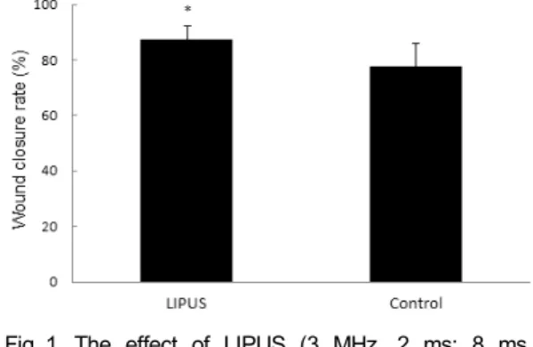
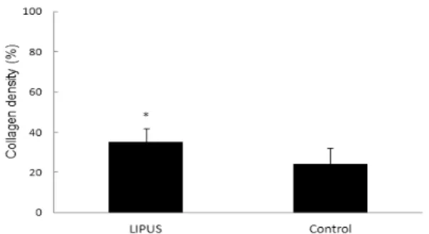
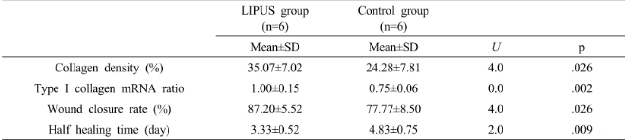
Low-Intensity Pulsed Ultrasound Promotes Healing with Increases Collagen Deposition and Collagen mRNA Expression in Skin Wound of Rat
Jae-Hyoung Lee †
⋅Seung-Joo Jekal 1 ⋅Pil-Seung Kwon 1
Dept. of Physical Therapy, Electrotherapy Research Laboratory for Tissue Growth & Repair, Wonkwang Health Science University,
1 Dept. of Clinical Laboratory Science, Wonkwang Health Science University
저강도 맥동초음파에의한 피부 상처 치유 촉진과 아교질 축적 및 아교질 mRNA 발현 증가
이재형 † , 제갈승주 1 , 권필승 1
원광보건대학교 물리치료과, 1 원광보건대학교 임상병리과
Received: July 31, 2013 / Revised: August 14, 2013 / Accepted: August 19, 2013
ⓒ 2013 Journal of the Korean Society of Physical Medicine
| 초록 |