은 나노입자 합성을 위한 Bacterial Cellulose 생산 세균의 분리 및 특성
유지연, 장은영, 손용준, 박수연, 손홍주*
부산대학교생명환경화학과/생명산업융합연구원
Received: February 21, 2018 / Revised: March 26, 2018 / Accepted: March 27, 2018
서 론
세균셀룰로오스(bacterial cellulose, BC)는미생물이생 산하는다당류의일종으로서, 나노크기의미세망상구조, 높 은결정성, 강한기계적강도, 높은수분함량및우수한생 체적합성과생분해성을가지고있다[1]. 또한, 식물성셀룰로 오스와달리 BC는리그닌, 헤미셀룰로오스, 펙틴과같은불
순물을함유하고있지않다[2]. 이러한독특한특성때문에
BC는식품산업, 하수정화, 음향및광학, 의료분야등에
서활용가능성이높은것으로인정되고있으며[3], 최근상
처치료드레싱재료, 인공피부, 인공혈관등의생체적합분
야에서활발히연구되고있다[4]. 특히, BC는구조적특성의
변화없이멸균할수있으며, 상처에서발생하는열을흡수하 고, 기계적손상으로부터상처를보호함과동시에상처부위 에습윤환경을제공함으로써치료효과를높이고, 통증을줄 일수있는것으로보고되었다[5]. 그러나 BC 자체는항균 활성을가지고있지않고, 결과적으로상처부위의 2차감염 을예방하기어렵기때문에항균성을지닌차세대 BC 개발
에대한연구가진행되고있다[6].
은화합물이나은이온은역사적으로위생이나감염성질 병치료에널리사용되어왔던항균제이다. 최근, 은나노입 자는그모재(parent material)와달리특이적으로향상된이
화학적및생물학적성질을가진다는것이밝혀졌고[7], 이
에따라은나노소재가새롭게주목받고있다. 은나노입자 Isolation and Characterization of Bacterial Cellulose-Producing Bacteria for Silver Nanoparticle Synthesis
Ji-Yeon Yoo, Eun-Young Jang, Yong-Jun Son, Soo-Yeun Park, and Hong-Joo Son*
Department of Life Science and Environmental Biochemistry/Life and Industry Convergence Research Institute, Pusan National University, Miryang 50463, Republic of Korea
As a basic study for environment-friendly production of bacterial cellulose (BC) dressing with antimicro- bial activity, we isolated and identified acetic acid bacteria which are resistant to silver ions and can bio- synthesize silver nanoparticles. Furthermore, conditions of BC production by selected strain were also investigated. Strain G7 isolated from decayed grape skin was able to grow in the presence of 0.1 mM AgNO3 which was identified as Acetobacter intermedius based on 16S rRNA gene analysis. BC production was the highest in a medium containing 2% glucose as a carbon source, 2% yeast extract as a nitrogen source, and 0.115% acetic acid as a cosubstrate. Structural properties of BC produced in optimal medium were studied using Fourier-transform infrared spectroscopy and X-ray diffractometer, and it was found that BC pro- duced was cellulose type I that was the same as a typical native cellulose. When strain G7 was cultured in an optimal medium containing 0.1 mM AgNO3, the color of the culture broth turned into reddish brown, indicating that silver nanoparticles were formed. As a result of UV-Vis spectral analysis of the culture, it was found that a unique absorption spectrum of silver nanoparticles at 425 nm was also observed. Scan- ning electron microscopic observations showed that silver nanoparticles were formed on the surface and pores of BC membrane.
Keywords: Bacterial cellulose, Acetobacter intermedius, silver nanoparticle, silver-resistance
*Corresponding author
Tel: +82-55-350-5544, Fax: +82-55-350-5904 E-mail: [email protected]
© 2018, The Korean Society for Microbiology and Biotechnology
는인체무독성, 무자극성이며, 은나노입자에대한내성균 의출현도아직보고된바없어약제내성균을제거하는데
활용될수있을것으로기대된다[8]. 이러한은나노입자는
주로화학적으로합성되고있는데, 독성이높은환원제와안 정제의사용, 유해성부산물의배출등여러가지문제점을 가지고있어[9] 다양한분야로의응용을위해서는무독성물 질을이용한환경친화적합성법이필요함을알수있다. 따 라서이러한문제점을극복하기위해근래수년동안미생 물이나식물추출물을이용한은나노입자합성연구가활
발히시도되어왔다[10−12]. 생물학적은나노입자의합성
기전은이들이생산하는환원효소(reductase) 또는환원물질
이은이온을환원하여은나노입자로전환시키는것이라고
추정되고있지만아직정확하게밝혀진것은아니다[13].
지금까지 BC에항균성을부여하기위하여질산은 (AgNO3) 용액에 BC를침지시킨후, 화학적으로은나노입자를합성 하는방법이보고되었으나이방법은 BC에독성용매가잔
류한다는단점이있다[14]. 따라서은나노입자를생합성할
수있는 BC 생산균주를확보할 수있다면 보다안전하게
2차감염을방지할수있는상처치료 BC 드레싱을제조할
수있을것이다. 본연구는환경친화적으로항균성이부여된 상처치료용 BC 드레싱개발을최종목적으로설계되었으며, 이에따라먼저은이온에대해내성이있으면서은나노입 자를생합성할수있는균주를분리및동정한후, 이균주 에의한 BC 생산최적조건및구조적특성을조사하였다.
재료 및 방법
실험균주의 분리 및 동정
가정에서전통적인방법에의하여제조된식초와감, 사과, 감귤, 포도등의과일을 BC 생산균주를분리하기위한시료 로사용하였다. 각시료를 HS 배지에접종하여 30℃에서배
양하면서막(membrane) 형태의 BC 생성유무를관찰하였
다. 생성된 BC를생리식염수에넣어강하게교반한후, 현 탁액을 HS 평판배지(0.0022% bromocresol green 함유)에 접종하여배양하면서배지의색깔을녹색에서노란색으로 변화시키는집락을 BC 생산균주로선정하였다. 이균주들을
대상으로다양한농도의 AgNO3가함유된 HS 배지에접종
하여배양함으로써 BC 생산이가능한 AgNO3의농도를확 인하였다. 실험에 사용된 HS 배지의 조성은 glucose 2%, yeast extract 0.5%, polypeptone 0.5%, Na2HPO4· 12H2O 0.675% 및 citric acid monohydrate 0.115% (pH 6)이었다. 분리균주를동정하기위하여 16S rRNA 유전자의염기서 열을분석하였다. 16S rRNA 유전자를증폭하기위하여사 용된 primers는 27F (5'-AGAGTTTGATCM TGGCTCAG-3') primer와 1492R (5'-TACGGY TACCTTGTTACGACTT-3')
primer이었다. 16S rRNA 유전자의염기서열을결정한후,
GenBank DB의 유사균주들과 비교하였으며, Clustal X
(version 1.81) program package를이용하여염기서열을정 렬한후, neighbor-joining method에의거하여분리균주의 계통분류학적위치를확인하였다[15].
BC 생산 조건 조사
BC 생산에영향을미치는배지성분을조사하기위하여탄 소원, 질소원및보조탄소원에따른 BC 생산량을조사하였 다. 전배양은 HS 평판배지에서계대배양후, 냉장보관중인 실험균주를한백금이(one loop) 취하여 50 ml의 HS 배지에 접종하여 30℃에서 4일동안정치배양하였다. 생성된 BC로 부터세포를유리시키기위하여 5분간강하게진탕한후, 멸 균된거즈로여과하여세균현탁액을조제하였다. 이세균
현탁액 5% (v/v)를상기에서보는바와같은배양변수들이
각각조절된 HS 배지 50 ml에접종하여 30℃에서 7일간정 치배양하였다.
BC의 구조적 특성 조사
동결건조된 BC를금으로코팅한후, 주사전자현미경(JEOL JSM-6390, JEOL TECHNIC Ltd., Japan)을이용하여 3차 원적미세구조를관찰하였다. BC의화학적구조를조사하기 위하여 FT-IR spectrophotometer (IRAffinity-1, Shimadzu Corp., USA)를이용하여 400−4000 cm-1에서 측정하였다. BC의결정구조는 X-ray Diffractometer (Rigaku III, Rigaku Corp., Japan)를 이용하여 측정하였다. 즉 BC를 40 kV, 30 mA 조건에서 Cu Kα radiation에의한반사식측정법으 로 2θ = 5°−40°, scan speed는 10°/min로측정하였다. 결정화 지수(crystallite index)는 [(I002−Iam)/I002] × 100에의하여 산출하였다. 여기서 I002는 2θ = 22.6°에서 (002)면에상응하 는회절 peak의최대강도(intensity)이며, Iam은 2θ = 18°에 서회절강도이다[16].
실험균주에 의한 은 나노입자 합성 확인
0.1 mM의 AgNO3가함유된최적배지에실험균주를접종
하여배양하면서배양액의색깔변화를관찰하였다. 7일동안 배양한후, 배양액을원심분리(9,200 ×g, 5분)하여회수된상 등액의은나노입자합성유무를조사하였다. 이때, 은나노 입자고유의색깔발현과고유흡수파장을조사하여은나 노입자의합성을확인하였다[10]. 또한, BC 막에은나노입 자가생성되었는지를주사전사현미경을통하여관찰하였다. 분석방법
배양액내에생성된 BC를거즈를이용하여여과하여회 수한후, 배양액성분을제거하기위하여수돗물로세척하
였다. 세척된 BC를 0.5 N NaOH 200 ml에넣어 100℃에 서 1시간 30분동안 가열하여세포를 용해시킨후, 0.5 N HCl을이용하여중성이될때까지중화시켰다. 최종적으로 회수된 BC를 105℃에서항량이될때까지건조한후, 데시 케이터에서방냉하여건조중량을측정하였다[17].
결과 및 고찰
은 내성 BC 생산균주의 분리 및 동정
전통식초, 사과, 감귤및포도시료로부터각각 23 균주, 11 균주, 7 균주및 16 균주에해당하는 BC 생산균을분리 하였다. 그중 0.02 mM AgNO3가함유된 HS 배지에서 BC 를생산하는 G7 균주를최종실험균주로선정하였다. 이균 주는부패된포도껍질에서분리되었다. 본균주의 AgNO3 농 도에따른 BC 생산유무를조사한결과, 0−0.1 mM AgNO3 범위에서 BC를생산하였으나 AgNO3 농도증가에따라 BC
생산량은감소하였다. 0.12 mM 이상의 AgNO3 농도에서는 BC가생산되지않았다(Fig. 1).
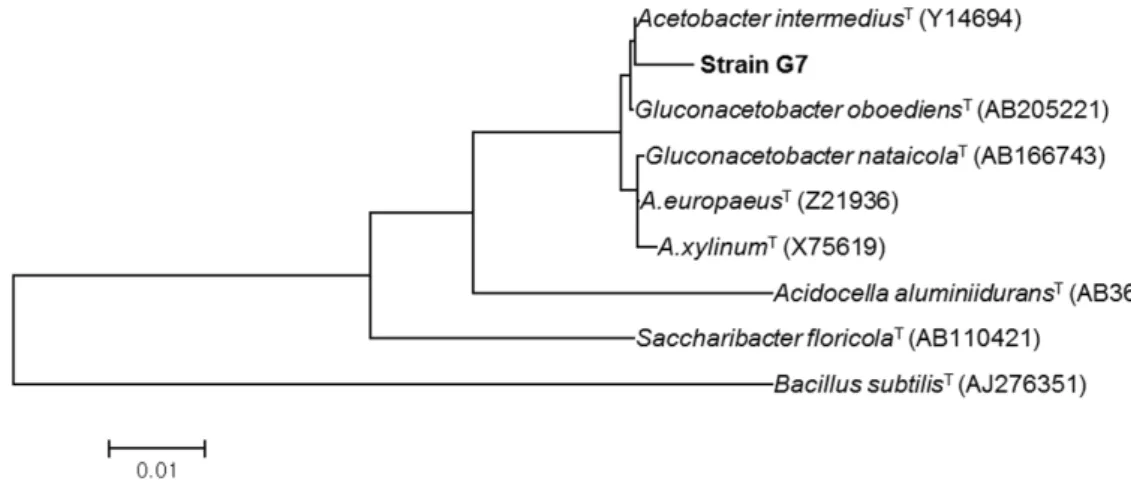
실험균주를동정하기위하여 16S rRNA 유전자의염기서 열을 분석한 결과, 본 균주는 Acetobacter intermdiusT와 99%의유전자간상동성을보였다. 또한 16S rRNA 유전자 의구조에근거하여유사균주들과의계통학적관련성을조 사하기 위하여 계통수를 작성한 결과, 실험균주는 A.
intermdiusT와동일한계통학적그룹에속함을알수있었
다(Fig. 2). 따라서실험균주를 A. intermdius G7로명명하 였다.
BC 생산 조건
탄소원의종류와농도가 BC 생산에미치는영향을조사 한결과, glucose를탄소원으로사용하였을때 BC 생산이가 장우수하였다(Table 1). Glucose의농도를 0%에서 3%까지 각각달리하여배양한결과, 2%까지탄소원의농도증가에 따라 BC 생산도증가하였으며, 그이상의농도에서는생산 량이일정하였다(Fig. 3). BC를생산하는초산균의특성은 glucose로부터 BC 합성을위한전구체를생성하는 uridine diphosphoglucose pyrophosphorylase 활성이높다는것이 다[18]. 따라서 glucose는생육을위한에너지생성에이용 되지않고, 주로 BC 생산에소비될수있다. 질소원의종류
와 농도가 BC 생산에 미치는 영향을 조사한 결과, beef
extract와 yeast extract를질소원으로사용하였을때 BC 생 산이가장좋았으며, 질소원의첨가는 BC 생산에필수적임 을알수있었다(Table 1). BC 생산에최적인 beef extract와 yeast extract의농도를조사한결과, 각각 1%와 2%로나타 났다(Fig. 3). 한편, Acetobacter xylinum을이용한 BC 생산 에 있어 다양한 질소원의 영향이 보고되었는데, casein hydrolyzate, peptone, yeast extract가적합한질소원임이 밝혀졌다[3].
Fig. 1. Effect of silver nitrate concentration on BC production by Acetobacter intermedius G7.
Fig. 2. Phylogenetic tree based on 16S rRNA gene sequences constructed by the neighbor-joining method.
보조탄소원의 첨가는 대사 구동력(metabolic driving
force) 측면에서 BC 합성에상당히중요한역할을수행한다
[3]. 따라서각종보조탄소원이 BC 생산에미치는영향을조
사한결과, acetic acid, citric acid, lactic acid 및 ethanol을 첨가했을때우수한 BC 생산을보여주었으며, 그중 acetic acid에서 가장높은생산량을나타내었다(Table 2). Acetic acid, lactic acid 및 ethanol은초산균의생육을촉진하고, 동 시에 BC 생산을 증가시킨다고 알려져 있다[19]. 특히, ethanol은 glucose의세포내대사즉, ATP 합성을억제함과 동시에 glucose가직접적으로 BC 생산에이용될수있게하
는것으로알려져있다[20].
BC의 구조적 특성
실험균주에 의하여 생산된 BC의 화학적조성을 FT-IR spectrophotometer를이용하여조사한결과는 Fig. 4A에서 보는바와같다. 생산된 BC는 cellulose I의 C-H 결합의흡
수스펙트럼에상응하는 2,900 cm-1부근의밴드를나타내 었다. 3,400 cm-1부근의밴드는 cellulose의 O-H stretching frequency에해당하며, 1,370 cm-1의밴드는 cellulose의 CH stretching vibration에해당한다. 실험균주에의하여생산된 BC의 X선 회절 패턴은 Fig. 4B에서 보는 바와 같다. Cellulose I의 (101)면및 (101)면에해당하는 2θ = 14.6° 및
16.4°에서회절피크가중첩되어나타났으며, (002)면의회
절피크가 2θ = 22.6에서나타났다. 그러나 cellulose II의회 절 peak (2θ = 12.1° 및 20.8°)는나타나지않았다. 이것은실 험균주에의하여생산된 BC는 cellulose I이라는것을의미 한다. 또한, 생산된 BC의결정화지수는 79.4%로나타났다.
실험균주에 의한 은 나노입자 합성
0.1 mM AgNO3가함유된최적배지에실험균주를접종하
여배양하면서배양시간경과에따른은나노입자합성유무 를조사하였다. Fig. 5A에서보는바와같이배양시간경과 에따라반응액의색깔이은나노입자특유의적갈색으로변 Table 1. Effect of carbon and nitrogen sources on BC production by Acetobacter intermedius G7.
Carbon source (2%) BC yield (g/l) Nitrogen source (1%) BC yield (g/l)
Fructose Galactose Glucose Glycerol Lactose Maltose Mannitol Sorbitol Starch Sucrose Xylitol None
0.244 0.042 0.304 0.256 0.048 0.188 0.148 0.116 0.191 0.258 0.232 0.170
Beef extract Casamino acid
Casein enzymatic hydrolysate Corn steep liquor
Malt extract Polypeptone
Proteose peptone NO. 3 Soytone
Tryptone Yeast extract
Polypeptone 0.5% + Yeast extract 0.5%
None
1.141 0.244 0.695 0.356 0.326 0.554 0.608 0.952 0.954 1.246 0.312 0.000
Fig. 3. Effect of glucose (●), yeast extract (○) and beef extract (■) concentrations on BC production by Acetobacter interme- dius G7.
Table 2. Effect of cosubstrate on BC production by Aceto- bacter intermedius G7.
Cosubstrate (0.115%) BC yield (g/l) 2-ketoglutaric acid
Acetic acid Citric acid Formic acid Fumaric acid Gluconic acid Lactic acid Maleic acid Malic acid Pyruvic acid Succinic acid Tartaric acid Ethanol None
0.956 1.402 1.352 0.676 0.873 0.944 1.366 0.760 0.968 0.967 0.831 0.949 1.387 0.852
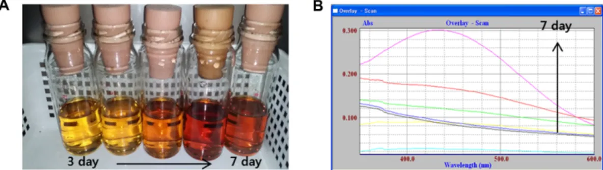
화됨을관찰할수있었는데, 이러한변화는은나노입자의 표면플라즈몬공명(surface plasmon resonance; SPR) 진동 의들뜸현상때문인것으로보고되어있으며, 이것은은이 온이은나노입자로환원되었다는것을의미한다[10−12]. 실
험균주에의한은나노입자합성은 UV-Vis 스펙트럼분석
을 통하여 재확인하였는데, Fig. 5B에서 보는 바와 같이
425 nm에서은나노입자의고유한흡수파장이관찰되었다.
Mie의이론에따르면, 구형나노입자의흡수스펙트럼에서
는한개의 SPR 밴드가나타나지만이방성입자는입자의
모양에따라둘이상의 SPR 밴드를발생시킬수있다[21].
본연구에서는반응혼합물의 SPR 밴드가한개나타났으 므로구형의은나노입자가합성되었음을알수있었다. 또 한본연구결과는은이온의환원은 A. intermdius G7에의 해생성된환원제에의해발생했다는것을시사한다. 0.1 mM AgNO3가함유된 HS 배지에서생성된 BC 막은표면과기 공에은나노입자가잘분산되어형성됐음을보여주는반면 (Fig. 6A), AgNO3가함유되지않은배지에서생성된순수한 BC 막은매끄럽고, 깨끗한표면을가진수많은 nanofibrils 로이루어져있음을알수있었다(Fig. 6B).
현재실험균주에의한은나노입자합성기전에대한실험 을수행중에있으며, 그결과를바탕으로효율적인상처치 료용드레싱개발을위한항균능등피부관련생리활성을확 인하고자한다.
요 약
환경친화적으로항균성이부여된상처치료용 BC 드레싱 을개발하기위한기초연구로서, 은이온에대해내성이있 으면서은나노입자를생합성할수있는초산균을분리및 동정하였다. 나아가실험균주에의한 BC 생산조건을조사 하였다. 부패된포도껍질로부터분리된 G7 균주는 0.1 mM AgNO3존재하에서생육할수있었으며, 16S rRNA 유전자 의염기서열분석에의거하여 Acetobacter intermdius로동 정되었다. 탄소원으로 2% glucose, 질소원으로 2% yeast extract, 보조탄소원으로 0.115% acetic acid가함유된배지 에서 BC 생산량이최대였다. 최적배지에서생성된 BC의구 조적특성을 FT-IR 및 XRD를사용하여조사한결과, 생성 된 BC는전형적인천연 cellulose와동일한 cellulose I인것 으로확인되었다. G7 균주를 0.1 mM AgNO3가함유된최적 배지에서배양한결과, 배양액의색깔이적갈색으로변하였 으며, 이것은은나노입자가생성되었음을의미한다. 은나 노입자의합성유무를 UV-Vis 스펙트럼분석에의하여확인
Fig. 5. Color change (A) and UV-visible spectrum (B) of culture supernatant over incubation time.
Fig. 4. FT-IR spectrum (A) and X-ray diffraction profile (B) of BC produced by Acetobacter intermedius G7.
한바, 425 nm에서은나노입자의고유한흡수스펙트럼이 관찰되었다. 또한, 생성된 BC를주사전자현미경으로관찰한 결과, 표면과기공에은나노입자가생성되어있음을재확인 하였다.
Acknowledgments
This research was supported by Basic Science Research Program through the National Research Foundation of Korea (NRF) funded by the Ministry of Education (NRF-2015R1D1A1A01056919).
Conflict of Interest
The authors have no financial conflicts of interest to declare.
References
1. Sutherland IW. 1998. Novel and estabilished applications of microbial polysaccharides. Trends Biotechnol. 16: 41-46.
2. Iguchi M, Yamanaka S, Budhiono A. 2000. Bacterial cellulose - a masterpiece of natural's arts. J. Mater. Sci. 35: 261-270.
3. Chawla PR, Bajaj IB, Survase SA, Singhal RS. 2009. Microbial cel- lulose: fermentative production and applications. Food Technol.
Biotechnol. 47: 107-124.
4. Torres FG, Commeaux S, Troncoso OP. 2012. Biocompatibility of bacterial cellulose based biomaterials. J. Funct Biomater. 3:
864-878.
5. Rajwade JM, Paknikar KM, Kumbhar JV. 2015. Applications of bacterial cellulose and its composites in biomedicine. Appl.
Microbiol. Biotechnol. 99: 2491-2511.
6. Sulaeva I, Henniges U, Rosenau T, Potthast A. 2015. Bacterial cellulose as a material for wound treatment: properties and modifications. A review. Biotechnol. Adv. 33: 1547-1571.
7. Lansdown AB. 2006. Silver in health care: antimicrobial effects and safety in use. Curr. Probl. Dermatol. 33: 17-34.
8. Rai MK, Deshmukh SD, Ingle AP, Gade AK. 2012. Silver nanopar- ticles: the powerful nanoweapon against multidrug-resistant bacteria. J. Appl. Microbiol. 112: 841-852.
9. Hebbalalu D, Lalley J, Nadagouda MN, Varma RS. 2013. Greener techniques for the synthesis of silver nanoparticles using plant extracts, enzymes, bacteria, biodegradable polymers, and microwaves. ACS Sustainable Chem. Eng. 1: 703-712.
10. Anthony KJP, Murugan M, Jeyaraj M, Rathinam NK, Sangili- yandi G. 2014. Synthesis of silver nanoparticles using pine mushroom extract: A potential antimicrobial agent against E.
coli and B. subtilis. J. Ind. Eng. Chem. 20: 2325-2331.
11. El-Baz AF, El-Batal AI, Abomosalam FM, Tayel AA, Shetaia YM, Yang S. 2016. Extracellular biosynthesis of anti-Candida silver nanoparticles using Monascus purpureus. J. Basic Microbiol. 56:
531-540.
12. Ramesh PS, Kokila T, Geetha D. 2015. Plant mediated green synthesis and antibacterial activity of silver nanoparticles using Emblica officinalis fruit extract. Spectrochim. Acta, Part A.
142: 339-343.
13. Rai M, Ingle AP, Gupta IR, Birla SS, Yadav AP, Abd-Elsalam KA.
2013. Potential role of biological systems in formation of nanoparticles: mechanism of synthesis and biomedical appli- cations. Curr. Nanosci. 9: 576-587.
14. Maneerung T, Tokura S, Rujiravanit R. 2008. Impregnation of sil- ver nanoparticles into bacterial cellulose for antimicrobial wound dressing. Carbohydr. Polym. 72: 43-51.
15. Saitou N, Nei M. 1987. The neighbor-joining method: a new method for reconstruction phylogenetic trees. Mol. Biol. Evol. 4:
406-426.
16. Focher B, Palma MT, Canetti M, Torri G, Cosentino C, Gastaldi G.
2001. Structural differences between non-wood plant cellulo- ses: evidence from solid state NMR, vibrational spectroscopy and X-ray diffractometry. Ind. Crops Prod. 13: 193-208.
17. Embuscado ME, BeMiller JN, Marks JS. 1996. Isolation and par- tial characterization of cellulose produced by Acetobacter xyli- num. Food Hydrocoll. 10: 75-82.
18. Masaoka S, Ohe T, Sakota N. 1993. Production of cellulose from glucose by Acetobacter xylinum, J. Ferment. Bioeng. 75: 18-22.
Fig. 6. Scanning electron micrographs of BC produced in media with (A) or without (B) silver nitrate. Circles show silver nanopar- ticles accumulated on the bacterial cellulose surface and pores.
19. Toda K, Asakura T, Fukaya M, Entani E, Kawamura Y. 1997. Cellu- lose production by acetic acid-resistant Acetobacter xylinum, J.
Ferment. Bioeng. 84: 228-231.
20. Yunoki S, Osada Y, Kono H, Takai M. 2004. Role of ethanol in
improvement of bacterial cellulose production: analysis using
13C-labeled carbon sources. Food Sci. Technol. Res. 10: 307-313.
21. Mie G. 1908. Contribution to the optics of turbid media, partic- ularly of colloidal metal solutions. Ann. Phys. 25: 377-445.