서 론
Genistein은 콩에 존재하는 대표적인 isoflavone으로서 유방 암 세포를 비롯한 많은 암세포에서 G2/M arrest와 세포자멸사를
조절하는 유전인자에 관여하여 암세포의 성장을 억제하는 등의 항암 효과가 있는 것으로 알려져 있다.(1-8) 1987년에 Akiyama 등(9)이 genistein이 protein tyrosine kinase (PTK)의 활성을 억제하는 효과가 있음을 처음으로 기술하였는데, PTKs는 종양 발생뿐 아니라 분화에도 결정적인 역할을 미치는 것으로 알려져 있어서(10) genistein이 암을 억제하는 역할을 설명하는 근거가 되고있다. 또다른연구는genistein에의한DNA topoisomerase II의 억제(11)와 ribosomal S6 kinase 활성의 억제를 보고하였 Purpose: To analyze the effect of the growth control on
human breast cancer cells with genistein treatment and to investigate the mechanism of genistein-induced G2/M arrest in T47D and MDA-MB231 breast carcinoma cells by Cdc25C expression. Methods: We analysed the proliferartion of the two cell lines by using MTT proliferation assay, flow cytometric analysis, real-time quantitative RT-PCR and western blotting and investigated the effect of genistein on cell survival, cel- lular toxicity, cell cycle progression-related genes and their mRNA and protein alterations. Results: The DNA flow cyto- metric analysis of both cell lines treated with genistein showed a dose-dependent growth inhibition and accumulation in the G2/M phase of cell cycle. The expression of p21 mRNA and protein increased in both cell lines following genistein treat- ment but p27 expression was unchanged. Furthermore, decreased Cdc25C expression with decreased polo-like kinase (PLK) 1 expression and increased PLK3 expression were observed after genistein treatment. The decreased level
of Cdc25C in the nucleus was associated with decreased phosphorylation of Cdc25C by PLK1. The expression of PLK3 was increased with a dose-dependent and a time-dependent manner and was associated with decreased Cdc25C expres- sion. Check point kinase (CHK) 1 and CHK2 revealed different expression patterns each other. The CHK1 expression was independent of the presence of genestein. CHK2 expression increased in MDA-MB231 cells associated with decreased Cdc25C expression but not in T47D. Conclusion: These results suggest that genistein induces a G2/M arrest in human breast cancer cells, the mechanism of which is due, in part, to decreased in Cdc25C phosphatase by a regulatory effect of PLK1, PLK3, and CHK2 as well as increased expression of the cyclin dependent kinase inhibitor p21(WAF1/CIP1).
Key Words: Breast neoplasms, Cell cycle, Genistein 중심단어: 유방암, 세포주기, 게니스틴
Molecular Mechanism of the G2/M Arrest in Breast Cancer Cell Lines (T47D and MDA-MB231) Induced by Genistein
Ji-Young Park, Ku-Seong Kang, Yoon Kyung Sohn
Department of Pathology, Kyungpook National University School of Medicine, Daegu, Korea
Breast Cancer
O R I G I N A L A R T I C L E
박지영ㆍ강구성ㆍ손윤경
경북대학교 의학전문대학원 병리학교실
유방암 세포주(T47D와 MDA-MB231)에서 Genistein의 세포주기 G2/M 정지 기전
책임저자: 손윤경
700-721 대구광역시 중구 동덕로 200, 경북대병원 병리과 Tel: 053-420-5241, Fax: 053-426-1525
E-mail: [email protected]
접수일: 2010년 6월 18일 게재승인일: 2010년 11월 12일
382
다.(12) 또한, 시험관 연구에서 genistein은 혈관 생성을 억제하 고,(13) 종양세포의 전이를 억제하였다.(14) Genistein의 작용에 p53 단백이나 에스트로겐 수용체와 안드로겐 수용체 등은 유의 한 관계가 없는 것으로 알려져 있다.(15) 지금까지 연구 결과들을 토대로 할 때, genistein이 암세포의 성장을 억제하고, 분화를 유도하며 세포자멸사를 초래하여 암세포의 성장을 억제하는 역 할을 하는 것으로 보이지만, 이러한 작용의 분자생물학적 기전은 아직 명확히 규명되지 않았다.
Genistein을 유방암 세포에 처리했을 때 G2/M arrest를 유 도하여 암세포의 성장을 억제하는데, 그 기전으로 genistein에 의한 p21 단백의 증가와 cyclin B1의 감소, 그리고 cdc2의 활성 억제를 들 수 있다.(1,4,15) 다양한 종류의 암세포주에 genistein 을 처리하였을 때 p21 단백의 증가와 G2/M arrest 유도가 대부 분 관련이 있었으나, p21 단백의 변화와 상관없이 G2/M arrest 가 일어나기도 하였다.(2,16) 따라서 genistein이 G2/M arrest 를 유도하는 데 있어서 p21 단백을 경유하는 경로 외에 다른 기전 이 관여하였을 것으로 생각된다. Cdc25C 단백은 CDK/cyclin 복합체를 탈 인산화하여 G2에서 M주기로 넘어가는 것을 촉진하 는 데 중요한 역할을 한다. Cdc25C의 활성은 인산화된 serine의 위치에 따라 달라지는데, 인산화를 조절하는 데에 polo-like kinase (PLK)와 check point kinase (CHK)가 역할을 하는 것 으로 알려져 있다.(17-22)
본 연구에서는 genistein이 유방암 세포에서 G2/M arrest를 유도하는 과정에서 p21과 같은 cyclin dependent kinase inhi- bitor를 통하지 않는 다른 경로, 특히 Cdc25C 단백과 그 조절인 자들과 관련된 경로와 연관되었는지를 알아보고자 하였다.
방 법
세포배양과genistein 처리
유방암 세포주는 p53 단백 돌연변이 세포주이면서 에스트로겐 수용체가 있는 T47D와 에스트로겐 수용체가 없는 MDA-MB231 (American Type Culture Collection, Rockville, USA)를 이 용하였고, genistein (Sigma Chemical Co., St. Louis, USA) 은 dimethyl sulfoxide (DMSO; Sigma Chemical Co., St.
Louis, USA)에 용해하여 사용하였다.
세포성장정도분석
Genistein 처리 후의 세포 성장 정도를 알아보기 위해서 T47D 와 MDA-MB231을 한 well당 2,000개의 세포를 24시간 동안 배양한 후 genistein을 각각 0, 0.1, 1, 10, 20, 50, 100 μM로 처 리하여 72시간 동안 배양하였다. Triazolyl blue (MTT; Sigma
Chemical Co., St. Louis, USA) 용액(2 mg/mL)을 한 well당 50 μL씩 첨가하여 4시간 동안 37℃에서 반응시킨 후 glycine:
DMSO (1:8) 혼합액을 한 well당 150 μL씩 첨가하여 반응을 종 료하고 570 nm에서의 흡광도를 측정하였다. 이때 대조군으로서 최종농도가 0.1%가 되도록 DMSO를 첨가하였다.
DNA 유세포분석
100 mm 배양용기에 2×10⁶개의 세포를 배양하고 24시간 후 최종 농도가 100 μM가 되게 genistein을 처리하였다. 3일 후 PBS 완충용액으로 수세하고 Trypsin/EDTA를 처리하여 세포 를 모은 후 70% 에탄올로 4℃에서 1시간 동안 처리하여 세포를 고정하고 PBS 완충용액으로 두 번 수세하였다. 50 μg/mL의 RNase A를 37℃에서 30분 동안 반응시킨 후 첨가한 RNase A 와 같은 부피로 50 μg/mL의 propidium iodide 용액을 첨가한 후 FACScan flow cytometer (Becton-Dickinson, San Jose, USA)를 이용하여 분석하였다.
세포주기관련유전자의mRNA 정량분석 Total RNA 분리
60 mm 배양용기에배양된각각의세포에TRIzol (Invitrogen, Carlsbad, USA)을 1.0 mL 넣고, 1.5 mL 튜브에 옮긴 후, 0.2 mL의 클로로포름을 넣고 잘 섞어준 후 3분간 상온에서 반응시켰 다. 12,000×g에서 7분간 원심 분리하여 생긴 상층 액을 새 튜브 로 옮긴 후 0.8배의 이소프로판올을 첨가하여 섞어주고 상온에서 10분간 반응시킨 후 12,000×g에서 10분간 원심 분리하였다. 원 심 분리 후 생긴 침전물을 75%의 알코올을 첨가하여 vortex한 후 7,500×g에서 5분간 원심 분리하여 용액을 덜어낸 후 침전물을 공기 중에서 말리고 각각의 튜브마다 30 μL의 RNase free water 를 첨가하여 녹인 후 원액을 100배 희석하여 260 nm 파장에서 정량을 하였다. RNA를 분리하는 데 사용된 용기와 용액은 모두 0.1% 4-diethyl pyrocarbonate를 처리하였다.
First strand cDNA 합성
First strand cDNA 합성 kit는 TaqMan Reverse Tran- scription Reagent (Applied Biosystems, Foster City, USA) 를 이용하였다. 각각의 튜브에 전체 RNA의 양이 5 μg, random hexamer를 2.5 μg, MgCl2을 5.5 mM, deoxy NTP를 500 μM, RNase inhibitor를 0.8 U, 10X RT buffer를 2 μL, multiscribe reverse transcriptase를 0.5 U 넣은 다음 최종 부피가 20 μL 가 되게 한 후, 25℃에서 10분, 48℃에서 30분, 95℃에서 5분 반응시켜 cDNA를 합성하였다.
실시간정량분석(Real time quantitative RT-PCR)
주형으로 사용된 cDNA는 위에서 만든 cDNA를 10배 희석하 여 사용하였다. 반응액의 조성은 2X SYBR green master mix 10.0 μL, 주형 4 μL, 각각의 primer를 200 nM 되게 넣고 물로 최종 부피를 20 μL 되게 하였다. 반응조건은 50℃에서 2분, 95℃ 에서 10분, 그리고 95℃에서 15초와 60℃에서 1분을 40회 반응 시켰다. 이때 각 cycle마다의 형광신호를 SDS 7000 (Applied Biosystems, Foster City, USA)로 모니터링하여 나타나는 threshold cycle (CT)을 분석한 후 2-△△CT방법을 이용하여 그래 프로 나타내었다.(23) 이때 사용한 각 유전자의 프라이머 염기서 열은 Table 1과 같다.
Western blot 분석
100 mm 배양용기에 2×10⁶개의 세포를 배양한 후 genistein 100 μM 되게 첨가하고 1일, 2일, 3일 후 세포를 모아서 lysis buffer (150 mM NaCl, 1.0% NP-40, 50 mM Tris [pH 8.0], 1 mM PMSF, 1 μg/mL aprotinin, 1 μg/mL Leupeptin, 0.1 mM sodium orthovanadate, 10 mM NaF)를 첨가하여 얼음 에서 10분간 반응시킨 후 12,000 rpm에서 10분간 원심 분리하 여 상층액을 모아서 BCA 방법으로 단백질의 양을 결정하였다.
약 30-100 μg의 단백질을 8-12% acrylamide gel에 전기영동 하고, nitrocellulose membrane에 블로팅한 후 Ponceau 용액 으로 단백질의 블로팅을 확인하고 TTBS 용액(25 mM Tris [pH 7.5], 150 mM NaCl, 0.1% Tween 20)으로 수세한 후 blocking 용액(25 mM Tris [pH 7.5], 150 mM NaCl, 0.1% Tween 20,
5% skim milk)으로 상온에서 2시간 동안 반응시킨 후 TTBS 용 액으로 수세하고 1차 항체 p21, p27, PLK1, PLK3, Cdc25C, p-S-216-Cdc25C, CHK1, alpha-tublin (Santa Cruz Bio- technology, Santa Cruz, USA), p-Serine-198 Cdc25C 항 체(Cell Signaling Technologies, Beverly, USA)을 1:200- 1:1,000으로 항체 반응 용액(25 mM Tris [pH 7.5], 150 mM NaCl, 0.1% Tween 20, 1% skim milk)으로 희석하여 4℃에서 12시간 동안 반응시켰다. TTBS 용액으로 수세한 후 2차 항체를 1:1,000으로 희석하여 상온에서 2시간 동안 반응시킨 후 TTBS 용 액으로 수세하고 Immobilon Western 키트(Millipore, Billerica, USA)를 이용하여 X-ray 필름에 감광시켰다.
결 과
Genistein에의한T47D와MDA-MB231의성장억제
Genistein이 유방암 세포주 T47D와 MDA-MB231의 성장에 미치는 영향을 알아보기 위해서 각각의 세포주에 genistein을 0.1, 1, 10, 20, 50, 100 μM 되게 처리하여 MTT 분석을 하였다.
각각의 세포는 10 μM까지는 성장에 변화를 보이지 않지만, 20 μM 부터 농도에 비례하여 세포의 성장이 억제되었다(Figure 1). 이때 대조군에비해genistein 처리후세포의밀도가줄었다(Figure 2).
Genistein에의한G2/M arrest 유도
Genistein이 각각의 세포분열주기에 미치는 영향을 알아보기 위해서 FACS 분석을 하였다. T47D는 G2/M에 해당하는 세포수 가 대조군은 21.94%이었고 genistein을 처리하였을 때 69.26%
로 약 3배 증가하였다. MDA-MB231은 대조군이 13.2%이었고 Table 1. Primer sequence of cell cycle regulatory genes
Gene Primer sequence (sense/antisense) GAPDH 5′-TGG GCT ACACTG AGC ACC AG-3′
5′-GGG TGT CGC TGT TGA AGT CA-3′ p21 5′-CAA AGG CCC GCT CTA CAT CTT-3′
5′-AGG AAC CTC TCA TTC AAC CGC-3′ p27 5′-GGA ATA AGG AAG CGA CCT GCA-3′
5′-TCC ACA GAA CCG GCA TTT G-3′ PLK1 5′-ATA GAG CGT GAC GGC ACT GAG T-3′
5′-TGC TCG CTC ATG TAA TTG CG-3′ PLK3 5′-TGT CAA AGT CAT CCC GCA GAG-3′
5′-TGC GAA AAA CGC ACG ATG T-3′ Cdc25C 5′-TTT TTC CAA GGT ATG TGC GCT G-3′
5′-TGG AAC TTC CCC GAC AGT AAG G-3′ CHK1 5′-CGT GAG CGT TTG TTG AAC AAG A-3′
5′-CAC AGG ACC AAA CAT CAA CTG G-3′ CHK2 5′-GTG CCT GTG GAG AGG TAA AGC T-3′ 5′-TGC CTC TCT TGC TGA ACC AAT A-3′ GAPDH=glyceraldehyde3-phosphatedehydrogenase; PLK=polo-like kinase; CHK=check point kinase.
Viable cells (%)
100 80
60 40 20 0
0 0.1 1 10 20 50 100
T47D MDA-MB-231
Figure 1. Concentration dependent growth inhibition of estrogen receptor (ER)-positive (T47D) and ER-negative (MDA-MB231) breast cancer cells by genistein. Twenty-four hours after inocu- lation, cells were cultured for 3 days in the absence or presence of genistein at concentratins ranging from 0.1 to 100 μM. The data represent the mean±SD of six independent experiments.
genistein을 처리하였을 때에는 55.55%로서 약 4배 증가하였다 (Figure 3).
Genistein 처리후세포주기관련유전자의mRNA 발현 p21 mRNA와p27 mRNA의발현
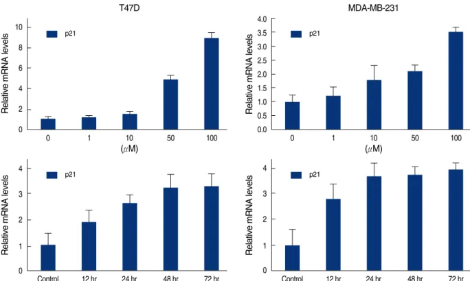
세포 분열주기에 관여하는 유전자들의 발현 양상을 알아보기 위하여 mRNA 실시간 정량분석을 하였다. CDK 저해제인 p21 의 mRNA는 처리한 genistein의 농도가 높을수록 발현이 두 세 포 주 모두에서 증가하였고, 100 μM의 genistein을 처리한 후 시 간에 비례하여 24시간에서 48시간 사이에 최대치로 발현하였다 (Figure 4). 반면 p27 mRNA는 genistein 처리를 한 군과 하지 않은군사이의발현이거의차이가 없었고, 배양시간과 genistein 의 농도와도 무관하였다(Figure 5).
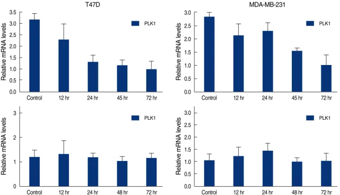
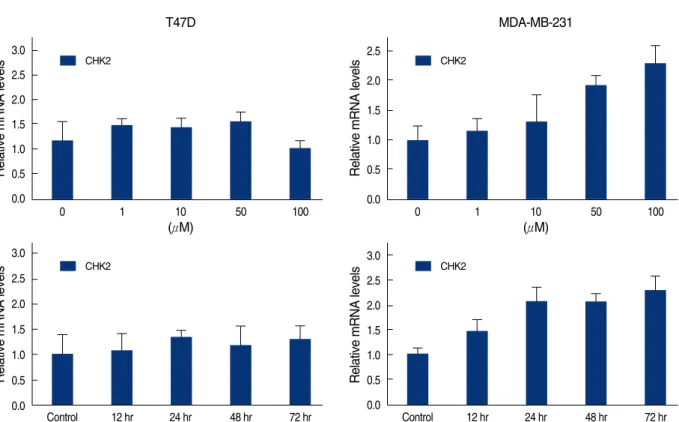
Cdc25C와polo-like kinase (PLK1, PLK3), check point kinase (CHK1, CHK2) mRNA의발현
Cdc25C의 활성을 증가시킨다고 알려져 있는 PLK1의 mRNA 는 genistein 처리 후 시간이 지남에 따라 두 세포주 모두에서 그 발현이 감소하였다(Figure 6). 그러나 Cdc25C의 활성을 억제하 는 데에 관여하는 것으로 알려진 PLK3, CHK1, CHK2의 mRNA 는 서로 다른 발현 양상을 나타내었다. PLK3은 genistein의 농 도와 genistein 처리 후 배양시간에 비례하여 mRNA의 발현이 두 세포주 모두에서 증가하였다(Figure 7). 반면 CHK1은 genis- tein 처리 후 시간에 관계없이 두 세포주 모두에서 mRNA의 발 현 양상에 변화가 없었고(Figure 6), CHK2는 MDA-MB231 Figure 2. Changes of cell densitiy in T47D cells (A, C) and MDA-
MB231cells (B, D). Each cells were treated with 0.1% DMSO (control) or 100 μM of genistein for 3 days. Genistein-treated cells are decreased in cellularity than control.
A B
C D
T47D MDA-MB231
Counts
450 360 270 180 90 0
0 200 400 600
FL2-A
3.57% 58.19% 12.04% 21.94% CON 11.22% 25.48% 3.52% 55.6% Gen 100 μM
3% 73.9% 7.02% 13.2% CON 5.63% 20.7% 10.06% 55.55% GEN 100 μM
Counts
450 360 270 180 90 0
0 200 400 600
FL2-A
Counts
400 320 240 160 80 0
0 200 400 600
FL2-A
Figure 3. DNA-fluorescence histogram of T47D and MDA-MB231 cells after genistein treatment. T47D (A) and MDA-MB231 (B) cells were treated with 0.1% DMSO (CON) or 100 μM of genistein (GEN) for 72 hr. Three days after genistein treatment, there was 3.1-fold increase in the percent of T47D cells in G2/M and 4.2-fold increase in the percent of MDA-MB231cells in G2/M, respectively.
A
B
Counts
200 160 120 80 40 0
0 200 400 600
FL2-A
Relative mRNA levels 10
8 6 4 2 0
0 1 10 50 100
(μM) T47D
p21
Figure 4. Real time quantitative analysis for p21 mRNA of genistein-treated T47D and MDA-MB231 cells. Each cell (5×10⁵cells) was treated with 0-100 μM genistein for 3 days or 100 μM of genistein for 0.5, 1, 2, 3 days.
Relative mRNA levels
4.0 3.5 3.0 2.5 2.0 1.5 1.0 0.5 0.0
0 1 10 50 100
(μM) MDA-MB-231
p21
Relative mRNA levels
4
3
2
1
0
Control 12 hr 24 hr 48 hr 72 hr p21
Relative mRNA levels
4
3
2
1
0
Control 12 hr 24 hr 48 hr 72 hr p21
Relative mRNA levels
1.5
1.0
0.5
0.0
0 1 10 50 100
T47D
p27
Figure 5. Real time quantitative analysis for p27 mRNA of genistein-treated T47D and MDA-MB231 cells. Each cell (5×10⁵cells) treated with 0-100 μM genistein for 3 days or 100 μM of genistein for 0.5, 1, 2, 3 days.
Relative mRNA levels
3.0 2.5 2.0 1.5 1.0 0.5 0.0
0 1 10 50 100
MDA-MB-231
p27
Relative mRNA levels
3.0 2.5 2.0 1.5 1.0 0.5 0.0
Control 12 hr 24 hr 48 hr 72 hr p27
Relative mRNA levels
2.5 2.0 1.5 1.0 0.5 0.0
Control 12 hr 24 hr 48 hr 72 hr p27
Relative mRNA levels 3.5 3.0 2.5 2.0 1.5 1.0 0.5 0.0
Control 12 hr 24 hr 45 hr 72 hr T47D
PLK1
Figure 6. Real time quantitative analysis for PLK1 mRNA and CHK1 mRNA of genistein-treated T47D and MDA-MB231 cells. Each cell (5×10⁵cells) treated with 100 μM of genistein for 0.5, 1, 2, 3 days.
Relative mRNA levels
3.0 2.5 2.0 1.5 1.0 0.5 0.0
Control 12 hr 24 hr 45 hr 72 hr
MDA-MB-231
PLK1
Relative mRNA levels
3
2
1
0
Control 12 hr 24 hr 48 hr 72 hr PLK1
Relative mRNA levels
3.0 2.5 2.0 1.5 1.0 0.5 0.0
Control 12 hr 24 hr 48 hr 72 hr
PLK1
Relative mRNA levels
6 5 4 3 2 1 0
0 1 10 50 100
(μM) T47D
PLK3
Figure 7. Real time quantitative analysis for PLK3 of genistein-treated T47D and MDA-MB231 cells. Each cell (5×10⁵cells) treated with 0-100 μM genistein for 3 days or 100 μM of genistein for 0.5, 1, 2, 3 days.
Relative mRNA levels
2.5 2.0 1.5 1.0 0.5 0.0
0 1 10 50 100
(μM) MDA-MB-231
PLK3
Relative mRNA levels
3.5 3.0 2.5 2.0 1.5 1.0 0.5 0.0
Control 12 hr 24 hr 48 hr 72 hr PLK3
Relative mRNA levels
2.5 2.0 1.5 1.0 0.5 0.0
Control 12 hr 24 hr 48 hr 72 hr
PLK3
세포에서는 genistein의 농도와 배양시간에 따라서 mRNA의 발현이 증가하였으나, T47D 세포에서는 유의한 변화가 없었다 (Figure 8). 그리고 G2/M 전환에 중심 역할을 하는 Cdc25C는 세포주의 종류와 상관없이 genistein 처리 후 배양시간에 비례 하여 mRNA의 발현이 줄었다(Figure 9).
Genistein 처리후세포주기관련유전자의단백발현양상
Genistein 100 μM을 처리한 후 시간에 따른 세포주기 관련
유전자의 단백 발현 양상을 비교하기 위하여 western blot을 수 행하였다(Figure 10). CDK 저해제인 p21은 두 세포주 모두에서 시간에 비례하여 단백의 발현이 증가하였고, p27은 시간에 관계 없이 단백의 발현 양상에 변화가 없었다. PLK1은 두 세포주 모두 시간에 비례하여 단백의 발현이 감소하였고, PLK3은 시간에 비 례하여 발현이 증가하였다. 그러나, CHK1은 발현 양상에 변화가 없었다. CDC25C는 두 세포 주 모두 시간에 비례하여 단백의 발현 이 감소하였는데, 198-serine과 216-serine 아미노산이 인산화
Relative mRNA levels
3.0 2.5 2.0 1.5 1.0 0.5 0.0
0 1 10 50 100
(μM) T47D
CHK2
Figure 8. Real time quantitative analysis for CHK2 mRNA of genistein-treated T47D and MDA-MB231 cells. Each cell (5×10⁵cells) treated with 0-100 μM genistein for 3 days or 100 μM of genistein for 0.5, 1, 2, 3 days.
Relative mRNA levels
2.5 2.0 1.5 1.0 0.5 0.0
0 1 10 50 100
(μM) MDA-MB-231
CHK2
Relative mRNA levels
3.0 2.5 2.0 1.5 1.0 0.5 0.0
Control 12 hr 24 hr 48 hr 72 hr CHK2
Relative mRNA levels
3.0 2.5 2.0 1.5 1.0 0.5 0.0
Control 12 hr 24 hr 48 hr 72 hr CHK2
T47D
Figure 9. Real time quantitative analysis for Cdc25C mRNA of genistein-treated T47D and MDA-MB231 cells. Each cell (5×10⁵cells) treated with 0-100 μM genistein for 3 days or 100 μM of genistein for 0.5, 1, 2, 3 days.
MDA-MB-231
Relative mRNA levels
3.0 2.5 2.0 1.5 1.0 0.5 0.0
Control 12 hr 24 hr 48 hr 72 hr cdc25c
Relative mRNA levels
3.5 3.0 2.5 2.0 1.5 1.0 0.5 0.0
Control 12 hr 24 hr 48 hr 72 hr cdc25c
된 Cdc25C 단백의 양도 각각 감소하였다.
고 찰
본연구에서genistein에의한세포성장의억제와G2/M arrest 는 세포의 에스트로겐 수용체의 유무와 상관없었다. Genistein 처리 후 CDK 저해제인 p21의 mRNA와 단백발현은 시간과 농도 에 비례하여 증가하였으며 이는 이미 보고된 다른 연구들과 일치 하는 결과이다.(3-5,15) 반면, genistein 처리 후 농도에 의존하 여 유의하게 증가함을 보고한 Yoon 등(24)의 연구 결과와는 달리 p27 단백은 변화가 거의 없었다. 이는 MDA-MB231과 MCF-7 유방암 세포주, MCF-10A 유방 상피세포주, 그리고 DU145 전 립선암 세포주에서 genistein 처리 후 p27 단백의 발현이 증가 하였다는 보고와는 다소 다르지만,(6,24-26) B16-F1 흑색종 세 포주에서 genistein 처리 후 p27 단백의 발현이 변화가 없었다 는 보고와는 일치한다.(8)
PLK1, PLK3, CHK1, CHK2는 Cdc25C를 인산화시키는 효 소로서 DNA 손상 후에 일어나는 G2/M arrest와 관계가 있다.
PLK1은 Cdc25C의 198번 serine을 인산화하여 핵 내로 이동하 는 것을 촉진하여 세포 분열을 활성화시키는 데 관여한다.(27,28) 본 연구에서는 genistein 처리 후 PLK1 단백의 발현이 낮아졌고, 이는 Cdc25C의 198번 serine의 인산화의 감소를 초래하여 결국 핵에 존재하는 Cdc25C의 양이 줄어들므로 G2/M arrest유도에 관여할 것으로 생각된다. 반면, genistein을 두 유방암 세포주에 처리하였을 때 시간과 농도에 비례하여 PLK3 단백 발현은 증가 하였다. Xie 등(29)의 보고에 의하면 암세포 주에 DNA 손상을 주었을 때, PLK1의 발현은 억제되었지만 PLK3의 발현은 오히려 증가하였다. 따라서 DNA 손상 후 일어나는 수복 과정에 PLK1과
PLK3은 서로 반대의 역할을 할 것이라고 주장하였다. 두 유방암 세포주에 genistein 처리 후 PLK1과 PLK3의 서로 다른 발현 양 상은 DNA 손상 후 일어나는 G2/M arrest와 관계되어 있음을 시 사한다. 하지만 PLK3이 Cdc25C를 유일한 기질로 사용하지 않기 때문에 genistein에 의해 증가된 PLK3이 어떻게 G2/M arrest를 유도하는가에대한기전은더많은연구가필요할것이다. Cdc25C 의 216번 serine을 인산화시켜 Cdc25C의 핵으로의 이동을 억제 하는 데 관여하는 CHK1과 CHK2는 서로 다른 발현 양상을 나타 내었다. Genistein 처리 후 두 세포주 모두 CHK1 단백 발현은 변화가 없었다. 반면 CHK2은 T47D 세포주에서는 단백 발현에 변화가 없었으나 MDA-MB231 세포주에서는 발현이 증가하였다.
Ismail 등(30)은 신경모세포암 세포주를 이용한 연구에서 genis- tein 처리 후 mediator of DNA damage check point protein 1 (MDC1)의발현이증가되고, 이와연관하여CHK2가 활성화되며, PLK-1의 발현의 저하와 연관된 세포자멸사가 유도되는 것을 보 고하였고, 이 과정에 MDC1이 중요한 역할을 한다고 하였다.
그러나 본 연구에서 유방암 세포주에 genistein을 처리하였을 때 Cdc25C 단백의 발현양도 줄어들었고 또한 216번 serine이 인산화된 Cdc25C의 양도 함께 감소하였다. 뿐만 아니라 198번 serine이 인산화된 Cdc25C의 양도 감소하였다. 이는 Cdc25C 의 전체 양이 줄어들었기 때문에 인산화된 Cdc25C의 양도 적은 것으로 생각한다. Genistein 처리에 따른 Cdc25C 단백 발현과 관련하여 Chang 등(3)은 genistein 처리에 의해 Cdc25C의 발 현은 줄어들지만, 216번 serine에 인산화된 Cdc25C는 증가한다 고 보고하였다. 이는 본 연구의 결과와 상반되는 결과이며, 어떻게 서로 다르게 발현될 수 있는지는 좀 더 많은 연구가 필요하지만, genistein 처리 후 CHK2, PLK1과 PLK3이 서로 복합적으로 상호작용하여 Cdc25C의 인산화를 조절하여 G2/M arrest를 일 으키는 데 관계할 것으로 생각한다.
결 론
지금까지의 결과를 요약하면 genistein 처리에 의해서 일어나 는 G2/M arrest는 p21 CDK 억제 단백질의 증가와 Cdc25C의 감소, 그리고 CDC25C를 인산화시키는 PLK3의 발현 증가와 PLK1의 감소가 관계되는 것으로 보이며, 여기에 CHK2와 CHK1 이 DNA 손상에 의한 checkpoint pathway에 영향을 미치는 것으로 보인다.
참고문헌
1. Cappelletti V, Fioravanti L, Miodini P, Di Fronzo G. Genistein blocks Figure 10. Western blot analysis of cell cycle-related genes. Each
cell was treated with 100 μM of genistein for 1, 2, 3 days. C; 0.1%
DMSO treated cells.
T47D C 1D 2D 3D p21
p27 p27 PLK1 PLK3 CHK1 cdc25C
a-tublin p-cdc25C- S198 p-cdc25C- S216
MB231 C 1D 2D 3D
breast cancer cells in the G(2)M phase of the cell cycle. J Cell Biochem 2000;79:594-600.
2. Casagrande F, Darbon JM. p21CIP1 is dispensable for the G2 arrest caused by genistein in human melanoma cells. Exp Cell Res 2000;258:
101-8.
3. Chang KL, Kung ML, Chow NH, Su SJ. Genistein arrests hepatoma cells at G2/M phase: involvement of ATM activation and upregulation of p21waf1/cip1 and Wee1. Biochem Pharmacol 2004;67:717-26.
4. Choi YH, Zhang L, Lee WH, Park KY. Genistein-induced G2/M arrest is associated with the inhibition of cyclin B1 and the induction of p21 in human breast carcinoma cells. Int J Oncol 1998;13:391-6.
5. Ding H, Duan W, Zhu WG, Ju R, Srinivasan K, Otterson GA, et al.
P21 response to DNA damage induced by genistein and etoposide in human lung cancer cells. Biochem Biophys Res Commun 2003;
305:950-6.
6. Frey RS, Li J, Singletary KW. Effects of genistein on cell proliferation and cell cycle arrest in nonneoplastic human mammary epithelial cells: involvement of Cdc2, p21(waf/cip1), p27(kip1), and Cdc25C expression. Biochem Pharmacol 2001;61:979-89.
7. Frey RS, Singletary KW. Genistein activates p38 mitogen-activated protein kinase, inactivates ERK1/ERK2 and decreases Cdc25C expression in immortalized human mammary epithelial cells. J Nutr 2003;133:226-31.
8. Kuzumaki T, Kobayashi T, Ishikawa K. Genistein induces p21(Cip1/
WAF1) expression and blocks the G1 to S phase transition in mouse fibroblast and melanoma cells. Biochem Biophys Res Commun 1998;
251:291-5.
9. Akiyama T, Ishida J, Nakagawa S, Ogawara H, Watanabe S, Itoh N, et al. Genistein, a specific inhibitor of tyrosine-specific protein kinases.
J Biol Chem 1987;262:5592-5.
10. Hunter T, Cooper JA. Protein-tyrosine kinases. Annu Rev Biochem 1985;54:897-930.
11. Markovits J, Linassier C, Fosse P, Couprie J, Pierre J, Jacquemin-Sablon A, et al. Inhibitory effects of the tyrosine kinase inhibitor genistein on mammalian DNA topoisomerase II. Cancer Res 1989;49:5111-7.
12. Linassier C, Pierre M, Le Pecq JB, Pierre J. Mechanisms of action in NIH-3T3 cells of genistein, an inhibitor of EGF receptor tyrosine kinase activity. Biochem Pharmacol 1990;39:187-93.
13. Fotsis T, Pepper M, Adlercreutz H, Fleischmann G, Hase T, Montesano R, et al. Genistein, a dietary-derived inhibitor of in vitro angiogenesis.
Proc Natl Acad Sci U S A 1993;90:2690-4.
14. Mueller SC, Yeh Y, Chen WT. Tyrosine phosphorylation of membrane
proteins mediates cellular invasion by transformed cells. J Cell Biol 1992;119:1309-25.
15. Shao ZM, Alpaugh ML, Fontana JA, Barsky SH. Genistein inhibits proliferation similarly in estrogen receptor-positive and negative human breast carcinoma cell lines characterized by P21WAF1/CIP1 induction, G2/M arrest, and apoptosis. J Cell Biochem 1998;69:44-54.
16. Upadhyay S, Neburi M, Chinni SR, Alhasan S, Miller F, Sarkar FH.
Differential sensitivity of normal and malignant breast epithelial cells to genistein is partly mediated by p21(WAF1). Clin Cancer Res 2001;
7:1782-9.
17. Bahassi el M, Myer DL, McKenney RJ, Hennigan RF, Stambrook PJ. Priming phosphorylation of Chk2 by polo-like kinase 3 (Plk3) mediates its full activation by ATM and a downstream checkpoint in response to DNA damage. Mutat Res 2006;596:166-76.
18. Boutros R, Dozier C, Ducommun B. The when and wheres of CDC25 phosphatases. Curr Opin Cell Biol 2006;18:185-91.
19. Eckerdt F, Yuan J, Strebhardt K. Polo-like kinases and oncogenesis.
Oncogene 2005;24:267-76.
20. Jiang N, Wang X, Jhanwar-Uniyal M, Darzynkiewicz Z, Dai W. Polo box domain of Plk3 functions as a centrosome localization signal, overexpression of which causes mitotic arrest, cytokinesis defects, and apoptosis. J Biol Chem 2006;281:10577-82.
21. Myer DL, Bahassi el M, Stambrook PJ. The Plk3-Cdc25 circuit.
Oncogene 2005;24:299-305.
22. van de Weerdt BC, Medema RH. Polo-like kinases: a team in control of the division. Cell Cycle 2006;5:853-64.
23. Livak KJ, Schmittgen TD. Analysis of relative gene expression data using real-time quantitative PCR and the 2(-Delta Delta C(T)) Method.
Methods 2001;25:402-8.
24. Yoon DS, Song KH, Kim ST. Different expression of p27(kip1) and p57(kip2) by genistein treatment in MDA-MB-231 human breast cancer cells. J Korean Breast Cancer Soc 2004;7:98-103.
25. Agarwal R. Cell signaling and regulators of cell cycle as molecular targets for prostate cancer prevention by dietary agents. Biochem Pharmacol 2000;60:1051-9.
26. Waite KA, Sinden MR, Eng C. Phytoestrogen exposure elevates PTEN levels. Hum Mol Genet 2005;14:1457-63.
27. Masuda Y, Nishida A, Hori K, Hirabayashi T, Kajimoto S, Nakajo S, et al. Beta-hydroxyisovalerylshikonin induces apoptosis in human leukemia cells by inhibiting the activity of a polo-like kinase 1 (PLK1).
Oncogene 2003;22:1012-23.
28. Zhu H, Chang BD, Uchiumi T, Roninson IB. Identification of promoter
elements responsible for transcriptional inhibition of polo-like kinase 1 and topoisomerase IIalpha genes by p21(WAF1/CIP1/SDI1). Cell Cycle 2002;1:59-66.
29. Xie S, Wu H, Wang Q, Cogswell JP, Husain I, Conn C, et al. Plk3 functionally links DNA damage to cell cycle arrest and apoptosis at
least in part via the p53 pathway. J Biol Chem 2001;276:43305-12.
30. Ismail IA, Kang KS, Lee HA, Kim JW, Sohn YK. Genistein-induced neuronal apoptosis and G2/M cell cycle arrest is associated with MDC1 up-regulation and PLK1 down-regulation. Eur J Pharmacol 2007;
575:12-20.