INTRODUCTION
Association of different cancers is classified depending on the timing of their detection. According to Gluckman’s defini- tion “synchronous malignancy” includes carcinomas that pres- ent either simultaneously or within a 6-month period of iden- tification of the original malignancy. Beyond the 6-month in- terval they are referred to as “metachronous malignancy” [1].
The presence of multiple synchronous malignancies in the head and neck area and the upper aerodigestive tract is well es- tablished [2] and has been explained by the concept of “field cancerization” [3]. Few cases of association of synchronous breast malignancy in males with other malignancies have been reported but they are far and few in between. Yaman et al. [4]
and Sun et al. [5] have reported synchronous bilateral breast cancer in males who had no family history of breast cancer.
Another case of synchronous chronic lymphocytic leukemia and breast malignancy in a male was reported by Dubashi et al.
[6] in a 69-year-old man who had a family history breast can- cer. Another case of synchronous breast malignancy and Large B cell Non-Hodgkin’s lymphoma was reported by Sordi et al.
[7]. This patient too had a family history of breast cancer. How- ever, association of synchronous breast malignancy with head and neck cancer is relatively rare. Strobel et al. [8] in a system- atic detection of synchronous primaries to squamous head and neck carcinomas on 589 patients noted only one case of second cancer in breast. Synchronous cancers were predominantly lo- cated in the aerodigestive tract, primarily in the lung, head and neck, and oesophagus. A case of synchronous undifferentiated nasopharyngeal carcinoma and infiltrating ductal breast carci- noma in a 47-year-old Moroccan female is on records by Mes- moudi et al. [9]. Our case is probably the first report of a ran- dom synchronous male breast carcinoma in a known case of head and neck malignancy which was detected on whole body 18-fluorine deoxyglucose positron emission tomography and computed tomography (18F-FDG PET/CT) study.
CASE REPORT
This 58-year-old male Indian male presented with an indu- rated lesion of size 2×2 cm in the posterior 1/3rd of right side of tongue, not crossing midline with normal tongue move- ments. Biopsy confirmed a poorly differentiated squamous cell carcinoma of base of tongue (Figure 1). Subsequently he underwent a contrast enhanced whole body 18F-FDG PET/CT (head to mid thigh) on our General Electric Discovery PET 8 slice CT scanner. Standardized uptake value (SUVmax) of the lesions were calculated for body weight and expressed in gram
Random Synchronous Malignancy in Male Breast: A Case Report
Manjit Sarma, Chaitanya Borde, Padma Subramanyam, Palaniswamy Shanmuga Sundaram
Department of Nuclear Medicine & PET CT, Amrita Institute of Medical Sciences (Amrita Vishwa Vidyapeetham), Cochin, Kerala, India CASE REPORT
J Breast Cancer 2013 December; 16(4): 442-446 http://dx.doi.org/10.4048/jbc.2013.16.4.442
We report here a case of a random synchronous male breast malignancy in a patient with a known base of tongue malignancy that was incidentally detected on a whole body 18-fluorine de- oxyglucose positron emission tomography and computed tomo- graphy (18F-FDG PET/CT). Patient was referred to us for PET/CT staging and radiotherapy planning for a poorly differentiated squamous cell carcinoma of base of tongue. Histopathologically, the incidentally detected breast lesion was proven to be an inva- sive ductal carcinoma. 18F-FDG PET/CT being a whole body im- aging modality is known to detect a considerable number of syn-
chronous primaries. Synchronous malignancies in the head and neck area and the upper aerodigestive tract are well established.
However, synchronous malignancy in male breast is reportedly uncommon. Our case is unique for the fact that a random syn- chronous dual malignancy of base of tongue and breast in a male patient was detected during a whole body 18F-FDG PET/CT imaging.
Key Words: Breast neoplasms, Male, Multiple primary neoplasms, Positron- emission tomography, Tongue neoplasms
Correspondence to: Manjit Sarma
Department of Nuclear Medicine & PET CT, Amrita Institute of Medical Sciences (Amrita Vishwa Vidyapeetham), Cochin 682041, India Tel: +91-484-2852001, Fax: +91-484-2852003
E-mail: [email protected]
Received: June 5, 2013 Accepted: December 11, 2013
Journal of
Breast
Cancer
per milliliter (g/mL). Apart from the known base of tongue le- sion (Figures 2 and 3) in the right posterior 1/3rd (SUVmax, 8.2), a right level II cervical lymph nodal metastases (SUVmax, 5.3). Incidental FDG avid enhancing subcutaneous soft tissue lesion with spiculated margins of size 15×14 mm, in retro areolar region of right breast was noted (SUVmax, 2.1) (Fig- ures 2 and 4). On subsequent physical examination, a right breast lump of size 2×2 cm at 9 o’clock position with sur- rounding periareolar nodularity was noted with no palpable axillary lymph nodes. Left breast and axilla were normal. Exci- sion biopsy revealed an invasive ductal carcinoma, not other- wise specified, modified Bloom Richardson grade 1 (tubule 2,
atypia 1, mitosis 1), tumor size of 1.5×1.3×1 cm. Immunohis- tochemistry revealed estrogen and progesterone receptor posi- tive (Figure 5). He underwent a right modified radical mastec- tomy. Histopathology showed no residual tumor while one lymph node was positive for metastases. After percutaneous endoscopic gastrostomy tube placement, concurrent chemora- diation for poorly differentiated squamous cell carcinoma of base of tongue (T1N1M0) was started. Patient received intensi- ty modulated radiation therapy with 69 Gy to the tumor in 30 fractions, 60 Gy to level II-IV lymphnodes and 54 Gy to low anterior neck and weekly chemotherapy with Cisplatin 50 mg.
Patient tolerated the concurrent chemoradiation well. On fol- low-up after 1 month of completion of concurrent chemoradi- ation, there was no visible base of tongue growth on endoscopy
A
B Figure 1. Base of tongue (right side) lesion. (A) Stratified squamous mu- cosa with cells arranged in nests, islands (arrows) and cords in a des- moplastic stroma (H&E stain, ×100). (B) Cells with moderate keratinized cytoplasm (arrow) and pleomorphic hyperchromatic nuclei (H&E stain,
×400).
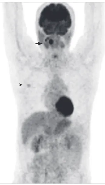
Figure 2. Positron emission tomography (PET) maximal intensity projec- tion image showing the whole body PET image wherein both the base of tongue lesion (arrow) and the right breast lesion (arrowhead) are seen.
444 Manjit Sarma, et al.
Figure 3. (A) Axial 18-Fluorine deoxyglucose positron emission tomography (18F-FDG PET), (B) axial computed tomography (CT) (contrast enhanced), (C) PET/CT fusion sections showing focal FDG uptake in right base of tongue with no corresponding CT lesion (triangulated).
A B C
Figure 4. (A) 18-fluorine deoxyglucose positron emission tomography (18F-FDG PET), (B) axial computed tomography (CT) (contrast enhanced), and (C) PET/CT fusion axial sections showing focal 18F-FDG uptake in the well defined 15×14 mm soft tissue subcutaneous lesion in retroareolar region on right side (triangulated).
A B C
Figure 5. Excision biopsy (right breast lump). (A) Infiltrating neoplasm composed of cells arranged in tubules, cords, singly and in islands (arrows) (H&E stain, ×100). Moderate cytoplasm and pleomorphic nuclei in cells. Invasive ductal carcinoma, not otherwise specified. Modified Bloom Richardson grade 1 (tubule 2, atypia 1, mitosis 1). (B) In higher magnification (H&E stain, ×400).
A B
or on palpapation. Subsequently, patient received six cycles of adriamycin, cyclophosphamide, and 5-flurouracil for carcino- ma breast (pT1cN1M0). Patient was subsequently on tamoxi- fen. Apart from a positive family history, patient does not have any known risk factors like alcoholism, liver cirrhosis, etc. One of his brother is known to have breast carcinoma. Genetic mu- tation testing was not done as this is locally not available to us in our hospital and outsourced testing for the same was not insisted upon taking in consideration the poor financial status of the patient.
DISCUSSION
The overall occurrence rate of multiple primary malignan- cies reported in literature is between 0.73% and 11.7% [10] . Three diagnostic criteria have been proposed for multiple pri- mary malignancy: 1) each tumor must present with definite features of malignancy; 2) each must be distinct; and 3) the chance of one being a metastasis of the other must be excluded [11]. Our patient was detected with the second malignancy within a month of detection of the first malignancy, fulfilling the criteria laid down by Gluckman’s definition of synchronous malignancy. Based on the location, multiple primary malig- nancies are classified into four types: 1) multicentric, if the two distinct carcinoma arise in the same organ or tissue; 2) system- ic, if they arise on anatomically or functionally allied organs of the same system (colon and rectum cancers); 3) paired organs, as in the breasts; and 4) random, if they occur as a coincidental or accidental association in unrelated sites [12]. Our case can be categorized into random synchronous malignancy as base of tongue and breast are unrelated sites. Mechanisms involved are yet to be clarified. However, some factors such as heredity, constitution, environmental, immunological, carcinogenic, vi- ral, radiological, and chemical factors have been implicated [13]. Genetic predisposition (e.g., higher racial prevalence in Ashkenazi Jews and African-Americans), exposure to female hormones (in cases of sex change operations), cirrhosis, other testicular dysfunction such as orchitis or undescended testis, soap and perfume workers, alcohol, etc. have been implicated for development of male breast cancer .While there was no family history of breast cancer in cases reported by Yaman et al. [4] and Sun et al. [5], family history was positive in cases re- ported by Dubashi et al. [6] and Sordi et al. [7]. A family his- tory is known to be a definite risk factor for breast malignan- cy-5% to 30% of male breast cancer patients report a family history of the disease. In view of this, the role of genetic testing for BRCA1 and BRCA2 and other molecular genetic markers assumes importance [14]. All histologic types of breast cancer have been identified in men, the most common type being in-
filtrating ductal adenocarcinoma (70%-95% of cases). Ductal carcinoma in situ is infrequent. The rate of receptor positivity is high, up to 85% (higher than in females) as in our patient [15].
This case report highlights the incremental value of whole body PET/CT in the oncological screening of patients. This case merits attention as it is relatively uncommon to have a synchronous male breast malignancy especially in a case of head and neck cancer. This case also corroborates the utility of
18F-FDG PET/CT in detecting known as well as unknown ma- lignant lesions and projects the need for a thorough screening with a whole body modality like 18F-FDG PET/CT in patients with family history of those malignancies which are seen in family members like breast carcinomas.
CONFLICT OF INTEREST
The authors declare that they have no competing interests.
REFERENCES
1. Gluckman JL, Crissman JD, Donegan JO. Multicentric squamous-cell carcinoma of the upper aerodigestive tract. Head Neck Surg 1980;3:90-6.
2. Rennemo E, Zätterström U, Boysen M. Synchronous second primary tumors in 2,016 head and neck cancer patients: role of symptom-direct- ed panendoscopy. Laryngoscope 2011;121:304-9.
3. Slaughter DP, Southwick HW, Smejkal W. Field cancerization in oral stratified squamous epithelium: clinical implications of multicentric or- igin. Cancer 1953;6:963-8.
4. Yaman E, Ozturk B, Coskun U, Buyukberber S, Kaya AO, Yildiz R, et al.
Synchronous bilateral breast cancer in an aged male patient. Onkologie 2010;33:255-8.
5. Sun WY, Lee KH, Lee HC, Ryu DH, Park JW, Yun HY, et al. Synchro- nous bilateral male breast cancer: a case report. J Breast Cancer 2012;
15:248-51.
6. Dubashi B, Jain A, Srinivasan K, Surendrakumar V, Vivekanandam S.
Chronic lymphocytic leukemia and breast cancer as synchronous pri- mary in a male-a rare combination. Curr Oncol 2011;18:e101-2.
7. Sordi E, Cagossi K, Lazzaretti MG, Gusolfino D, Artioli F, Santacroce G, et al. Rare case of male breast cancer and axillary lymphoma in the same patient: an unique case report. Case Rep Med 2011;2011:940803.
8. Strobel K, Haerle SK, Stoeckli SJ, Schrank M, Soyka JD, Veit-Haibach P, et al. Head and neck squamous cell carcinoma (HNSCC): detection of synchronous primaries with (18)F-FDG-PET/CT. Eur J Nucl Med Mol Imaging 2009;36:919-27.
9. Mesmoudi M, Mahfoud T, Ismaili N, Rami K, Kamouni M, Jroundi L, et al. A synchronous undifferentiated nasopharyngeal carcinoma and infiltrating ductal carcinoma of the breast successfully treated with in- duction chemotherapy followed by local control of both tumours: a case report. BMC Ear Nose Throat Disord 2011;11:6.
10. Demandante CG, Troyer DA, Miles TP. Multiple primary malignant neoplasms: case report and a comprehensive review of the literature.
Am J Clin Oncol 2003;26:79-83.
446 Manjit Sarma, et al.
11. Warren S, Gates O. Multiple primary malignant tumors: a survey of the literature and a statistical study. Am J Cancer 1932;16:1358-414.
12. Moertel CG. Multiple primary malignant neoplasms: historical per- spectives. Cancer 1977;40(4 Suppl):1786-92.
13. Tamura M, Shinagawa M, Funaki Y. Synchronous triple early cancers occurring in the stomach, colon and gallbladder. Asian J Surg 2003;26:
46-8.
14. Meguerditchian AN, Falardeau M, Martin G. Male breast carcinoma.
Can J Surg 2002;45:296-302.
15. Goss PE, Reid C, Pintilie M, Lim R, Miller N. Male breast carcinoma: a review of 229 patients who presented to the Princess Margaret Hospital during 40 years: 1955-1996. Cancer 1999;85:629-39.