논문접수일:2007년 4월 30일 채택일:2007년 5월 18일 교신저자:김병기, 135-710 서울시 강남구 일원동 50
성균관대학교 의과대학 삼성서울병원 산부인과 전화:02) 3410-3513․전송:02) 3410-0630 E-mail:[email protected]
본 연구는 삼성생명과학연구소(SBRI)의 연구비 지원으로 시행됨
(#SBRI C-A5-303-1).
자궁경부암 환자에서 MRI와 microarray 검사법의 골반 림프절 전이 예측 정확도 비교
성균관대학교 의과대학 삼성서울병원 산부인과1, 영상의학과2
김태중1․차현화1․최정주1․김우영1․최철훈1․이정원1․배덕수1․박병관2․김병기1
목적:자궁경부암에서 microarray 법을 이용하여 골반 림프절 전이 예측이 가능한지 조사하고, 이 검사법을 기존의 자기공 명영상술(MRI)과 비교하는 것이다.
연구 방법:근치적 자궁적출술 및 골반 림프절 절제술을 시행받은 43명의 환자들을 대상으로, Macrogen사의 10K oligonucleotide 유전자 chip 검사를 시행하였다. 연구대상자들을 31예의 training set과 12예의 test set으로 무작위 구분하여, 골반 림프절 전이 예측 모델을 통계기법을 이용하여 고안하였다. 또한, 특별히 고안한 판정기준을 적용하여 MRI 검사법의 림프절 전이를 판정하고, 두 검사법을 비교하였으며, 두 검사법을 병합한 새로운 접근방식을 고안하였다.
결과:Microarray의 림프절 예측 정확도는 83%, MRI는 69%의 정확도를 나타내어, 두 검사법 간에는 통계적 차이를 보이 지 않았다. 또한, 두 검사법을 병합한 새로운 모델은 74%의 정확도를 나타내었으나, MRI 단독과 비교하여 통계적 의미는 없었다.
결론:자궁경부암에서 DNA microarray 법은 단독 또는 MRI와 병합하여 골반 림프절 전이를 예측할 수 있으며, 예측 타당도는 MRI와 차이를 보이지 않았다.
중심단어:자궁경부암, 자기공명영상술, 림프절, 유전자 칩
서 론
자궁경부암은 전세계적으로 여성에서 발생하는 암 중 두 번째로 흔한 암으로, International Federation of Gy- necology and Obstetrics (FIGO)의 권고안에 따라 임상적 으로 병기가 결정되는 유일한 부인과적 악성 종양이다.1 본 질환에서 골반 림프절(LN) 전이는 근치적 수술 후의 가장 중요한 예후 인자로 알려져 있으나, 임상적 병기에 는 LN 전이 여부가 포함되지 않는 문제점이 있다.2,3이 러한 문제점을 보완하기 위해 LN 전이를 치료 전에 알 아내기 위한 많은 연구가 있어 왔지만, 이는 주로 ma- gnetic resonance imaging (MRI), computed tomography
(CT), 또는 positron emission tomography (PET)과 같은 영 상의학검사에 국한되어 있다. 이러한 영상의학검사는 그 유용성에도 불구하고, 작은 LN 전이를 발견하지 못 하는 한계가 있으며, 전이 양성 판정 기준에 따라 연구 자마다 LN 전이 진단의 타당도에 있어 서로 상이한 결 과를 보여왔다.4-8
그동안 자궁경부암의 생물학적 특성에 관한 분자유전 학 연구는 활발이 이루어져 온 반면에, LN 전이에 중점 을 둔 분자기전 연구는 드물었으며, LN 전이 기전에 관 하여 밝혀진 바는 거의 없는 실정이다. 자궁경부암의 골 반 LN 전이에는 focal adhesion kinase의 발현 저하,9자멸 유도(proapoptotic) 단백질인 Apaf-1의 down-regulation이 관련되어 있다고 알려져 있으며,10 cyclooxygenase-211 및 RCAS112의 과발현이 골반 LN 전이와 연관성이 있다고 보고되었다. 또한 종양주변 림프혈관밀도 및 VEGF-C 발현의 증가가 LN 전이와 관련된다는 보고도 있었다.13 DNA microarray는 동시에 많은 수의 유전자 발현을 분석하여, 암과 정상 조직, 또는 암 조직간의 유전자 발
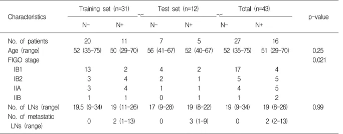
Table 1. Patient characteristics
Training set (n=31) Test set (n=12) Total (n=43)
Characteristics }} p-value
N- N+ N- N+ N- N+
No. of patients 20 11 7 5 27 16
Age (range) 52 (35-75) 50 (29-70) 56 (41-67) 52 (40-67) 52 (35-75) 51 (29-70) 0.25
FIGO stage 0.021
IB1 13 2 4 2 17 4
IB2 3 4 2 1 5 5
IIA 3 4 1 1 4 5
IIB 1 1 0 1 1 2
No. of LNs (range) 19.5 (9-34) 19 (11-26) 17 (9-28) 19 (8-22) 19 (9-34) 19 (8-26) 0.99 No. of metastatic
0 2 (1-13) 0 3 (1-9) 0 2 (2-13)
LNs (range)
FIGO; International Federation of Gynecology and Obstetrics 현 양상의 차이를 알아내는 유용한 방법이다.14,15이 기 법을 통하여 원발 암 조직에서 전이 유무에 따라 다르게 발현되는 백 여 개의 유전자가 보고되었으며 원발 암 조 직과 전이 암 조직에서 공통으로 발현되는 17개의 유전 자가 확인되었다.16이와 같은 DNA microarray의 유용성 을 이용하여, 자궁경부암에서 원발 암 조직의 유전자 발 현 양상을 분석하여 LN 전이를 예측할 수 있으리라 기 대할 수 있으며, LN 전이에 따라 발현 차이를 보이는 유전자들을 발견하여 LN 전이 관련 분자 기전을 연구하 는 기초로 삼을 수 있을 것이다.
본 연구의 목적은 DNA microarray를 이용하여 자궁경 부암 조직의 유전자 발현 양상이 골반 LN 전이를 예측 할 수 있는지를 알아보고, 이를 임상적으로 널리 이용되 는 MRI 검사법의 골반 LN 전이 예측도와 비교 분석하 여, 본 질환에서 LN 전이에 관한 microarray의 임상적 가 치를 살펴보는 것이다.
연구 대상 및 방법
1. 연구 대상
2002년 1월부터 2003년 10월까지 본원 산부인과에서 원발성 자궁경부암으로 근치적 자궁적출술 및 골반림프 절 절제술을 시행 받은 43예를 연구 대상으로 삼았다.
43예 중 16예는 수술 후 병리조직검사에서 골반 LN 전 이가 확인되어 N+군으로 분류되었으며, 나머지 27예는
LN 전이가 없어 N-군으로 분류되었다. 본 연구는 병원 윤리위원회의 규정에 따라 시행되었다.
수술 시 채취한 경부암 조직은 병리학 전문의가 즉각 적으로 조사하여, 괴사가 없는 암 조직의 가장자리 부위 에서 2-3개의 조직을 확보하여 즉시 -80oC에서 냉동 보 관하였다. 그 중 경부암 세포가 약 70% (범위 50-90%)를 차지하는 조직을 선택하여 TRIZOL 방식(Invitrogen Corporation, Carlsbad, CA)으로 RNA를 추출하였다. N+
및 N- 두 군 간에 암세포가 차지하는 비율에 의미 있는 차이는 없었다.
2. DNA microarray
각 조직으로부터 추출된 약 5-10μg의 RNA를 oli- gonucleotide microarray 사용하여 분석하였다. 본 연구에 서 사용된 Macrogen사의 Oligo Human 10K Microarrays 는 50-mer oligonucleotide probe로 구성되어 10,416개의 인간유전자를 알아 보는 것으로, 이 중 8,032개는 유전자 가 알려진 것이고, 2,076개는 expressed sequence tags (ESTs)이다.17
3. Supervised classification
Microarray 사진은 Imagene ver. 5.5 software (BioDis- covery, El Segundo, CA)를 사용하여 분석하였으며, lowess 방식으로 normalization하였다. N- 및 N+군 간 에 유전자 발현 양상이 구별될 수 있는지 확인하기 위해
Fig. 1. Prediction flow of LN me - tastasis. G ene select and cons- truct model using 31 training samples, measures model perfo- rmance for test set (12 patients).
supervised hierarchical clustering를 시행하였으며, permu- tation test를 통해 발현에 차이를 보이는 유전자를 선택 하였다. p-value 보정을 위하여 Westfall-Young method를 시행하였다.18 LN 전이 예측 모델은 10-fold cross-vali- dation을 이용하여 support vector machine 방식으로 제작 되었다. 예측 모델 제작을 위하여 43예를 무작위로 나누 어, 31예의 training set와 12예의 test set을 설정하였다 (Fig. 1). Training set에서 LN 전이를 구별할 수 있는 예측 모델을 제작하였으며, 이 모델의 예측 타당도를 독립적 인 test set에 적용하였다.
4. MRI의 골반 LN 전이 예측
1예를 제외한 42명의 환자들은 수술 전에 복부 및 골 반 MRI (General Electric Medical System, Milwaukee, WI) 검사를 받았다. 한 명의 영상의학과 전문의가 LN의 병 리 검사결과를 모르는 상태에서, 연구 대상 환자들의 MRI 사진을 판독, 골반 LN 전이 유무를 평가하였다. 이 때 사용한 골반 LN 전이 판독 기준은 다음과 같았다.
1) definitive positive: short axis dimension ≥1 cm; 2) probable positive: 0.5 cm ≤ short axis dimension <1 cm and long axis dimension ≥1 cm; 3) possible positive: 0.5 cm ≤ short axis dimension <1 cm and 0.5 cm ≤ long axis dimension <1 cm; 4) probable negative: short axis dimension <0.5 cm; 5) definitive negative: unmeasurable short or long axis dimension. 위의 분류 기준 중 1), 2), 3)에 해당하는 경우 골반 LN 전이 양성으로 판정하였다.
5. 골반 LN 전이 판정에 있어 MRI와 microarray의 병합 본 연구자들은 작은 LN 전이를 놓치는 MRI의 단점을 극복하고, 이러한 상황에서 microarray의 임상적 가치를
알아보고자 MRI와 microarray를 병합한 새로운 예측 모 델을 고안하였다. 만약 MRI에서 LN 전이 양성(mN+) 으로 판독되면, microarray 결과에 상관없이 환자는 LN 전이 양성(nN+)으로 분류된다. 반면에, MRI에서 LN 전 이 음성(mN-)으로 판독되면, microarray 예측 결과에 따 라서 환자는 LN 전이 양성 혹은 음성으로 분류된다. 이 와 같이 고안된 새로운 모델의 LN 전이 예측 정확도를 MRI의 정확도와 비교해 보았다.
6. 통계적 분석
통계적 분석에는 SPSS 13.0 (SPSS Inc., Chicago, IL)이 사용되었다. N-와 N+군 간의 평균값 비교를 위하여, 연속 변수는 t-test, 범주형 변수는 chi-square test를 하였 다. Microarry와 MRI 간의 예측 정확도 비교를 위하여 McNemar test를 적용하였다. 모든 분석에 있어 p<0.05 인 경우를 통계적 의미가 있는 것으로 간주하였다.
결 과
1. 임상병리학적 특성
연구 대상자들의 임상병리학적 특성을 Table 1에 제시 하였다. 수술 시 환자 일인당 평균 19 (범위 8-34)개의 LN가 적출되어, 전체 연구 대상에서 총 816개의 LN가 조사되었고, 이 중 54개가 병리조직검사에서 전이성 LN 로 판정되었다. N-와 N+군 간에 FIGO 임상적 병기가 유의한 차이를 보여서, microarray 결과가 LN 전이를 예 측하는데, FIGO 병기가 어떠한 영향을 미치는지 확인하 기 위하여, 회귀분석을 시행하였다. 그 결과 microarray 결과(p=0.006)가 FIGO 병기(p=0.234)보다 강력하게 LN 전이를 예측함을 확인하였다.
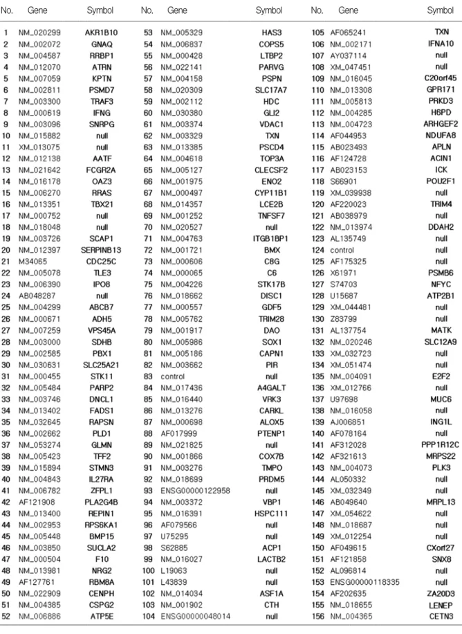
Table 2. Differentially expressed 156 genes from DNA microarray (127 known genes and 29 ESTs, p<0.01)
No. Gene Symbol No. Gene Symbol No. Gene Symbol
A
Sensitivity: 63.6%
Specificity: 80.0%
PPV: 63.6%
NPV: 80.0%
Accuracy: 74.2%
B
Sensitivity: 60.0%
Specificity: 100%
PPV: 100%
NPV: 77.8%
Accuracy: 83.3%
pN- pN+ Total pN- pN+ Total
cN- cN+
16 4
4 7
20 11
cN- cN+
7 0
2 3
9 3
Total 20 11 31 Total 7 5 12
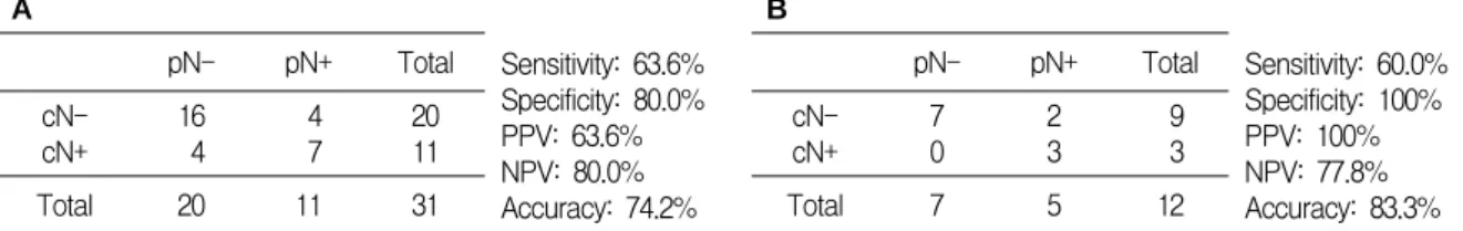
cN-; N- by a classifier, cN+; N+ by a classifier, pN-; N- by pathologic evaluation, pN+; N+ by pathologic evaluation Fig. 2. Performance of “LN metastasis prediction model” using 156 G enes. (A) represented the validity of a classifier from 31 training set and (B) showed from 12 test set.
A
Sensitivity: 66.7%
Specificity: 70.4%
PPV: 55.6%
NPV: 79.2%
Accuracy: 69.0%
B
Sensitivity: 62.5%
Specificity: 85.2%
PPV: 71.4%
NPV: 79.3%
Accuracy: 76.7%
pN- pN+ Total pN- pN+ Total
mN- mN+
19 8
5 10
24 18
cN- cN+
23 4
6 10
29 14
Total 27 15 42 Total 27 16 43
C
p value Sensitivity: 1.000 Specificity: 0.289 Accuracy: 0.454
cN-; N- by a classifier, cN+; N+ by a classifier, pN-; N- by pathologic evaluation, pN+; N+ by pathologic evaluation
*One patient with cN- and pN+ was exclude for comparison analysis because of not taking MRI scan
pN- pN+ Total
mN- cN-
cN+
17 2
1
4 24
mN+ cN-
cN+
6 2
4
6 18
Total 27 15 42*
Fig. 3. Performance of pelvis M RI and a classifier on the prediction of LN metastasis. (A) showed the validity of pelvis M RI and (B) showed a classifier. (C) represented the rearranged date from 42 patients who took both pelvis M RI and microarray test for comparison. There is no significant difference between two prediction models.
2. Microarray에서 나타난 유전자 발현 양상 및 예측 모델
LN 전이와 관련하여 microarray 검사 결과 통계적 차 이를 보이는 유전자는 모두 156개이다(p<0.01, Table 2).
이들 유전자와 support vector machine을 이용하여, LN 예 측 모델을 만들어, 12명의 test set에 적용한 결과, microarray의 LN 예측도는 민감도 60%, 특이도 100%, 정 확도 83%를 나타내었다(Fig. 2).
3. MRI의 LN 전이 예측도 및 microarray와의 비교 MRI는 LN 전이를 지닌 15예 중 10예를 정확하게 예측 하였다. 또한, 8예를 위양성으로 예측하여, 결국 70% 의 특이도를 나타내었다. MRI에서 LN 전이 양성으로 판정 받은 18예를 판독 기준에 따라 세분해 보면, 10예는 1), 2)에 속했으며, 8예는 3)에 해당되었다(Fig. 3). MRI 의
예측 정확도는 69% (29/42)이고, microarray의 정확도는 77% (33/43)였다. 두 검사법의 차이를 검정하기 위하여, MRI 검사를 받지 않은 한 예를 제외하고, 분석을 한 결 과 통계적 차이는 보이지 않았다(Fig. 3).
4. MRI와 microarray의 병합 예측 모델
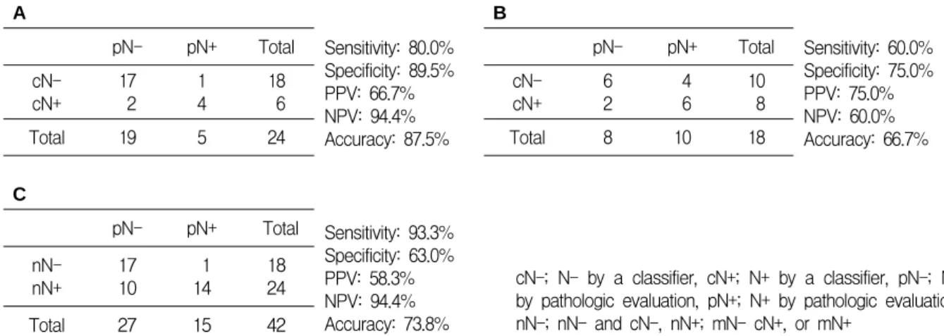
MRI에서 LN 전이 음성으로 판정 받은 환자들만을 대 상으로, microarray의 LN 전이 예측도를 확인해 보니, 민 감도, 특이도 및 정확도가 각각 80%, 90% 및 88%로 조 사되었다(Fig. 4). 이러한 수치에 근거하여, 우리가 고안 해 낸 새로운 병합 예측 모델은 민감도 93%, 특이도 63%
와 정확도 74%를 나타내었다. 하지만, 병합 모델의 예측 도는 MRI 단독과 비교하여 통계적 차이를 나타내지는 않았다(Fig. 4).
A
Sensitivity: 80.0%
Specificity: 89.5%
PPV: 66.7%
NPV: 94.4%
Accuracy: 87.5%
B
Sensitivity: 60.0%
Specificity: 75.0%
PPV: 75.0%
NPV: 60.0%
Accuracy: 66.7%
pN- pN+ Total pN- pN+ Total
cN- cN+
17 2
1 4
18 6
cN- cN+
6 2
4 6
10 8
Total 19 5 24 Total 8 10 18
C
Sensitivity: 93.3%
Specificity: 63.0%
PPV: 58.3%
NPV: 94.4%
Accuracy: 73.8%
cN-; N- by a classifier, cN+; N+ by a classifier, pN-; N- by pathologic evaluation, pN+; N+ by pathologic evaluation, nN-; nN- and cN-, nN+; mN- cN+, or mN+
pN- pN+ Total nN-
nN+
17 10
1 14
18 24
Total 27 15 42
Fig. 4. LN prediction by “new model” combining pelvis M RI and microarray. (A) showed the prediction validity of microarray in the mN- patients. (B) showed that in the mN+. (C) represented the performance of “new model”.
고 찰
자궁경부암에서 microarray를 이용하여 정상 경부 조 직과 경부암 조직 간에 발현 차이가 있는 유전자를 발견 해 낸 몇몇 보고가 있었지만,19-23 경부암 조직의 유전자 발현 양상을 분석하여 LN 전이를 예측하는 연구는 거의 없는 실정이다.24
Microarray 기법으로 여러 암에서의 LN 전이를 연구 한 결과 chemokines, chemokine receptors와 interferon 관 련 유전자들이 특히 관련되어 있다고 보고되었다.16,25본 연구는 경부암에서 LN 전이에 관계할 가능성이 높은 유 전자로 127개를 제시하였는데(Table 2), 이들 중 IFNγ, IL27RA, TNFSF7, GDF5, IFNA10와 PLK3는 chemokine 관련 유전자로 알려져 있다.
일반적으로, 영상의학과 전문의들은 MRI에서 단경 1 cm 이상의 LN를 전이 양성으로 판정한다.26이러한 기준 으로 대규모 meta-analysis를 시행한 결과 MRI의 LN 전 이 예측의 민감도와 특이도는 비교적 높지 않은 것으로 보고되었고, 주된 이유는 작은 전이 LN를 진단하지 못 하기 때문이다.5그러므로, 본 연구에서는 작은 전이 LN 를 놓치지 않기 위하여 새롭게 고안된 판정 기준을 사용 하게 되었다.
Microarray와 같은 유전자적 접근 방식은 이론적으로 임상적으로 발현되기 전의 분자기전을 확인할 수 있는 장점을 지니고 있다. 즉, 이론적으로 LN 전이가 MRI에 서 확인되기 이전 단계를 발견할 가능성이 있다. 이러한
특성과 MRI의 제한점을 고려하여, 우선 MRI 결과에 따 라 연구 대상을 세분하여 각 군에서 microarray의 LN 전 이 예측 타당도를 평가하고, 두 검사법을 조합한 새로운 예측 모델을 고안하였다(Fig. 4). 그렇지만, 새 모델의 예 측 정확도는 74%로 MRI의 정확도 69%와 통계적 차이 를 보이지 않았다(p=0.688).
본 연구는 다음과 같은 제한점을 지니고 있다. 첫째, microarray 기법을 임상에 적용하는 데에는 비용-효과 측 면에서 문제가 된다. 더군다나 두경부암 또는 폐암과 달 리 자궁경부암에서 수술 전에 LN 전이를 파악하는 것은 치료 방침 결정에 큰 도움을 주지 못한다. 그렇지만, 적 은 수의 유전자들로 DNA chip을 개발하여, MRI에서 LN 전이가 없다고 알려진 고위험 환자들에게 microarray 로 확인해 보는 것은 가치가 있다고 생각된다. 둘째, 본 연 구는 비교적 적은 수의 환자들을 대상으로 시행되었다.
향후에 보다 많은 환자 조직과 임상 정보를 가지고 분석 한 연구가 필요하리라 생각된다.
요약하면, 본 연구는 자궁경부암 환자에서 경부암 조 직으로 시행한 microarray 검사법이 골반 LN 전이를 77%의 정확도로 예측할 수 있음을 증명하였으며, MRI 와 조합하여 microarray를 임상에 적용할 수 있는 하나의 접근 방식을 제시하였다. 또한, 경부암에서 LN 전이 분 자 기전에 관련한 많은 유전자들을 발굴하였다.
참고문헌
1. Parkin DM, Bray F, Ferlay J, Pisani P. Estimating the world
cancer burden: Globocan 2000. Int J Cancer 2001; 94: 153-6.
2. Waggoner SE. Cervical cancer. Lancet 2003; 361: 2217-25.
3. Lai CH, Hong JH, Hsueh S, Ng KK, Chang TC, Tseng CJ, et al. Preoperative prognostic variables and the impact of postoperative adjuvant therapy on the outcomes of Stage IB or II cervical carcinoma patients with or without pelvic lymph node metastases: An analysis of 891 cases. Cancer 1999; 85:
1537-46.
4. Yang WT, Lam WW, Yu MY, Cheung TH, Metreweli C.
Comparison of dynamic helical CT and dynamic MR imaging in the evaluation of pelvic lymph nodes in cervical carcinoma.
AJR Am J Roentgenol 2000; 175: 759-66.
5. Scheidler J, Hricak H, Yu KK, Subak L, Segal MR. Radio- logical evaluation of lymph node metastases in patients with cervical cancer. A meta-analysis. Jama 1997; 278: 1096-101.
6. Grigsby PW, Siegel BA, Dehdashti F. Lymph node staging by positron emission tomography in patients with carcinoma of the cervix. J Clin Oncol 2001; 19: 3745-9.
7. Belhocine T, Thille A, Fridman V, Albert A, Seidel L, Nickers P, et al. Contribution of whole-body 18FDG PET imaging in the management of cervical cancer. Gynecol Oncol 2002; 87:
90-7.
8. Choi HJ, Roh JW, Seo SS, Lee S, Kim JY, Kim SK, et al.
Comparison of the accuracy of magnetic resonance imaging and positron emission tomography/computed tomography in the presurgical detection of lymph node metastases in patients with uterine cervical carcinoma: A prospective study. Cancer 2006; 106: 914-22.
9. Gabriel B, zur Hausen A, Stickeler E, Dietz C, Gitsch G, Fischer DC, et al. Weak expression of focal adhesion kinase (pp125FAK) in patients with cervical cancer is associated with poor disease outcome. Clin Cancer Res 2006; 12: 2476-83.
10. Leo C, Richter C, Horn LC, Schutz A, Pilch H, Hockel M.
Expression of Apaf-1 in cervical cancer correlates with lymph node metastasis but not with intratumoral hypoxia. Gynecol Oncol 2005; 97: 602-6.
11. Manchana T, Triratanachat S, Sirisabya N, Vasuratna A, Termrungruanglert W, Tresukosol D. Prevalence and prog- nostic significance of COX-2 expression in stage IB cervical cancer. Gynecol Oncol 2006; 100: 556-60.
12. Sonoda K, Miyamoto S, Hirakawa T, Yagi H, Yotsumoto F, Nakashima M, et al. Invasive potency related to RCAS1 expression in uterine cervical cancer. Gynecol Oncol 2005; 99:
189-98.
13. Gombos Z, Xu X, Chu CS, Zhang PJ, Acs G. Peritumoral lymphatic vessel density and vascular endothelial growth factor C expression in early-stage squamous cell carcinoma of
the uterine cervix. Clin Cancer Res 2005; 11: 8364-71.
14. Golub TR, Slonim DK, Tamayo P, Huard C, Gaasenbeek M, Mesirov JP, et al. Molecular classification of cancer: Class discovery and class prediction by gene expression monitoring.
Science 1999; 286: 531-7.
15. van't Veer LJ, Dai H, van de Vijver MJ, He YD, Hart AA, Mao M, et al. Gene expression profiling predicts clinical outcome of breast cancer. Nature 2002; 415: 530-6.
16. Ramaswamy S, Ross KN, Lander ES, Golub TR. A molecular signature of metastasis in primary solid tumors. Nat Genet 2003; 33: 49-54.
17. Kono T, Obata Y, Wu Q, Niwa K, Ono Y, Yamamoto Y, et al. Birth of parthenogenetic mice that can develop to adulthood. Nature 2004; 428: 860-4.
18. Westfall P, Young S. Resampling-based multiple testing. New York: Wiley;1993.
19. Cheng Q, Lau WM, Tay SK, Chew SH, Ho TH, Hui KM.
Identification and characterization of genes involved in the carcinogenesis of human squamous cell cervical carcinoma. Int J Cancer 2002; 98: 419-26.
20. Kitahara O, Katagiri T, Tsunoda T, Harima Y, Nakamura Y.
Classification of sensitivity or resistance of cervical cancers to ionizing radiation according to expression profiles of 62 genes selected by cDNA microarray analysis. Neoplasia 2002;
4: 295-303.
21. Chen Y, Miller C, Mosher R, Zhao X, Deeds J, Morrissey M, et al. Identification of cervical cancer markers by cDNA and tissue microarrays. Cancer Res 2003; 63: 1927-35.
22. Ahn WS, Bae SM, Lee JM, Namkoong SE, Han SJ, Cho YL, et al. Searching for pathogenic gene functions to cervical cancer. Gynecol Oncol 2004; 93: 41-8.
23. Wong YF, Cheung TH, Tsao GS, Lo KW, Yim SF, Wang VW, et al. Genome-wide gene expression profiling of cervical cancer in Hong Kong women by oligonucleotide microarray.
Int J Cancer 2006; 118: 2461-9.
24. Lyng H, Brovig RS, Svendsrud DH, Holm R, Kaalhus O, Knutstad K, et al. Gene expressions and copy numbers asso- ciated with metastatic phenotypes of uterine cervical cancer.
BMC Genomics 2006; 7: 268.
25. Huang E, Cheng SH, Dressman H, Pittman J, Tsou MH, Horng CF, et al. Gene expression predictors of breast cancer outcomes. Lancet 2003; 361: 1590-6.
26. Kim SH, Choi BI, Han JK, Kim HD, Lee HP, Kang SB, et al. Preoperative staging of uterine cervical carcinoma: Com- parison of CT and MRI in 99 patients. J Comput Assist Tomogr 1993; 17: 633-40.
Microarray versus magnetic resonance imaging prediction of lymph node metastasis in patients with cervical squamous cell carcinoma
Tae-Joong Kim1, Hyun Hwa Cha1, Jung-Joo Choi1, Woo Young Kim1, Chel Hun Choi1, Jeong-Won Lee1, Duk-Soo Bae1, Byung Kwan Park2, Byoung-Gie Kim1 Departments of Obstetrics and Gynecology1and Radiology2, Samsung Medical Center,
Sungkyunkwan University School of Medicine, Seoul, Korea
Objective:We investigated whether microarray-based gene expression profiling of primary tumor biopsy material could be used to predict lymph node (LN) metastasis in patients with uterine squamous cell carcinoma by comparing this approach with magnetic resonance imaging.
Methods:Forty three primary cervical cancer samples (16 with LN metastasis and 27 without LN metastasis) from radical hysterectomy with pelvic LN dissection were obtained, RNA was isolated, and oligonucleotide gene chips (Macrogen, Seoul, Korea) were hybridized. The samples were randomly divided into training (31 samples) and test (12 samples) sets. A prediction model for LN metastasis from the training set was developed by support vector machine methods using a 10-fold cross-validation and it was tested for its prediction accuracy by applying it to the test set. We evaluated pelvic LN status by MRI with newly designed criteria in these patients and compared the accuracy of MRI with microarray. In addition, we created a new approach by a combination of both.
Results:The “LN prediction model” derived from the signature of 156 distinctive genes had a prediction accuracy of 83% when applied to the independent test set. MRI showed an accuracy (69%) for the prediction of LN metastasis. The combination model with MRI findings and microarray improved prediction accuracy over MRI alone but the improvement was not statistically significant (74% and 69%, respectively; p=0.688).
Conclusion:Current data show that the prediction of LN metastasis can be allowed by DNA microarray of the primary tumor biopsy, alone or in combination with MRI.
Key Words : Uterine cervical neoplasms, Microarray, MRI, Lymph node