http://dx.doi.org/10.3988/jcn.2012.8.2.130 J Clin Neurol 2012;8:130-138
Introduction
Primary insomniacs (PIs) have a chronic clinical condition that is characterized by the subjective experience of chroni- cally disturbed sleep and sleep loss, and they show evidence of conditioned sleep difficulties and/or heightened arousal in bed.1 Although the long-term consequences of hyperarousal
in PIs has not been fully evaluated, animal studies have shown that persistent exposure to stress and adrenal steroids may result in the degeneration or inhibition of neurogenesis in the hippocampus.2,3
There have been several controversial findings concerning hippocampal volume (HV) in human insomnia studies. A previous pilot study found that the bilateral HV was signifi- cantly lower in PIs than in good sleepers (GSs).4 In contrast, a recent study did not find any objective differences in the HVs of PIs, even though some patients with sleep maintenan- ce problems were shown to have smaller HVs, as determined by wrist actigraphy.5
The Relationship between Hippocampal Volume and Cognition in Patients with Chronic Primary Insomnia
Hyun Jin Noh,a Eun Yeon Joo,a Sung Tae Kim,b So Mee Yoon,c Dae Lim Koo,a Daeyoung Kim,a Geun-Ho Lee,d Seung Bong Honga
aDepartments of Neurology and bRadiology, Samsung Medical Center, Sungkyunkwan University School of Medicine, Seoul, Korea
cDepartment of Psychology, The Catholic University of Korea College of Medicine, Seoul, Korea
dDepartment of Neurology, Medical College, Dankook University, Cheonan, Korea
Received June 21, 2011 Revised August 25, 2011 Accepted August 25, 2011 Correspondence Eun Yeon Joo, MD Department of Neurology, Samsung Medical Center, Sungkyunkwan University School of Medicine, 50 Irwon-dong, Gangnam-gu, Seoul 135-710, Korea Tel +82-2-3410-3597 Fax +82-2-3410-0052 E-mail [email protected]
Background and PurposezzDifferences in hippocampal volume (HV) were compared be- tween chronic primary insomniacs (PIs) and good sleepers (GSs), and the relationship between HV and memory function in PIs was investigated to clarify the effect of chronic sleep depriva- tion on brain structure and cognition.
MethodszzTwenty PIs (mean age, 50 years; 18 females) and 20 age-, gender-, and education- matched GSs were enrolled. Brain magnetic resonance imaging (MRI) was performed on a 1.5- T MRI scanner. Left and right HV and intracranial volume (ICV) were measured manually.
Nighttime polysomnography and neuropsychological testing were also applied to all subjects.
Group differences in HV were analyzed and the relationships between HV and sleep question- naire data, nighttime polysomnography, and neuropsychological findings were evaluated.
ResultszzCompared to GSs, PIs exhibited significantly increased sleep latency and arousal in- dex and a decreased percentage of REM sleep in nighttime polysomnography, as well as im- paired verbal and visual memory, and frontal lobe function. Absolute HV and ICV did not differ significantly between PIs and GSs. In the PIs, right and left HVs were negatively correlated with the duration of insomnia and the arousal index, and positively correlated with the recognition of visual memory. In addition, free recall in verbal memory was positively correlated with left HV in PIs.
ConclusionszzThese findings suggest that chronic sleep deprivation impairs memory and fron- tal lobe function, and that a long duration of insomnia and poor sleep quality contribute to a bi-
lateral reduction in HV. J Clin Neurol 2012;8:130-138
Key Wordszz primary insomnia, hippocampus, volumetry, memory, sleep.
Open Access
cc This is an Open Access article distributed under the terms of the Cre- ative Commons Attribution Non-Commercial License (http://creative- commons.org/licenses/by-nc/3.0) which permits unrestricted non-com- mercial use, distribution, and reproduction in any medium, provided the ori- ginal work is properly cited.
It has been well documented that prolonged sleep depriva- tion has a negative influence on cognition, even in normal sleepers.6 Experimental sleep curtailment can result in a de- terioration of neurocognitive performance by disrupting the nocturnal consolidation of hippocampal-dependent memo- ries, and previous studies have demonstrated that consider- able sleep loss worsens structural brain plasticity by attenuat- ing neurogenesis in the hippocampus.7,8 Moreover, it has been demonstrated that cognitive dysfunction occurs in PIs.9 This might be related to altered hippocampal function or structure, since the hippocampus is critically involved in cognitive per- formance and memory formation.10 However, the relationship between cognitive dysfunction and reduction of HV in PIs has not been elucidated.
The aims of the present study were 1) to compare HV and cognitive function between PIs and age-, gender-, and educa- tion-matched GSs, and 2) to determine whether HV is associa- ted with clinical profiles and memory scores in PIs.
Methods
Subjects
Twenty-five patients who complained of sleep onset and/or maintenance insomnia or unrefreshing sleep were enrolled between October 2009 and June 2010. They were diagnosed with PI according to the following inclusion criteria: 1) aged 20-65 years, 2) satisfying the definition of PI according to the International Classification of Sleep Disorders revision 2, and 3) an insomnia duration of ≥1 year. Thirty-one age-, gen- der-, and education status-matched healthy volunteers who were GSs were recruited through an advertisement in a local community.
The history of medical and sleep disorders was evaluated by clinical interview, and the history of psychiatric disorders was evaluated with the Structured Clinical Interview for the Diagnostic and Statistical Manual of Mental Disorders, Fourth Edition in all PIs and healthy GSs. The subjects com- pleted a sleep diary for at least 2 weeks, along with the fol- lowing self-completed questionnaires: the Pittsburgh Sleep Quality Index (PSQI), the Beck Depression Inventory (BDI), the Insomnia Severity Index (ISI), the Epworth Sleepiness Scale (ESS), and the Stanford Sleepiness Scale. In addition, PIs and healthy GSs underwent a physical examination, and laboratory testing to measure several factors, including elec- trolytes, complete blood counts, urine toxic screening tests for barbiturates, benzodiazepine, methamphetamine, canna- binoids, cocaine, and opiates, liver, thyroid, and renal func- tion tests, nighttime polysomnography (PSG), and neuropsy- chological tests. GSs with obstructive sleep apnea syndrome (apnea-hypopnea index >5/hour) or periodic limb movement
disorders were excluded from the study.
The general criteria for excluding PIs and GSs included the following parameters: 1) abnormal sleep-wake rhythms, 2) other sleep disorders, 3) hypertension, diabetes, heart, or respiratory diseases, 4) history of cerebrovascular disease, 5) other neurological (neurodegenerative diseases, epilepsy, or head injury) or psychiatric (psychosis or current depression) diseases, 6) alcohol or illicit drug abuse, or current intake of psychoactive medications, and 7) a structural lesion on brain magnetic resonance imaging (MRI). Volunteers with a mean daily sleep duration of <7 hours were also excluded from the GS group. To exclude the possible effect of hypnotics on cognition, these drugs were prohibited for at least 1 month before the study.
Six patients whose brain MRI revealed diffuse brain atro- phy or lacunar infarctions were excluded from the study. Fi- nally, 20 PI patients and 20 GSs were included as the study cohort. Informed consent was obtained from all PIs and GSs for PSG and brain MRI, and the institutional review board of our hospital authorized the study protocol.
Nighttime PSG
Subjects were asked not to drink alcohol or caffeinated bev- erages on the day before the sleep studies. Sleep data were recorded using a Remlogic (Embla Systems, Denver, CO, USA). Nighttime PSG was performed using a four-channel electroencephalogram (C3/A2, C4/A1, O1/A2, and O2/A1), a four-channel electrooculogram, an electromyogram of the submental, intercostal, and anterior tibialis muscles, and an electrocardiogram with surface electrodes. A thermistor (for monitoring nasal airflow), a nasal air pressure monitor, an oximeter (for measuring oxygen saturation), piezoelectric bands (for determining thoracic and abdominal wall mo- tions), and a body position sensor were attached to all pa- tients. Subjects were recorded on videotape using an infrared video camera and were monitored continuously by a PSG technician. Sleep architecture was scored in 30-s epochs, and sleep staging was interpreted according to the standard crite- ria of Rechtschaffen and Kales.11 Apnea and hypopnea epi- sodes were defined by previously established criteria. Ob- structive apnea was defined as a reduction in airflow greater than 90% that lasted at least 10 s, during which there was ev- idence of a persistent respiratory effort. Central apnea was defined as a reduction in airflow of more than 90% that last- ed at least 10 s, during which there was no evidence of respi- ratory effort. Hypopnea was defined as a reduction in airflow of 30% for more than 10 s that was accompanied by oxygen desaturation of at least 4%. According to the American Sleep Disorders Association Task Force criteria, arousals were classified as breathing-related arousals (occurring within 3 s
following apnea, hypopnea, or snoring) and other types of arousal (spontaneous arousal or arousal associated with peri- odic limb movements).
Brain MRI
MRI scanning was performed with a GE Signa 1.5-tesla scan- ner (GE Medical Systems, Milwaukee, WI, USA). Fluid-at- tenuated inversion recovery MRI was performed with the following parameters: 1.0-mm gap, 4.0-mm thickness, repeti- tion time/echo time (TR/TE)=10002/127.5 ms, number of exci- tations (NEX)=1, and oblique coronal. T1-weighted and T2- weighted MRI was performed with the following parameters:
0.3-mm gap, 3.0-mm thickness, TR/TE=5300/99 ms, flip an- gle (FA)=95º, NEX=3, and oblique coronal. Coronal spoiled gradient recalled (SPGR) MRI was performed with the fol- lowing scanning variables: 1.6-mm thickness, no gap, 124 slic- es, TR/TE=30/7 ms, FA=45°, NEX=1, matrix size=256×192, and field of view=22×22 cm. The voxel dimensions in the SPGR MRI images were 0.86×0.86×1.6 mm.
Intracranial volume measurement
Preprocessing of T1-weighted MRI data and manual volume measurements for intracranial volume (ICV) and the hippo- campus were performed using a Unix-based Sun Ultra 1 Creator workstation (Sun Microsystems, Santa Clara, CA, USA) and Analyze 7.5 (Biomedical Imaging Resource, Mayo Foundation, Rochester, MN, USA). ICV was used to normalize HV and to correct for variations in individual brain sizes.12 The ICV was measured by reconstructing the original T1-weighted MRI data in order to create 5-mm-thick sagittal images, which were then magnified twofold. The ce- rebrum, cerebellum, and midbrain were included in the ICV volume with the outer boundary of dura mater.13-15 The lateral limits of the ICV were defined as the right- and leftmost slic- es of the brain parenchyma on sagittal images, and the lower tip of the cerebellum was defined as the lower limit. We in- creased the brightness of the image in order to improve the visual clarity of the boundary of the dura mater. Using the established measurement criteria,15 the dura mater of the ce- rebrum, the cerebellum, and the midbrain (except for the in- ferior boundary) were traced manually. In order to establish the inferior boundary on the head tilt-corrected sagittal im- ages, a horizontal line that included the lower tip of the cere- bellum was drawn across the midbrain.14 The ICV measure- ments have been illustrated previously.16
Hippocampal volumetry
The methodological details of hippocampal volumetry are described elsewhere.17 The entire HV was measured from the anterior head to the posterior tail, including the cornu ammo-
nis, gyrus dentatus, hippocampus, and subiculum. The ante- rior boundary of the hippocampus was identified as the alve- us. The lateral border of the hippocampus was delineated against the entorhinal cortex by the upper margin of the white matter of the subiculum. The posterior end of the hip- pocampus was taken as the point at which the tail of the hip- pocampus disappeared. The rater manually traced the alveus according to the defined hippocampal boundary criteria.
The hippocampus was traced manually by one neuroimag- ing analyst (ST Kim) who was blind to the patient diagnosis.
The HV in each slice was calculated by multiplying the number of voxels contained within each trace by the voxel volume (1 mm3) and dividing by the magnification factor.
The total volume of each structure was the sum of all in-slice volumes. In order to calculate the intrarater correlation coef- ficients, the brain images of each subject were manually traced twice. The intrarater correlation coefficients were 0.96 and 0.95 for the left and right HVs, respectively, and 0.94 for both ICVs.
Neuropsychological assessments
A battery of neuropsychological tests and an individual stan- dardized intelligence test were administered to all subjects.
The neuropsychological tests, as described below, took 2.5 hours to perform.
Memory tests
Verbal memory was evaluated using the Korean California Verbal Test (K-CVLT).18 Nonverbal (visual) memory was as- sessed using the Rey Complex Figure Test (RCFT), which comprises an immediate recall test, a delayed recall test, and a recognition test.19 The K-CVLT involves 5 learning steps and 16 words, and immediate recall, immediate hint recall, delayed free recall, delayed hint recall, and recognition were tested 20 min after the 5 learning steps. The RCFT involves immediate recall after copying a complex figure, and free re- call and recognition tested 20 min later.
Attention and working memory tests
The digit span tests from the Wechsler Memory Scale-Re- vised were administered using a standard protocol,20 both for- ward and backward, in order to assess verbal attention and wo- rking memory. The Corsi block-tapping tests (forward and back- ward) were used to examine visual attention and working me- mory.21 In addition, we administered Trail-Making Tests A and B and the digit symbol test.22,23
Other tests
Subjects also completed the Wisconsin Card-Sorting Test,24 the Stroop Test,25 the Controlled Oral Word Association Test,26
and Raven’s colored progressive matrices27 to assess execu- tive function, the Korean Boston Naming Test28 to evaluate verbal function, and the BDI21 to test emotional status.
Statistics
The demographic factors (age, education, and body mass in- dex) and the sleep questionnaire data were compared be- tween PIs and GSs using the Mann-Whitney U-test. Group differences in HV were analyzed two ways: 1) by analysis of covariance with the covariate of ICV29-31 and 2) by normaliz- ing the absolute HV relative to ICV.32 In order to evaluate the relationships between HV and the sleep questionnaire data and nighttime PSG, a multilinear regression analysis was performed after controlling for ICV, age, and education. The differences in the neuropsychological results between PIs and GSs were analyzed using multiple regressions with the covariates of age and education.
For the relationship between HV and neuropsychological re- sults, a multiple regression analysis was performed with the covariates of education, age, and ICV. Statistical analysis was performed using SPSS version 18.0 (IBM, Chicago, IL, USA). All tests were two-tailed, and the level of significance was set at p<0.05.
Results
Clinical characteristics and sleep studies
All subjects were right-handed. The mean age, gender ratio,
and BMI did not differ between PIs and GSs. The mean du- ration of insomnia was 7.6 years, and PIs had been taking an average of two kinds of sleeping pill for a mean duration of 3.7 years. The patients had not taken any hypnotic agents, including zolpidem, alprazolam, or flurazepam, for more than 1 month before the study. Sleep quality was determined by the PSQI, which was assessed during the 2 weeks before the study, and was found to be significantly worse in PIs than in GSs. PIs reported severe insomnia symptoms (mean ISI=
19.1). The ESS score was higher in PIs than in GSs (p=0.02), but it did not indicate that PIs experienced daytime sleepi- ness (mean ESS score=2.9). PIs reported significantly more depressed symptoms than did GSs (BDI scores: 13.8 vs. 3.5, p=0.04), and they experienced the general depressive symp- toms (10.6 vs. 1.5, p=0.025) but not the somatic symptoms (3.2 vs. 2.0, p=0.510) of the BDI subscale.
None of the PIs had taken antidepressants or had been pre- viously diagnosed with major depressive disorder. According to the self-completed questionnaires, PIs reported a statisti- cally shorter total sleep time, decreased sleep efficiency, and increased wake after sleep onset (WASO) compared to GSs.
In nighttime PSG, PIs had a longer sleep latency and more frequent arousals during sleep than did GSs.
The details of the clinical demographic characteristics and nighttime PSG data are summarized in Tables 1 and 2.
Neuropsychological tests
PIs had significantly lower scores on tests of attention and Table 1. Clinical demographic characteristics in primary insomniacs and good sleepers
Primary insomniacs Good sleepers p
Age (years) 50.8 (10.8) 50.4 (11.7) 0.960
Males : Females 2 : 18 2 : 18 -
Duration of insomnia (years) 7.6 (6.1) - -
Body mass index (kg/m2) 23.4 (2.6) 22.9 (2.5) 0.216
Education (years) 11.8 (3.1) 11.2 (2.9) 0.190
Pittsburgh Sleep Quality Index 19.1 (4.3) 3.2 (1.2) 0.001*
Insomnia Severity Index 19.1 (4.3) 2.5 (1.4) 0.045*
Epworth Sleepiness Scale 2.9 (1.3) 0.7 (0.5) 0.020*
Stanford Sleepiness Scale 3.2 (1.8) 1.1 (0.9) 0.042*
Beck Depression Inventory 13.8 (8.6) 3.5 (2.5) 0.040*
General depressive symptoms 10.6 (5.6) 1.5 (1.4) 0.025*
Somatic symptoms 3.2 (3.0) 2.0 (1.1) 0.510
Sleep log
Time in bed (hours) 6.2 (1.2) 6.9 (0.9) 0.150
Total sleep time (hours) 4.6 (1.0) 6.0 (0.6) 0.030*
Sleep efficiency (%) 74.1 (5.4) 89.5 (3.6) 0.035*
Sleep latency (min) 19.5 (4.5) 14.2 (5.2) 0.070
Wake after sleep onset (min) 66.5 (4.5) 39.8 (3.2) 0.003*
Data are mean (SD) values.
*Mann-Whitney U-test, p<0.05.
SD: standard deviation.
frontal lobe function and impaired verbal (recognition) and visual memories (copying and free recall) relative to GSs (Table 3).
Group differences in ICV and HV
The ICV did not differ between the two groups (1506.8±
111.1 cm3 in PIs vs. 1487.9±331.5 cm3 in GSs, mean±SD; p=
0.110, two-tailed t-test). The absolute volumes of the left and right hippocampi did not differ between PIs and GSs (left:
3000.3±214.7 mm3 vs. 2897.4±390.5 mm3, p=0.265; right:
3265.2±255.1 mm3 vs. 3151.3±295.7 mm3, p=0.185). After controlling for the ICV, the left and right HVs also did not differ between PIs and GSs (left: p=0.499; right: p=0.570).
Relationships between HV and demographic characteristics, sleep study data, and
neuropsychological tests
In PIs, left or right HV was negatively correlated with a lon- ger insomnia duration (left HV: r=-0.872, p<0.001; right HV:
r=-0.868, p<0.001) and with a higher arousal index in night- time PSG (left HV: r=-0.435, p=0.045; right HV: r=-0.409, p=0.026) (Fig. 1).
A multiple linear regression analysis showed that HV was negatively correlated with the duration of insomnia (left HV:
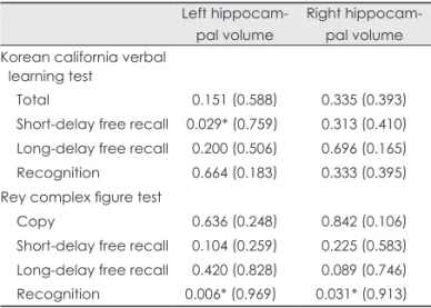
β=-0.669, p<0.001; right HV: β=-0.545, p=0.001) and the arousal index (left HV: β=-0.296, p=0.028; right HV: β=-0.431, p=0.015). A positive correlation was found between short- delay free recall in the K-CVLT and the left HV (r=0.759, p=
0.029) and between recognition in the RCFT and the left (r=0.969, p=0.006) and right (r=0.913, p=0.031) HV in the PIs (Table 4).
The GSs exhibited no significant correlations between HV and any of the other parameters, including demographic characteristics, PSG data, or neuropsychological results.
Discussion
We compared HV and cognitive function between chronic PIs and GSs and investigated the correlation between HV and demographic characteristics and various factors mea- sured from sleep studies and neuropsychological tests.
Table 3. Results of neuropsychological tests in primary insomni- acs and good sleepers
Primary insomniacs vs.
good sleepers Coefficient (SD) p Attention and frontal lobe function
Digit span forward -3.4 (0.8) 0.001*
Digit span backward -2.0 (0.4) 0.001*
Corsi block forward -2.4 (0.7) 0.007*
Corsi block backward -3.8 (0.5) 0.001*
Controlled oral word association test
Semantic word -14.1(3.6) 0.002*
Trail-making test -2.5 (0.6) 0.065
Trail-making test -1.5 (1.1) 0.455
Digit symbol test -1.1 (0.2) 0.325
Rey Complex Figure Test
Copy -3.3 (1.1) 0.001*
Short-delay free recall -6.8 (2.3) 0.001*
Long-delay free recall -6.8 (2.7) 0.002*
Recognition -2.1 (1.1) 0.070
Korean California Verbal Test
Total -7.6 (4.6) 0.122
Short-delay free recall -1.9 (1.2) 0.176 Long-delay free recall -1.9 (1.2) 0.128
Recognition -2.1 (0.9) 0.036*
Korean Boston Naming Test -3.6 (1.9) 0.074
*Multiple regressions, p<0.05.
Table 2. Results of nighttime polysomnography in primary insomniacs and good sleepers
Primary insomniacs Good sleepers p
Total sleep time (min) 359.1 (51.6) 387.5 (42.1) 0.500
Sleep efficiency (%) 83.6 (7.1) 88.0 (5.2) 0.095
Sleep latency (min) 22.9 (9.7) 8.8 (5.6) 0.015*
REM sleep latency (min) 124.9 (69.9) 87.02 (60.2) 0.007*
Apnea-hypopnea index (/hour) 3.3 (3.2) 1.33 (1.1) 0.600
Arousal index (/hour) 13.7 (6.0) 5.2 (3.1) 0.005*
Periodic limb movement during sleep index (/hour) 0.64 (0.6) 1.82 (0.5) 0.125
NREM1 (%) 10.7 (4.9) 13.7 (5.1) 0.250
NREM2 (%) 63.2 (10.0) 54.2 (15.2) 0.060
NREM3 (%) 7.3 (6.6) 7.9 (5.5) 0.085
REM (%) 18.8 (7.1) 24.1 (5.1) 0.040*
Data are mean (SD) values.
*Mann-Whitney U-test, p<0.05.
NREM: nonrapid eye movement, REM: rapid eye movement, SD: standard deviation.
Hippocampal volumetry in chronic PIs
We found no definite difference in ICV and in absolute and ICV-normalized HV between PIs and GSs. However, the du- ration of insomnia and the arousal index in nighttime PSG exhibited significant negative correlations with HV in PIs.
There have been inconsistent reports on HV changes in PIs compared with GSs. One previous pilot study found that HV was significantly smaller in PIs than in GSs,4 but a sub- sequent cross-sectional study did not.5 Several factors could have contributed to this discrepancy. First, the studies used different anatomical landmarks to delineate the boundary of the hippocampus. Riemann et al.4 included the fimbra, the al- veus, and the hippocampus-amygdala transition area (HATA) when determining HV, but these regions were not included in the work of Winkelman et al.5 The present study included
the alveus but excluded the fimbra and the HATA because the fimbra is regarded as white matter and the HATA is not easy to delineate consistently between subjects.
Second, the different subsets of patients examined in the studies may also play a role. The mean duration of insomnia was longer of the study by Reiman et al. (11.6 years) than in our study (7.6 years) and that of Winkelman et al.5 A longer duration of insomnia might negatively influence hippocam- pal function and HV. The finding of no difference in HVs be- tween PIs and GSs in the present study was similar to the findings of Winkelman et al.5 However, our patients reported much more severe insomnia symptoms and a longer duration of disease than did those of Winkelman et al.,5 which is at- tributable to our study involving physician-referred patients with chronic insomnia while the other study selecting pa- tients from community recruits who complained of insomnia symptoms. The mean PSQI (19.1 vs. 12.1) and ISI (19.1 vs.
16.7) seemed to be higher in the present study than in that of Winkelman et al. We observed that left and right HVs in PIs were significantly and negatively correlated with the dura- tion of insomnia, a finding that was not reported by Winkel- man et al.5
Winkelman et al.5 reported that smaller HVs were associ- ated with a greater amount of WASO and a reduced sleep ef- ficiency, as measured by actigraphy. We found that a higher arousal index-which was measured in nighttime PSG-was related to a smaller HV. These findings suggest that sleep quality affects the anatomy of the hippocampus even if the absolute HV did not differ from that of normal controls. It has been shown that reduced sleep efficiency is correlated with higher cortisol levels in the evening and nighttime peri- A
4500
4000
3500
3000
2500
2000
0 5 10 15 20 25
Hippocampal volume (mm3)
Duration of insomnia (year)
Right hippocampus Left hippocampus
B
4500
4000
3500
3000
2500
2000
0 10 20 30 40
Hippocampal volume (mm3)
Arousal index (/hr)
Right hippocampus Left hippocampus
Fig. 1. Scatter plots of left or right hippocampal volume and duration of insomnia and arousal index in patients with chronic insomnia. Higher values on the arousal index correspond to poor sleep quality. Left or right hippocampal volume was negatively correlated with the insomnia du- ration (left: r=-0.872, p<0.001; right: r=-0.868, p<0.001) (A) and with the arousal index in nighttime polysomnography (left: r=-0.435, p=0.045;
right: r=-0.409, p=0.026) (B).
Table 4. Relationship between hippocampal volume and memory scores in primary insomniacs
Left hippocam- pal volume
Right hippocam- pal volume Korean california verbal
learning test
Total 0.151 (0.588) 0.335 (0.393)
Short-delay free recall 0.029* (0.759) 0.313 (0.410) Long-delay free recall 0.200 (0.506) 0.696 (0.165) Recognition 0.664 (0.183) 0.333 (0.395) Rey complex figure test
Copy 0.636 (0.248) 0.842 (0.106)
Short-delay free recall 0.104 (0.259) 0.225 (0.583) Long-delay free recall 0.420 (0.828) 0.089 (0.746) Recognition 0.006* (0.969) 0.031* (0.913) Data are rho (coefficient) values.
*ANCOVA covariate with intracranial volume, p<0.05.
ods.33 Furthermore, patients with Cushing’s disease have been reported to have hippocampal atrophy, with the degree of atrophy being correlated with the mean cortisol level.34 Although serum cortical levels were not obtained during nig- httime in the present study, our findings of a close relation- ship between poor sleep quality and smaller HVs in PIs may support a functional impairment of the hypothalamus-pitu- itary-adrenal gland axis in PIs.
It was well known that HVs are significantly smaller in patients with major depression than in normal controls.35 In the present study, PIs reported more depressive symptoms than GSs (BDI scores: 13.8 vs. 3.5, p=0.04). Although we excluded patients with major depression from the patient group, they might have had dysphoria, which could affect the reduced HVs. However, the BDI score was not correlated with HV in PIs (as assessed by multiple regression analyses).
Cognitive deficits in chronic PIs
Cognition-especially for attention and frontal lobe function- was significantly worse in PIs than in GSs in the present study. Task assessments of shifting attention and working memory generally reveal deficits among insomnia groups.36-40 When a task of sustained attention required a response choice, insomniacs exhibit consistent attention deficits.36-39,41,42 Neu- roimaging data have revealed a close relationship between insomnia and functional and anatomical damage of various brain regions. A significant association was found between insomnia duration and left dorsomedial prefrontal damage in 192 patients with focal brain lesions.43 The gray-matter vol- ume in the left orbitofrontal cortex was found to be a reduced in chronic insomniacs, with this being strongly correlated with the subjective severity of insomnia.44 The cerebral glu- cose metabolic rates during wakefulness in the bilateral pre- frontal and left superior temporal cortices, the thalamus, the hypothalamus, and the brainstem reticular were found to be lower in chronic insomniacs than in controls.45 These find- ings may support the impaired attention and frontal lobe function found in PIs compared to GSs in this study.
Another finding in the present study was that both verbal and nonverbal memory functions were significantly worse in PIs than in GSs. It was previously reported that PIs did not have a performance deficit on tasks of visual and verbal memory tests.46 Some researchers have argued that the word- list and word-pair paradigms used in most studies are not very sensitive to subtle memory deficits.47,48 In the present study, the direct comparison of memory scores with age-, gender-, and education level-matched normal controls, and the significantly positive correlation between HV and verbal and visual memory scores may support objective memory de- ficits in chronic insomniacs.
It is well known that sleep plays an important role in the formation and consolidation of memories49 and that memory consolidation is impaired in patients with chronic insomnia.50 The significantly negative correlation between arousal index and HV in the present study suggests that fragmented sleep is closely related to HV reduction.
A meta-analysis of results from 33 studies had shown wi- dely varying results regarding the relationship between memo- ry performance and HVs in participants without neurological or psychiatric disorders.51 Further study is necessary to reach a consensus regarding the relationship between memory and hippocampal volume in GSs.
Our study was subject to some limitations. PIs have more depressive symptoms than GSs, and their depression or dys- phoria may have affected the neuropsychological results.52 Memory deficit in PIs might be directly influenced by im- paired attention and frontal lobe function53 rather than via hippocampal dysfunction. We are aware that the sample in this study was too small to allow us to draw a definitive con- clusion, but there was a trend toward HV being smaller in PIs than in GSs. Studies with larger samples should evaluate more accurately the effect of insomnia on brain structure.
In summary, we found that HV in PIs was negatively cor- related with the duration of insomnia and the arousal index.
PIs exhibited significantly impaired attention, frontal lobe function, and memory, and their verbal and nonverbal mem- ory scores were positively correlated with HV. These find- ings suggest that chronic sleep deprivation associated with insomnia impairs memory and frontal lobe function, and that insomnia duration and poor sleep quality contribute to a bi- lateral reduction in HV.
Conflicts of Interest
The authors have no financial conflicts of interest.
Acknowledgements
This study was supported by a Grant (2010K000817) from Brain Re- search Center of the 21st Century Frontier Research Program funded by the Ministry of Education, Science, and Technology, and by a Grant of the Korean Health Technology R&D Project, Ministry for Health, Wel- fare & Family Affairs, Republic of Korea (No. A110097), and by the Global Frontier R&D Program on <Human-centered Interaction for Co- existence> funded by the National Research Foundation of Korea grant funded by the Korean Government (MEST) (NRF-M1AXA 003-2011- 0031688).
REFERENCES
1. Roth T, Roehrs T, Pies R. Insomnia: pathophysiology and implica- tions for treatment. Sleep Med Rev 2007;11:71-79.
2. Gould E, Tanapat P. Stress and hippocampal neurogenesis. Biol Psy- chiatry 1999;46:1472-1479.
3. Montaron MF, Drapeau E, Dupret D, Kitchener P, Aurousseau C, Le Moal M, et al. Lifelong corticosterone level determines age-related decline in neurogenesis and memory. Neurobiol Aging 2006;27:645-
4. Riemann D, Voderholzer U, Spiegelhalder K, Hornyak M, Buysse 654.
DJ, Nissen C, et al. Chronic insomnia and MRI-measured hippocam- pal volumes: a pilot study. Sleep 2007;30:955-958.
5. Winkelman JW, Benson KL, Buxton OM, Lyoo IK, Yoon S, O’Connor S, et al. Lack of hippocampal volume differences in pri- mary insomnia and good sleeper controls: an MRI volumetric study at 3 Tesla. Sleep Med 2010;11:576-582.
6. Johnson LC, Chernik DA. Sedative-hypnotics and human perfor- mance. Psychopharmacology (Berl) 1982;76:101-113.
7. Guzman-Marin R, Suntsova N, Methippara M, Greiffenstein R, Szy- musiak R, McGinty D. Sleep deprivation suppresses neurogenesis in the adult hippocampus of rats. Eur J Neurosci 2005;22:2111-2116.
8. Mirescu C, Peters JD, Noiman L, Gould E. Sleep deprivation inhibits adult neurogenesis in the hippocampus by elevating glucocorticoids.
Proc Natl Acad Sci U S A 2006;103:19170-19175.
9. Fortier-Brochu E, Beaulieu-Bonneau S, Ivers H, Morin CM. Insom- nia and daytime cognitive performance: a meta-analysis. Sleep Med Rev 2012;16:83-94.
10. Bird CM, Burgess N. The hippocampus and memory: insights from spatial processing. Nat Rev Neurosci 2008;9:182-194.
11. Kruggel F. MRI-based volumetry of head compartments: normative values of healthy adults. Neuroimage 2006;30:1-11.
12. Lemieux L, Salek-Haddadi A, Krakow K. The nature of MR signal changes. Radiology 2003;226:922-923; author reply 923-925.
13. MacLullich AM, Ferguson KJ, Deary IJ, Seckl JR, Starr JM, Ward- law JM. Intracranial capacity and brain volumes are associated with cognition in healthy elderly men. Neurology 2002;59:169-174.
14. Eritaia J, Wood SJ, Stuart GW, Bridle N, Dudgeon P, Maruff P, et al.
An optimized method for estimating intracranial volume from mag- netic resonance images. Magn Reson Med 2000;44:973-977.
15. Chey J, Na DG, Tae WS, Ryoo JW, Hong SB. Medial temporal lobe volume of nondemented elderly individuals with poor cognitive functions. Neurobiol Aging 2006;27:1269-1279.
16. Tae WS, Kim SS, Lee KU, Nam EC, Kim KW. Validation of hippo- campal volumes measured using a manual method and two automat- ed methods (FreeSurfer and IBASPM) in chronic major depressive disorder. Neuroradiology 2008;50:569-581.
17. Kim JK, Kang Y. Normative study of the Korean-California Verbal Learning Test (K-CVLT). Clin Neuropsychol 1999;13:365-369.
18. Lu PH, Boone KB, Cozolino L, Mitchell C. Effectiveness of the Rey-Osterrieth Complex Figure Test and the Meyers and Meyers recognition trial in the detection of suspect effort. Clin Neuropsychol 2003;17:426-440.
19. Wechsler D. Wechsler Memory Scale--Revised Manual. San Antonio:
The Psychological Corporation, Harcourt Brace Jovanovich, 1987.
20. Zola-Morgan S, Squire LR, Amaral DG. Human amnesia and the medial temporal region: enduring memory impairment following a bilateral lesion limited to field CA1 of the hippocampus. J Neurosci 1986;6:2950-2967.
21. Reitan RM. Trail Making Test: Manual for Administration and Scor- ing. Tucson: Reitan Neuropsychology Laboratory, 1992.
22. Lee YH, Song JY. A study of the reliability and the validity of the BDI, SDS, and MMPI-D scales. Korean J Clin Psychol 1991;10:98- 23. Heaton RK, Chelune GJ, Talley JL, Kay GG, Curtiss G. Wisconsin 113.
Card Sorting Test Manual Revised and Expanded. Odessa, FL: Psy- chological assessment resources, Inc., 1993.
24. Lee JH, Kang YW, Na DL. Efficiencies of stroop interference index- es in healthy older adults and dementia patients. Korean J Clin Psy- chol 2000;19:807-818.
25. Kang YW, Na DL. Seoul Neuropsychological Screening Battery.
Seoul: Haman Brain Research & Consulting, 2003.
26. Raven JC. Coloured Progressive Matrices Sets A, Ab, B. Manual Sections 1 & 2. Oxford: Oxford Psychological Press, 1995.
27. Kim HH, Na DL. Korean Boston Naming Test (K-BNT). Seoul:
Hakjisa, 1997.
28. Free SL, Bergin PS, Fish DR, Cook MJ, Shorvon SD, Stevens JM.
Methods for normalization of hippocampal volumes measured with MR. AJNR Am J Neuroradiol 1995;16:637-643.
29. O’Brien LM, Ziegler DA, Deutsch CK, Kennedy DN, Goldstein JM, Seidman LJ, et al. Adjustment for whole brain and cranial size in volumetric brain studies: a review of common adjustment factors and statistical methods. Harv Rev Psychiatry 2006;14:141-151.
30. Tae WS, Hong SB, Joo EY, Han SJ, Cho JW, Seo DW, et al. Struc- tural brain abnormalities in juvenile myoclonic epilepsy patients:
volumetry and voxel-based morphometry. Korean J Radiol 2006;
7:162-172.
31. Jack CR Jr, Twomey CK, Zinsmeister AR, Sharbrough FW, Petersen RC, Cascino GD. Anterior temporal lobes and hippocampal forma- tions: normative volumetric measurements from MR images in young adults. Radiology 1989;172:549-554.
32. Vgontzas AN, Bixler EO, Lin HM, Prolo P, Mastorakos G, Vela- Bueno A, et al. Chronic insomnia is associated with nyctohemeral activation of the hypothalamic-pituitary-adrenal axis: clinical impli- cations. J Clin Endocrinol Metab 2001;86:3787-3794.
33. Brown ES, Rush AJ, McEwen BS. Hippocampal remodeling and damage by corticosteroids: implications for mood disorders. Neuro- psychopharmacology 1999;21:474-484.
34. Campbell S, Marriott M, Nahmias C, MacQueen GM. Lower hippo- campal volume in patients suffering from depression: a meta-analy- sis. Am J Psychiatry 2004;161:598-607.
35. Edinger JD, Means MK, Carney CE, Krystal AD. Psychomotor per- formance deficits and their relation to prior nights’ sleep among indi- viduals with primary insomnia. Sleep 2008;31:599-607.
36. Varkevisser M, Kerkhof GA. Chronic insomnia and performance in a 24-h constant routine study. J Sleep Res 2005;14:49-59.
37. Altena E, Van Der Werf YD, Strijers RL, Van Someren EJ. Sleep loss affects vigilance: effects of chronic insomnia and sleep therapy. J Sleep Res 2008;17:335-343.
38. Hauri PJ. Cognitive deficits in insomnia patients. Acta Neurol Belg 1997;97:113-117.
39. Edinger JD, Glenn DM, Bastian LA, Marsh GR. Slow-wave sleep and waking cognitive performance II: Findings among middle-aged adults with and without insomnia complaints. Physiol Behav 2000;
70:127-134.
40. Sugerman JL, Stern JA, Walsh JK. Daytime alertness in subjective and objective insomnia: some preliminary findings. Biol Psychiatry 1985;20:741-750.
41. Schneider-Helmert D. Twenty-four-hour sleep-wake function and personality patterns in chronic insomniacs and healthy controls.
Sleep 1987;10:452-462.
42. Koenigs M, Holliday J, Solomon J, Grafman J. Left dorsomedial frontal brain damage is associated with insomnia. J Neurosci 2010;
30:16041-16043.
43. Altena E, Vrenken H, Van Der Werf YD, van den Heuvel OA, Van Someren EJ. Reduced orbitofrontal and parietal gray matter in chron- ic insomnia: a voxel-based morphometric study. Biol Psychiatry 2010;67:182-185.
44. Nofzinger EA, Buysse DJ, Germain A, Price JC, Miewald JM, Kup- fer DJ. Functional neuroimaging evidence for hyperarousal in insom- nia. Am J Psychiatry 2004;161:2126-2128.
45. Shekleton JA, Rogers NL, Rajaratnam SM. Searching for the day- time impairments of primary insomnia. Sleep Med Rev 2010;14:47- 46. Uttl B. Measurement of individual differences: lessons from memory 60.
assessment in research and clinical practice. Psychol Sci 2005;16:
460-467.
47. Uttl B, Graf P, Richter LK. Verbal Paired Associates tests limits on validity and reliability. Arch Clin Neuropsychol 2002;17:567-581.
48. Walker MP, Stickgold R. Sleep-dependent learning and memory con- solidation. Neuron 2004;44:121-133.
49. Backhaus J, Junghanns K, Born J, Hohaus K, Faasch F, Hohagen F.
Impaired declarative memory consolidation during sleep in patients with primary insomnia: Influence of sleep architecture and nocturnal cortisol release. Biol Psychiatry 2006;60:1324-1330.
50. Van Petten C. Relationship between hippocampal volume and mem-
ory ability in healthy individuals across the lifespan: review and me- ta-analysis. Neuropsychologia 2004;42:1394-1413.
51. Gotlib IH, Joormann J. Cognition and depression: current status and future directions. Annu Rev Clin Psychol 2010;6:285-312.
52. Takahashi H, Kato M, Hayashi M, Okubo Y, Takano A, Ito H, et al.
Memory and frontal lobe functions; possible relations with dopamine D2 receptors in the hippocampus. Neuroimage 2007;34:1643-1649.